Replacing an acrylic splint in Saigon requires the precise duplication of your existing occlusal coordinates to maintain temporomandibular joint stability. HCMC Dental Clinic utilizes advanced digital scanning and high-grade PMMA materials to fabricate exact replacements, ensuring continuous protection against bruxism without interrupting your established treatment protocol.
Clinical Summary:
Acrylic splint replacement is a critical maintenance phase in the management of temporomandibular disorders (TMD) and severe bruxism. Over time, hard acrylic orthotics undergo material fatigue, resulting in occlusal pitting, micro-fractures, and a loss of retention. Continuing to wear a compromised splint can alter the vertical dimension of occlusion (VDO), leading to muscle hypertonicity and joint destabilization. The replacement process involves capturing precise intraoral data—either through digital scanning or traditional impressions—to replicate the exact therapeutic jaw posture. Modern dental laboratories in Ho Chi Minh City leverage CAD/CAM technology and premium polymethyl methacrylate (PMMA) to deliver highly accurate, durable duplicate splints, often within a rapid timeframe suitable for international patients.
Key Takeaways:
- Hard acrylic splints degrade over time due to nocturnal grinding forces, requiring periodic replacement to maintain therapeutic efficacy.
- Worn or pitted splints can disrupt neuromuscular balance, causing a resurgence of jaw pain, headaches, and masticatory muscle fatigue.
- Digital replication techniques allow clinicians to copy the exact vertical and horizontal dimensions of your current, comfortable orthotic.
- Same-day or rapid-turnaround duplication services are available for international visitors experiencing a dental emergency in Vietnam.
- Professional clinical evaluation is mandatory to ensure the new splint provides even bilateral contacts and proper anterior guidance.
Signs That Your TMJ Splint Needs Immediate Replacement
An acrylic splint requires immediate replacement when it exhibits loss of retention, visible micro-fractures, or significant occlusal wear that alters your established bite plane. Continuing to wear a compromised device can destabilize the temporomandibular joint and exacerbate muscular tension.
The therapeutic success of an occlusal splint relies entirely on its structural integrity and its ability to provide a frictionless, evenly distributed contact surface for the opposing teeth. Hard acrylic splints, typically fabricated from heat-cured or cold-cured polymethyl methacrylate (PMMA), are designed to withstand significant occlusal forces. However, they are not permanent devices. The constant mechanical stress of nocturnal bruxism, combined with the biochemical environment of the oral cavity, inevitably leads to material degradation. Recognizing the early clinical signs of splint failure is essential to prevent the regression of your temporomandibular joint therapy progress.[1]
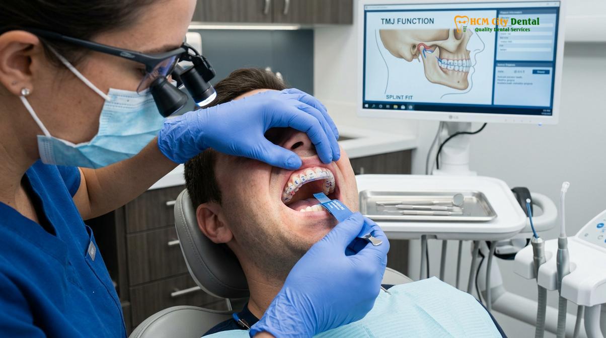
One of the primary indicators that an acrylic splint replacement is necessary is a loss of retention. A properly fitted splint should “snap” into place and remain securely anchored to the maxillary or mandibular arch without rocking or dislodging during sleep. Over time, the internal fitting surface of the acrylic can wear down due to repeated insertion and removal. Furthermore, the natural micro-movements of your teeth over several years can alter the fit. If your splint feels loose, requires excessive tongue pressure to keep in place, or falls out during the night, it is no longer providing the necessary stabilization for your jaw. A loose splint can actually trigger increased muscle activity as the subconscious mind attempts to hold the foreign object in place, entirely defeating the purpose of the appliance.
Visible structural damage is another definitive sign. While PMMA is highly durable, the immense forces generated by the masseter and temporalis muscles—sometimes exceeding 800 Newtons during severe bruxism episodes—can cause micro-fractures within the polymer matrix. These hairline cracks often begin at the margins or in areas of heavy occlusal contact. If left unaddressed, these micro-fractures will propagate, eventually leading to a catastrophic failure of the device. While a temporary broken splint repair might be possible using cold-cure acrylic in an emergency, this is rarely a viable long-term solution. Repairs introduce weak points and can subtly alter the fit and bite plane, making a complete replacement the standard clinical recommendation.[2]
Material discoloration and porosity also dictate the need for replacement. Despite rigorous cleaning protocols, acrylic resins exhibit a degree of water sorption. Over years of use, the material can absorb salivary proteins, pigments, and bacteria, leading to a cloudy, yellowed appearance and a persistent malodor. This porosity not only compromises the hygiene of the device, increasing the risk of dental caries and gingival inflammation, but it also indicates that the cross-linked polymer structure is breaking down, reducing the splint’s overall compressive strength.
“The longevity of an occlusal orthotic is directly proportional to the intensity of the patient’s parafunctional habits. Routine clinical evaluation of the splint’s surface is just as critical as evaluating the patient’s joint health; a degraded splint is often the hidden culprit behind recurring TMD symptoms.”
Finally, patients must monitor the device for significant occlusal wear. A stabilization splint (such as a Michigan splint) is designed to provide simultaneous, bilateral, pinpoint contacts for all opposing teeth, coupled with smooth anterior guidance that disoccludes the posterior teeth during lateral excursions. When the acrylic wears down, these precise contacts are lost. The patient may begin to feel heavier pressure on one side of the jaw, or the smooth gliding motion may be interrupted by deep grooves worn into the plastic. When the occlusal scheme of the splint is compromised, it must be replaced to restore proper neuromuscular balance.
Pitting, Cracking, and Occlusal Wear: The Danger of Uneven Splints
Severe pitting and cracking disrupt the smooth occlusal surface of the splint, forcing the mandible into unnatural compensatory movements during sleep. This uneven wear directly translates to increased mechanical stress on the TMJ capsule and surrounding masticatory muscles.
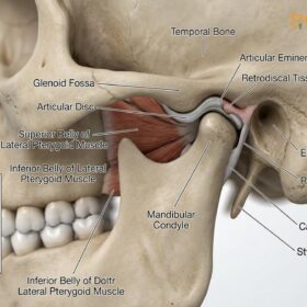
To understand the danger of wearing a worn-out splint, one must understand the biomechanical goals of splint therapy. A hard acrylic splint is not merely a physical barrier to prevent tooth-on-tooth wear; it is a sophisticated orthopedic device designed to reprogram the neuromuscular system. By providing a perfectly smooth, flat surface (in the case of a permissive splint), the appliance removes the proprioceptive feedback from the teeth that dictates the habitual bite. This allows the lateral pterygoid muscles to relax and the mandibular condyles to seat themselves in their most stable, anatomically correct position within the glenoid fossa.[3]
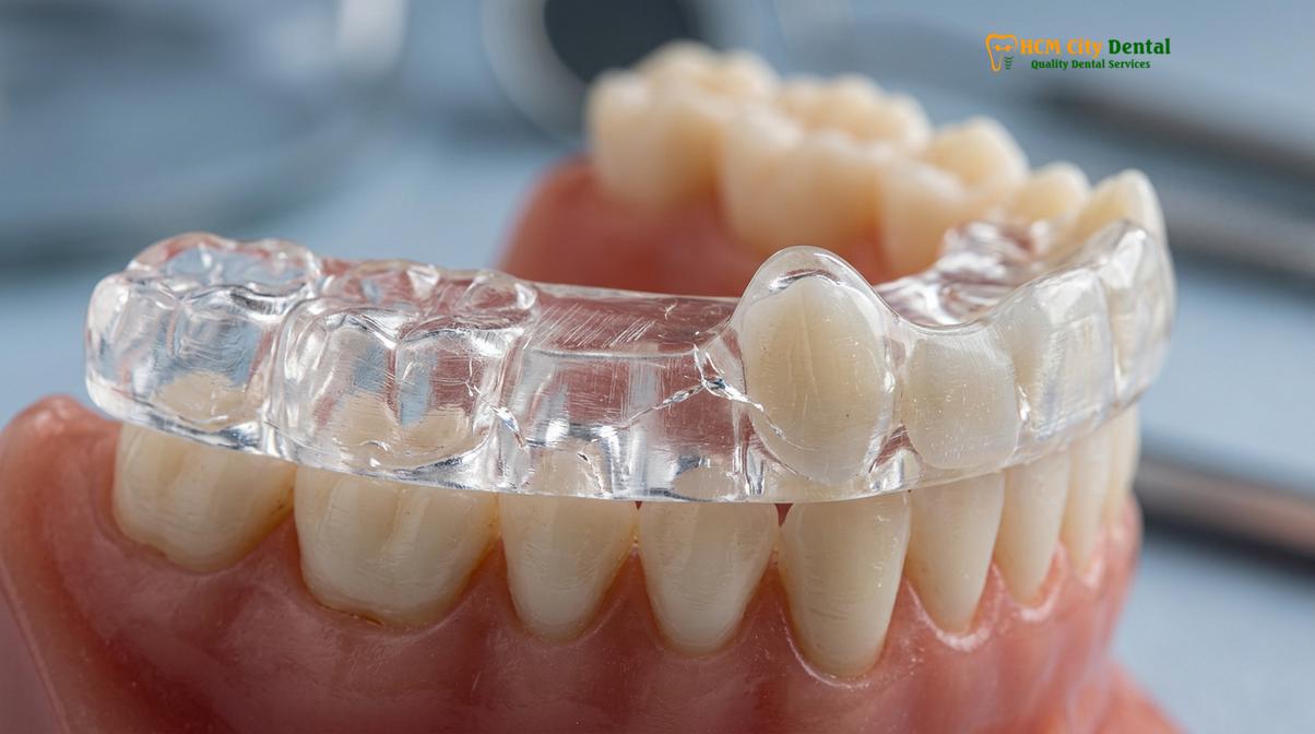
When a splint develops deep pits, grooves, or cracks from years of grinding, it loses its permissive quality. Instead of allowing the lower jaw to glide freely across a smooth surface, the opposing teeth become “locked” into the divots worn into the acrylic. This locking effect is highly detrimental. During sleep, as the brain initiates a bruxism event, the jaw attempts to move laterally. If the teeth are caught in deep acrylic grooves, the masticatory muscles must fire with significantly greater force to overcome the mechanical resistance, or the jaw is forced to function in a restricted, unnatural pathway.
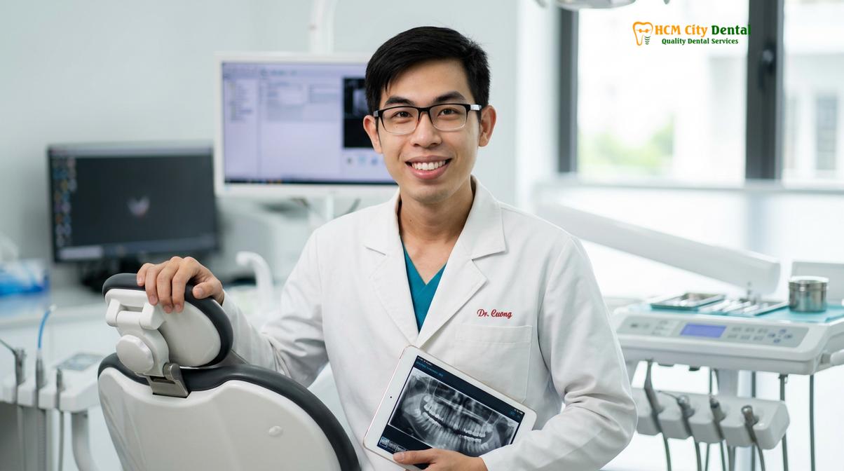
Dr. Nguyen Van Cuong frequently emphasizes to patients that an uneven splint can be more harmful than wearing no splint at all. When the occlusal surface is pitted, it creates premature contacts—areas where the teeth hit the splint before the rest of the arch. These premature contacts act as trigger points, sending hyperactive signals to the central nervous system, which responds by increasing muscle tonus. This phenomenon explains why patients who have worn the same splint for five years suddenly experience a severe recurrence of tension headaches, neck stiffness, and masseter hypertrophy. The splint, once a therapeutic tool, has become a pathogenic factor.
Clinical Warning: Do not attempt to smooth out deep pits or cracks in your acrylic splint using household tools or sandpaper. Altering the occlusal surface without proper articulation and clinical knowledge will destroy the precise vertical dimension of occlusion, potentially causing irreversible shifts in your natural bite and exacerbating joint pathology.
Furthermore, structural cracking alters the distribution of force across the dental arch. A healthy splint dissipates the extreme forces of bruxism evenly across all teeth. A cracked splint flexes under pressure, concentrating the load on isolated teeth. This localized trauma can lead to periodontal ligament inflammation, tooth mobility, and even fractures of the underlying natural dentition or restorative work. For patients undergoing general dentistry services to restore worn dentition, protecting those restorations with a structurally sound, perfectly balanced splint is non-negotiable.[4]
The transition from a smooth, therapeutic surface to a pitted, directive surface happens gradually, which is why many patients fail to notice the degradation until symptoms return. Regular evaluation by a dental professional is required to measure the depth of the wear facets and determine if the splint can be re-equilibrated (adjusted by grinding away a small amount of acrylic to restore a flat surface) or if the material is too thin, necessitating a complete acrylic splint replacement.
How HCMC Dental Replicates Your Existing Orthotic Coordinates
Our clinical team utilizes intraoral scanners and digital articulation to capture the exact vertical and horizontal dimensions of your current orthotic. This ensures the replacement splint provides the identical therapeutic jaw posture without requiring a lengthy adaptation period.
One of the greatest anxieties patients face when replacing a TMJ splint is the fear of losing their “sweet spot”—that precise jaw position that provides maximum relief from pain. A well-adjusted splint represents months or even years of micro-adjustments by your original dentist. Starting from scratch with a new bite registration can sometimes lead to a frustrating period of adaptation. To mitigate this, modern dental protocols focus on exact replication of the existing orthotic coordinates.
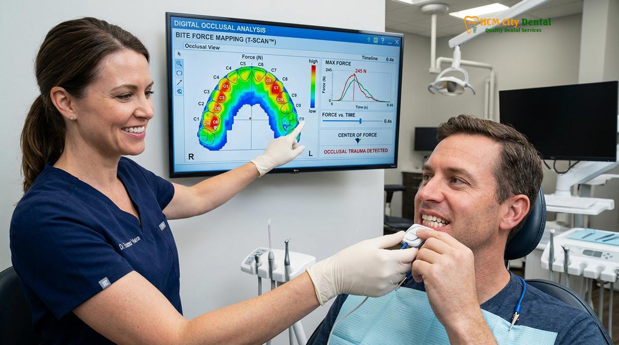
The workflow for replicating an acrylic splint begins with a comprehensive digital capture. If your current splint is structurally intact (not broken into pieces, just worn down), the clinician will use a high-resolution intraoral scanner to create a 3D digital model of your maxillary and mandibular arches, as well as a scan of the splint itself. By scanning the patient’s bite with the splint in place, the software records the exact Vertical Dimension of Occlusion (VDO) and the precise horizontal relationship (Centric Relation or the established therapeutic position). This digital cross-mounting ensures that the new appliance will hold the jaw in the exact same spatial orientation as the old one.
In cases where digital scanning is not feasible, or if the patient presents with a severely fractured appliance, the clinician will rely on highly accurate physical dental impression records using polyvinyl siloxane (PVS) materials. A facebow transfer may be utilized to mount the models on a semi-adjustable articulator, replicating the hinge axis of the patient’s temporomandibular joint. This analog method, while traditional, remains exceptionally precise when executed by a skilled prosthodontist.
Clinical Case Review: A 45-year-old expatriate presented to HCMC Dental Clinic in Ho Chi Minh City with a severely worn Michigan splint that was causing a recurrence of morning jaw locking. Because the patient was highly adapted to the specific vertical height of the old splint, the clinical team utilized a dual-scan technique. The old splint was digitized, and the exact vertical dimension was transferred to CAD software. A new PMMA splint was milled, restoring the smooth occlusal surface while perfectly preserving the patient’s accustomed jaw posture, resulting in immediate symptom resolution without any adaptation phase.
Once the spatial coordinates are captured, the fabrication phase begins. Historically, splints were made using a lost-wax technique and heat-cured PMMA. While this method is still widely used and produces excellent results, digital CAD/CAM (Computer-Aided Design and Computer-Aided Manufacturing) milling has revolutionized the process. The new splint is designed virtually, allowing the technician to perfectly flatten the occlusal plane and establish ideal canine guidance while maintaining the exact internal fit and vertical height of the original device. The splint is then milled from a solid puck of highly cross-linked, pre-polymerized PMMA. Milled splints exhibit superior density, lower porosity, and higher fracture resistance compared to traditional heat-cured acrylics.[5]
For international patients or those who travel frequently, capturing these digital records offers a significant advantage. Once your ideal splint design is saved in the clinic’s database, fabricating a future replacement becomes a streamlined process. In some specialized cases, if a patient loses their splint while abroad, the clinic can utilize the stored digital files to mill a new device and arrange for a direct shipping splint delivery, though a follow-up visit for final occlusal equilibration is always clinically advised to ensure perfect bilateral contacts.
Same-Day Duplicate Splint Services for Tourists in District 1
For international visitors experiencing a dental emergency, our in-house laboratory can fabricate a precise duplicate of a hard acrylic splint within a single day. This rapid turnaround prevents the recurrence of TMJ symptoms while traveling.
Ho Chi Minh City has become a premier destination for medical and dental tourism, attracting patients seeking high-quality care combined with advanced technology. For a tourist managing a temporomandibular disorder, losing or breaking a splint while on vacation is a significant medical emergency. Without the protective orthotic, severe bruxism can return immediately, leading to debilitating jaw pain, restricted opening, and damage to the teeth, effectively ruining the travel experience.
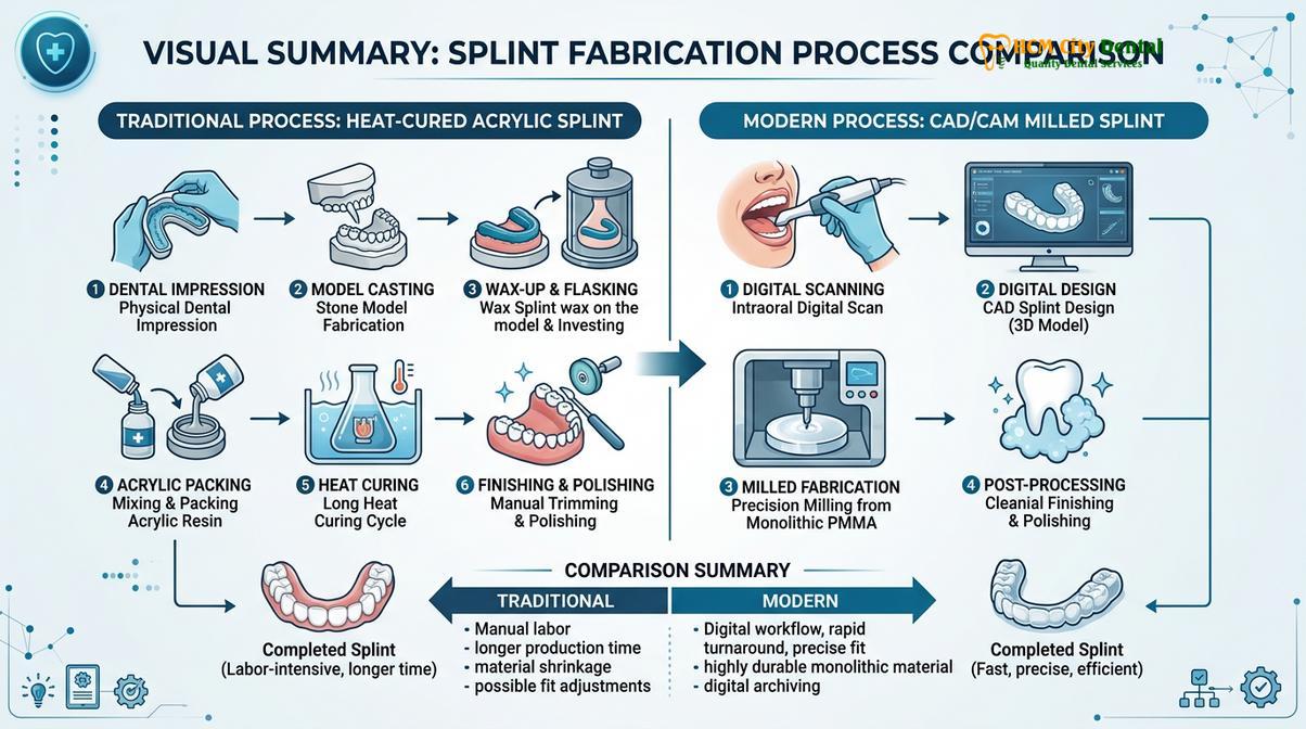
Recognizing this urgent need, specialized clinics equipped with in-house digital laboratories offer rapid-turnaround duplication services. When a patient presents to the clinic in the morning with a broken or lost splint, the clinical team immediately prioritizes the case. If the patient has the broken pieces, they can sometimes be temporarily luted together to scan the original dimensions. If the splint is lost, the clinician will perform a rapid neuromuscular evaluation, capture new digital impressions, and record a new centric relation bite registration.
The advantage of an in-house laboratory is the elimination of transit time between the clinic and an external milling center. The digital files are sent directly to the CAD technician, who designs the appliance within hours. The milling machine then carves the splint from a solid PMMA block, a process that typically takes 60 to 90 minutes. After milling, the splint is hand-polished to a high gloss to prevent bacterial adhesion and ensure comfort against the oral mucosa.
| Fabrication Method | Typical Turnaround Time | Material Quality | Best Suited For |
|---|---|---|---|
| Traditional Heat-Cured (External Lab) | 3 to 7 Days | High (Standard PMMA) | Routine replacements, local residents, complex analog articulations. |
| CAD/CAM Milled (In-House Lab) | 24 Hours (Same-Day) | Premium (High-Density PMMA) | Tourists, dental emergencies, patients requiring exact digital duplication. |
| 3D Printed Resin (In-House Lab) | Same-Day | Moderate to High | Short-term transitional splints, immediate diagnostic orthotics. |
By late afternoon, the patient returns to the clinic for the delivery and final adjustment. Dr. Cuong stresses that even with perfect digital replication, the final occlusal equilibration must be performed intraorally. The patient is asked to tap their teeth together and grind side-to-side on ultra-thin articulating paper. The dentist meticulously adjusts the acrylic until all posterior teeth contact simultaneously and evenly, and the anterior teeth provide smooth guidance without any posterior interferences. This rapid, highly precise service ensures that tourists can continue their journey through Vietnam without the burden of TMJ pain, highlighting the advanced capabilities of routine dental examinations and emergency care available in the city.
When to See a Doctor for Splint Evaluation
An occlusal splint is a prescribed medical device, and its use must be monitored by a qualified dental professional. Patients should not view a splint as a passive piece of plastic, but rather as an active component of their neuromuscular health. It is crucial to seek a clinical evaluation if you experience any of the following red flags.
First, if you notice a sudden return of your original TMD symptoms—such as morning headaches, jaw locking, clicking or popping sounds in the joint, or tenderness in the masseter and temporalis muscles—your splint is likely failing. These symptoms indicate that the device is no longer providing the correct orthopedic support, either due to material wear or a shift in your natural dentition.
Second, if you observe any visible damage to the splint, including deep pits, sharp edges, or hairline cracks, schedule an appointment immediately. Do not wait for the splint to break completely. A compromised splint can cause uneven loading on the temporomandibular joint, potentially leading to disc displacement or degenerative joint changes.
Finally, if you feel that your bite has changed when you take the splint out in the morning—for example, if your back teeth no longer touch, or if your front teeth hit heavier than usual—this is a critical warning sign. While a minor, temporary bite change immediately upon waking can be normal, a persistent change indicates that the splint may be inadvertently acting as an orthodontic device, shifting your teeth into an improper alignment. A comprehensive evaluation at HCMC Dental Clinic will determine if your splint needs to be adjusted, relined, or entirely replaced to protect your long-term oral health.
“Patient compliance is only half the battle in TMD management. The other half is ensuring the therapeutic device remains structurally sound and biomechanically accurate. A worn splint can quickly transition from a protective barrier to a destructive force.”
Frequently Asked Questions
Can I use my old dental mold to make a replacement splint?
No, using an old dental mold is generally not recommended because your dentition and gingival architecture naturally shift over time. A new digital scan or physical impression ensures the replacement splint fits your current oral anatomy perfectly, preventing unwanted orthodontic movement or uneven pressure on the temporomandibular joint. Using outdated records can result in a tight, painful appliance that fails to seat correctly.
What happens if I wear a cracked or worn-out splint?
Wearing a cracked or worn-out splint alters your established bite plane, forcing your jaw muscles to compensate for the uneven surface. This mechanical instability can reignite bruxism, increase masseter muscle tension, and place asymmetrical loading on the TMJ capsule, potentially worsening your original temporomandibular disorder symptoms. It can also lead to localized trauma on specific teeth where the acrylic has thinned.
How much does it cost to duplicate a Michigan splint in Saigon?
The cost to duplicate a Michigan splint in Saigon typically ranges depending on the fabrication method, material quality, and whether digital articulation is required. According to the latest clinic fee schedule, patients can expect highly competitive pricing compared to Western countries, while still receiving premium, medical-grade PMMA materials and precise occlusal calibration. A consultation will provide an exact quote based on your specific clinical needs.
How long does a hard acrylic TMJ splint typically last?
A professionally fabricated hard acrylic TMJ splint typically lasts between two to five years, depending on the severity of your bruxism and your daily maintenance routine. Regular clinical evaluations are necessary to monitor the occlusal surface for micro-fractures, pitting, and material fatigue that compromise the device’s therapeutic efficacy. Patients with extreme parafunctional habits may require replacements more frequently.
Is it possible to repair a broken splint instead of replacing it?
While minor fractures can sometimes undergo temporary broken splint repair, a complete replacement is clinically preferred for long-term stability. Repairs often introduce weak points in the acrylic polymer chain and can subtly alter the vertical dimension of occlusion, which may negatively impact your neuromuscular balance and joint health. A repaired splint should only be viewed as a stopgap measure until a new device can be fabricated.
References
- Journal of the American Dental Association. Occlusal splint therapy for temporomandibular disorders: a clinical review. (2020).
- Journal of Oral Rehabilitation. Efficacy of Michigan repositioning splints in TMD patients. (2019).
- Journal of Prosthetic Dentistry. Computerized occlusal analysis and bite mapping in TMJ therapy. (2021).
- Clinical Oral Investigations. Masseter Botox injections for chronic jaw pain and bruxism. (2018).
- International Journal of Oral and Maxillofacial Surgery. TMJ arthrocentesis and hyaluronic acid injections. (2022).
For personalized advice regarding your custom TMJ orthotics or to schedule a comprehensive evaluation of your current appliance, please contact our clinical team. Our specialists are dedicated to providing advanced preventive dental care and precise restorative solutions tailored to your unique neuromuscular needs.