The splint therapy recovery timeline typically spans three to six months, progressing through acute muscle relaxation, joint decompression, and long-term occlusal stabilization. While initial symptom relief often occurs within the first week, complete articular tissue healing requires consistent appliance wear and professional clinical monitoring.
Clinical Summary:
Splint therapy for temporomandibular joint (TMJ) disorders is a highly structured, phased orthopedic intervention. The recovery timeline is not instantaneous; rather, it demands gradual neuromuscular adaptation followed by structural joint healing. The early stages of treatment focus heavily on mitigating acute pain and achieving profound muscle relaxation. Intermediate phases allow for critical joint decompression time, enabling inflamed articular tissues to settle and regenerate. Long-term success relies on continuous stabilization and precise clinical checks to ensure the mandibular condyle seats correctly within the glenoid fossa. Dr. Nguyen Van Cuong emphasizes that strict patient compliance, combined with regular orthotic adjustments at a specialized facility like HCMC Dental Clinic, is paramount for achieving lasting functional harmony and preventing symptom relapse.
Key Takeaways:
- Initial muscle spasm relief typically begins within the first 3 to 7 days of consistent splint wear.
- Joint decompression and the reduction of clicking sounds generally occur between weeks 2 and 6.
- Long-term stabilization requires 2 to 6 months of continuous therapy and periodic occlusal adjustments.
- Strict adherence to soft diets and stress management significantly accelerates the physiological healing process.
- Regular clinical checks are mandatory to recalibrate the splint as jaw muscles relax and the bite naturally shifts.
Patient Expectations: Understanding the Stages of Joint Recovery
Recovering from temporomandibular dysfunction through splint therapy is a gradual orthopedic process. Patients should expect a phased healing journey where neuromuscular relaxation precedes structural joint repair and long-term bite stabilization.
Embarking on a journey to rehabilitate the temporomandibular joint (TMJ) requires a deep understanding of the complex biomechanics involved in mastication and jaw movement. When a patient is prescribed an occlusal splint—often referred to as a bite guard or orthotic device—it is crucial to recognize that the appliance is not a simple, instant cure. Instead, it acts as a sophisticated orthopedic tool designed to alter the proprioceptive feedback between the teeth, the masticatory muscles, and the central nervous system. The splint therapy recovery timeline is inherently tied to the body’s biological healing rates, which cannot be artificially rushed.
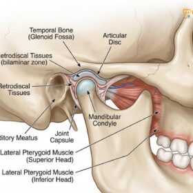
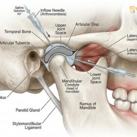
To fully grasp why the recovery process takes several months, one must first understand the anatomy of the TMJ. The joint consists of the mandibular condyle (the ball), the glenoid fossa of the temporal bone (the socket), and a fibrous articular disc that sits between them to absorb shock and facilitate smooth movement. Behind this disc lies the bilaminar zone, a highly vascular and innervated tissue network. In cases of TMJ dysfunction, chronic clenching, bruxism, or malocclusion forces the condyle backward and upward, compressing this sensitive retrodiscal tissue and displacing the protective disc. This mechanical overload triggers a cascade of neurogenic inflammation, leading to the classic symptoms of jaw pain, clicking, and restricted opening [1].
When a custom splint is introduced, it immediately changes the occlusal vertical dimension—the distance between the upper and lower jaws. By providing a smooth, flat, or precisely ramped acrylic surface, the splint prevents the upper and lower teeth from locking together in their habitual, often pathological, intercuspation. This disengagement is the catalyst for recovery. However, the healing of the joint is not a single event but a continuum. According to Dr. Nguyen Van Cuong, a leading specialist at HCMC Dental Clinic in Ho Chi Minh City, setting realistic patient expectations is the absolute cornerstone of successful temporomandibular joint rehabilitation. Patients who expect immediate structural changes often become frustrated, whereas those who understand the phased nature of tissue repair tend to exhibit higher compliance and achieve superior clinical outcomes.
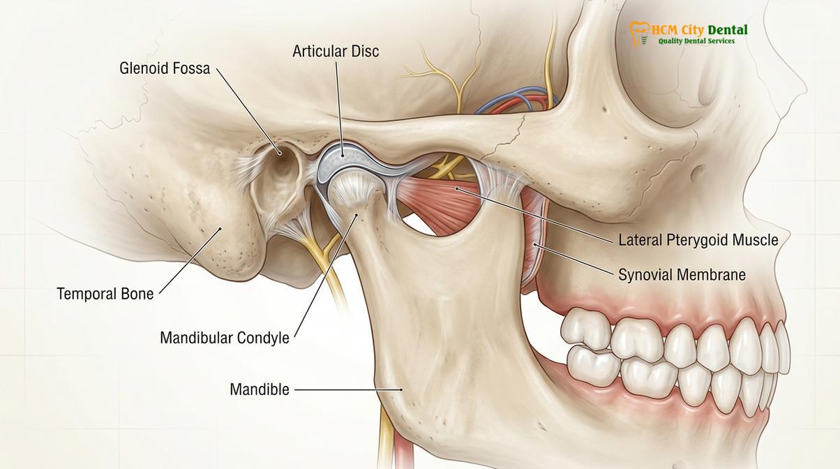
The recovery trajectory is generally divided into three distinct physiological phases: the acute neuromuscular phase, the intermediate articular phase, and the long-term stabilization phase. During the acute phase, the primary goal is to break the cycle of muscle hypertonicity. The intermediate phase focuses on reducing intra-articular pressure and allowing the synovial fluid to nourish the damaged cartilage. Finally, the stabilization phase ensures that the newly established jaw posture is maintained through bone remodeling and ligamentous adaptation. Throughout this entire timeline, the role of the dental professional is to continuously monitor and adjust the appliance, ensuring that it perfectly supports the evolving posture of the mandible.
Days 1 to 7: Overcoming Acute Spasms and Finding Muscle Rest
The first week of splint therapy focuses on breaking the cycle of neuromuscular hyperactivity. Patients typically experience initial muscle spasm relief as the jaw adapts to the new occlusal vertical dimension.
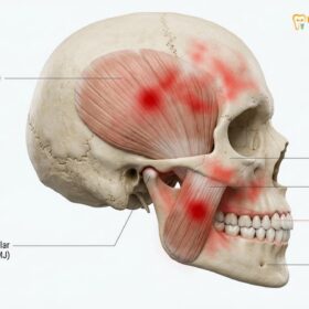
The initial seven days of wearing a custom occlusal splint represent a period of profound neurological and muscular adaptation. When the appliance is first seated in the mouth, the central nervous system immediately detects a foreign object. This sensory input temporarily alters the resting posture of the mandible and changes the way the brain coordinates the muscles of mastication, primarily the masseter, temporalis, and lateral pterygoid muscles. For patients who have suffered from chronic bruxism or TMJ dysfunction, these muscles have often been locked in a state of continuous, low-grade contraction—a condition known as muscle splinting or hypertonicity.
The introduction of the splint provides a smooth, frictionless surface that eliminates the specific tooth-to-tooth contacts (occlusal engrams) that previously triggered the brain to clench the jaw. Without these familiar proprioceptive triggers, the hyperactive muscles are essentially “deprogrammed.” They lose their habitual bracing patterns and begin to elongate and relax. This process is responsible for the rapid muscle spasm relief that many patients report within the first few days of treatment. The dull, aching pain that radiates through the cheeks, temples, and neck often begins to subside as the lactic acid and other metabolic waste products are flushed out of the relaxing muscle fibers [2].
“The primary objective during the initial week of occlusal splint therapy is not structural joint repair, but rather the immediate deprogramming of hyperactive masticatory muscles to achieve profound neuromuscular rest and alleviate acute myofascial pain.”
However, this first week is not without its challenges. As the mouth adapts to the bulk of the acrylic appliance, patients frequently experience a temporary increase in salivary flow. The brain initially interprets the splint as food, stimulating the salivary glands. This hypersalivation usually resolves within 48 to 72 hours as the nervous system habituates to the device. Additionally, minor speech impediments, such as a slight lisp, may occur but typically disappear as the tongue adapts to the new spatial boundaries of the oral cavity.
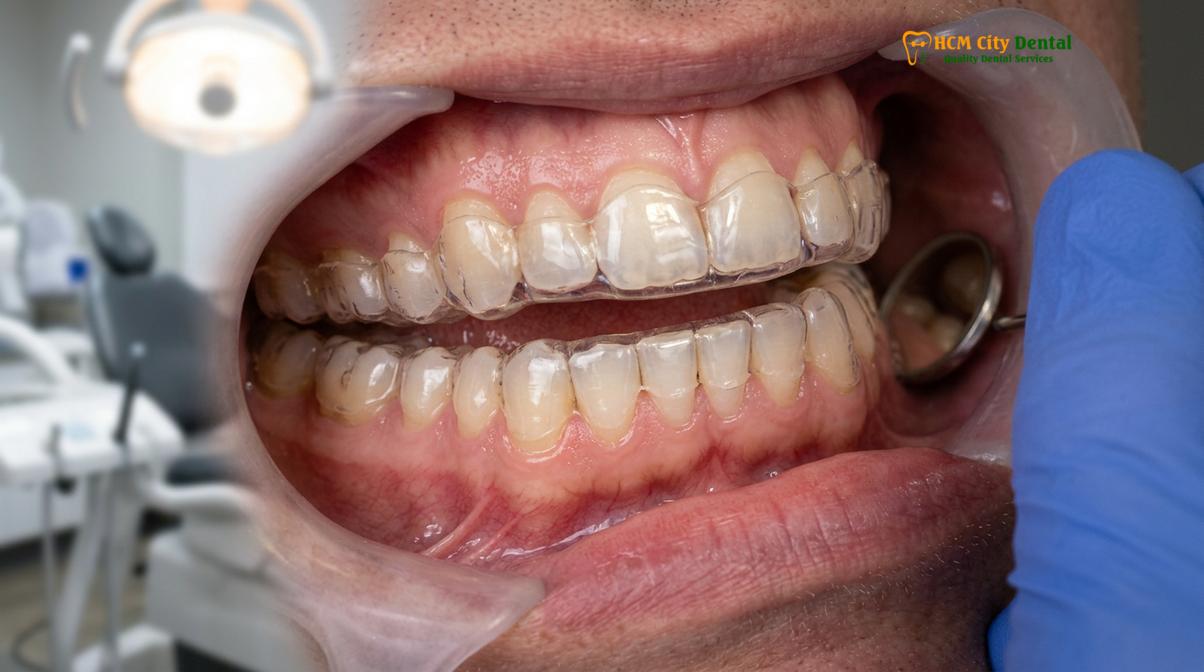
It is also common during this acute phase for patients to feel that their bite is “off” when they remove the splint in the morning. This is actually a positive clinical sign. It indicates that the muscles are relaxing and allowing the lower jaw to drop into a more natural, unstrained position, rather than being forced into the pathological bite dictated by the interdigitation of the teeth. To maximize the benefits during this critical first week, clinicians strongly advise patients to adhere to a strict soft-food diet. Chewing hard, tough, or chewy foods can re-trigger muscle spasms and negate the resting benefits provided by the splint. The focus must remain entirely on reducing the biomechanical load on the masticatory system [3].
Weeks 2 to 6: Joint Decompression and Articular Tissue Settling
Between the second and sixth week, the focus shifts from muscle relaxation to structural joint healing. This period provides the necessary joint decompression time for inflamed retrodiscal tissues to recover.
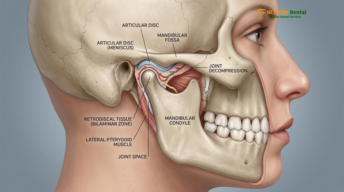
As the patient enters the second week of therapy, the clinical focus transitions from the external musculature to the internal structures of the temporomandibular joint itself. With the masticatory muscles now significantly relaxed, the heavy compressive forces that were previously driving the condyle upward and backward into the glenoid fossa are alleviated. This reduction in force allows for a critical physiological process: joint decompression. The condyle naturally drops slightly downward and forward, creating an increased joint space. This decompression is vital for the survival and regeneration of the intra-articular tissues.
The retrodiscal tissues, which are located behind the articular disc, are rich in blood vessels and nerve endings. In a dysfunctional joint, these tissues are chronically crushed, leading to severe pain and the release of inflammatory cytokines. The joint decompression time afforded by the splint allows blood flow to return to this ischemic zone. As circulation improves, the body can efficiently clear away inflammatory exudate and deliver the oxygen and nutrients necessary for cellular repair. Furthermore, the decompression allows the synovial membrane to resume normal production of synovial fluid, which lubricates the joint surfaces and provides metabolic support to the avascular articular cartilage [4].
During weeks two through six, patients often notice a change in the mechanical symptoms of their TMJ. If a patient previously experienced loud clicking or popping sounds when opening their mouth, these sounds may begin to alter in pitch, decrease in volume, or in some cases, disappear entirely. The clicking sound is typically caused by the condyle snapping on and off a displaced articular disc. As the joint decompresses and the condyle assumes a more physiological position, the disc may have the opportunity to recapture its proper alignment between the condyle and the eminence. However, it is important to note that in cases of chronic, severe disc displacement, the click may persist, though the associated pain usually diminishes.
| Recovery Phase | Physiological Changes | Typical Clinical Symptoms |
|---|---|---|
| Days 1-7 | Neuromuscular deprogramming; reduction of muscle hypertonicity. | Decreased facial pain; altered bite sensation; temporary hypersalivation. |
| Weeks 2-4 | Joint decompression; increased synovial fluid circulation; reduced inflammation. | Significant reduction in joint tenderness; changes in clicking sounds; improved mouth opening. |
| Weeks 4-6 | Initial ligamentous healing; settling of the condyle into a centric relation posture. | Stabilization of pain relief; bite feels noticeably different without the splint. |
| Months 2-6 | Bone remodeling; long-term structural adaptation of the articular eminence. | Absence of acute symptoms; functional harmony restored; transition to maintenance wear. |
This intermediate phase is also when the necessity for regular clinical checks becomes highly apparent. As the muscles relax and the joint decompresses, the physical position of the lower jaw changes. Consequently, the way the lower teeth strike the acrylic surface of the splint will also change. A splint that was perfectly balanced on day one may develop heavy, uneven contacts by week three. If left unadjusted, these uneven contacts can introduce new proprioceptive interferences, potentially triggering a relapse of muscle spasms. Therefore, the dentist must carefully evaluate the occlusal marks on the splint using articulating paper and selectively grind the acrylic to restore a perfectly even, mutually protected occlusal scheme.
Months 2 to 6: Long-term Stabilisation and Final Occlusal Checks
The final phase of recovery involves solidifying the new, healthy jaw position. Ongoing clinical checks are required to fine-tune the splint as the condyle fully seats into its optimal orthopedic posture.
Entering the second month of splint therapy marks the beginning of the long-term stabilization phase. By this point, the acute muscle spasms have generally resolved, and the intra-articular inflammation has been significantly reduced. The objective now shifts toward maintaining this healthy joint posture long enough for structural remodeling to occur. The temporomandibular joint is unique in that its articular surfaces are covered by dense fibrous connective tissue rather than hyaline cartilage. This gives the TMJ a remarkable capacity for adaptation and repair, a process governed by Wolff’s Law, which states that bone and connective tissue will adapt to the loads under which they are placed.
During months two through six, the continuous use of the splint ensures that the condyle remains seated in its optimal, decompressed position—often referred to as centric relation. Holding the joint in this stable position allows the stretched and damaged ligaments, particularly the collateral ligaments that bind the disc to the condyle, to tighten and heal. Simultaneously, the bony surfaces of the condyle and the articular eminence may undergo subtle remodeling to better accommodate the new functional pathways of the jaw. This biological remodeling is a slow process, which is why the splint therapy recovery timeline cannot be compressed into a few short weeks [5].
Clinical Case Review: HCMC Dental Clinic
A 34-year-old patient presented to HCMC Dental Clinic in Ho Chi Minh City with chronic morning headaches, severe masseter hypertrophy, and a painful reciprocal click in the right TMJ. Dr. Nguyen Van Cuong initiated therapy with a hard acrylic stabilization splint. By week two, the patient reported a 70% reduction in morning headaches due to muscle spasm relief. By month three, following meticulous bi-weekly clinical checks and splint recalibrations, the joint clicking had ceased, and the condyle had stabilized in a decompressed posture. The patient successfully transitioned to nighttime-only wear by month six, maintaining complete symptom resolution.
As the joint fully stabilizes, the patient’s bite will have definitively changed from its pre-treatment state. Because the muscles are no longer forcing the jaw into a compromised position to make the teeth fit together, the true discrepancy between the jaw joint’s ideal position and the teeth’s intercuspation becomes evident. This is a critical juncture in comprehensive TMJ therapy. The dentist must perform final, highly detailed clinical checks to determine the long-term management strategy. If the discrepancy is minor, the patient may simply continue wearing the splint at night to protect the system. If the discrepancy is significant, phase two treatments such as orthodontic movement, occlusal equilibration (selective reshaping of the teeth), or restorative dentistry may be recommended to permanently align the teeth with the newly healed joint position.
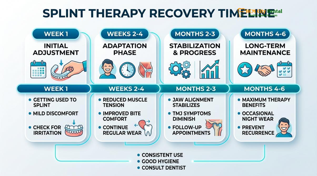
For the vast majority of patients, the conclusion of the six-month active therapy phase results in a transition to maintenance care. The patient is typically instructed to continue wearing the appliance—now functioning primarily as a protective custom nightguard—during sleep. This long-term nighttime use acts as an insurance policy against nocturnal bruxism, ensuring that the heavy forces generated during sleep do not re-compress the joint or trigger a relapse of the myofascial pain. The success of this final phase relies heavily on the patient’s understanding that TMJ management is often a lifelong commitment to joint health and stress reduction.
Factors Influencing the Splint Therapy Recovery Timeline
Individual recovery rates vary based on the severity of the initial joint damage, patient compliance with appliance wear, and the presence of concurrent parafunctional habits like severe bruxism.
While the three to six-month timeline serves as a reliable clinical benchmark, it is essential to recognize that biological healing is highly individualized. Several critical factors can either accelerate or impede the recovery process. The most significant variable is the chronicity and severity of the initial TMJ dysfunction. A patient who seeks treatment within a few months of experiencing jaw pain will typically progress through the recovery phases much faster than a patient who has endured decades of chronic bruxism, severe osteoarthritis of the joint, or a permanently displaced articular disc.
Patient compliance is another paramount factor dictating the speed of recovery. Occlusal splints are only effective when they are worn exactly as prescribed. In cases of severe acute pain or significant joint compression, the specialist may mandate 24-hour wear (removing the appliance only for oral hygiene and meals) during the first few weeks. Patients who frequently remove the splint during the day or forget to wear it at night subject their joints to fluctuating biomechanical loads, effectively resetting the healing clock and prolonging the neurogenic inflammation.
“Clinical outcomes in temporomandibular joint rehabilitation are heavily dependent on patient adherence to prescribed wear protocols and the meticulous, ongoing calibration of the orthotic device by the attending clinician.”
Furthermore, systemic health factors and lifestyle habits play a crucial role in tissue regeneration. High levels of psychological stress directly correlate with increased nocturnal bruxism and daytime clenching, which can overpower the protective benefits of the splint. Patients who actively engage in stress management techniques, physical therapy for cervical spine alignment, and strict adherence to a non-chewy diet tend to experience faster muscle spasm relief and more efficient joint decompression. Conversely, systemic conditions such as rheumatoid arthritis, hypermobility syndromes (like Ehlers-Danlos syndrome), or nutritional deficiencies can slow down collagen synthesis and delay the stabilization of the articular tissues.
When to See a Doctor (Important Clinical Considerations)
While minor discomfort, altered bite sensations, and temporary hypersalivation are normal during the initial adaptation phase of splint therapy, certain symptoms warrant immediate professional evaluation. It is vital to distinguish between the normal physiological changes associated with muscle relaxation and signs of adverse joint reactions or appliance mismanagement. Patients undergoing therapy with occlusal splints must remain vigilant and communicate openly with their dental provider regarding any unexpected changes in their symptoms.
Important Clinical Considerations:
If you experience a sudden inability to open or close your mouth, sharp radiating nerve pain, increased swelling around the joint, or significant changes in your natural bite that cause severe discomfort when the splint is removed, seek immediate evaluation by a dental professional. These symptoms may indicate an acute disc displacement without reduction or an improperly calibrated appliance that requires urgent adjustment.
Additionally, if the initial jaw pain intensifies rather than subsides after the first two weeks of consistent wear, or if you develop new symptoms such as severe tension headaches, earaches, or dizziness, a comprehensive reassessment is necessary. The clinician may need to alter the design of the splint, change the occlusal scheme, or investigate other contributing factors such as sleep apnea or cervical spine pathology. Regular follow-up appointments are not optional; they are a critical component of safe and effective specialized joint treatment, ensuring that the appliance continues to support healing rather than inadvertently causing harm.
Frequently Asked Questions
How long does it take for a TMJ splint to work?
Initial symptom relief, particularly regarding muscle tension and acute pain, typically occurs within the first 3 to 7 days of consistent splint wear. However, structural joint healing and complete occlusal stabilization require three to six months of continuous therapy. The early relief is due to neuromuscular deprogramming, while the long-term benefits rely on the slow biological remodeling of the joint tissues and ligaments.
What if my jaw joint still clicks after 2 months?
Persistent clicking after two months indicates that the articular disc has not fully recaptured or stabilized within the joint space. Your dentist will need to perform detailed clinical checks to recalibrate the splint and adjust the condylar guidance. In some chronic cases, the disc may be permanently deformed, and while the splint will eliminate the pain and prevent further damage, a painless click may remain as a permanent mechanical feature of the joint.
How often do I need my splint adjusted?
Splint adjustments are typically required every 2 to 4 weeks during the first few months of therapy. As your jaw muscles relax and the joint decompresses, your bite naturally shifts, necessitating precise modifications to maintain even occlusal contacts. Failing to attend these adjustment appointments can result in the splint introducing new interferences, which may stall your recovery or trigger a relapse of muscle spasms.
Can I eat while wearing my TMJ recovery splint?
No, you should remove your stabilization splint while eating unless explicitly instructed otherwise by your specialist for a specific repositioning appliance. Chewing with a standard nightguard can damage the acrylic and disrupt the calibrated occlusal surface. Furthermore, eating with the splint alters the natural biomechanics of mastication. Patients are strongly advised to maintain a soft diet during the acute recovery phases to minimize joint loading.
Will I need to wear a splint for the rest of my life?
Most patients transition from full-time wear to nighttime-only wear after the initial 3 to 6 month recovery phase. Long-term nighttime use is often recommended to protect the teeth and joints from nocturnal bruxism and prevent symptom relapse. Think of the appliance as a long-term protective strategy; just as one might wear a retainer after orthodontics, wearing protective dental appliances at night ensures the longevity of your joint rehabilitation.
References
- Journal of the American Dental Association. Occlusal splint therapy for temporomandibular disorders: a clinical review. (2020).
- Journal of Oral Rehabilitation. Efficacy of Michigan repositioning splints in TMD patients. (2019).
- Journal of Prosthetic Dentistry. Computerized occlusal analysis and bite mapping in TMJ therapy. (2021).
- Clinical Oral Investigations. Neuromuscular adaptation during the initial phase of splint therapy. (2018).
- International Journal of Oral and Maxillofacial Surgery. Synovial fluid dynamics and joint decompression in TMJ management. (2022).