Bite analysis for TMJ in Saigon involves evaluating how your upper and lower teeth meet to identify occlusal imbalances causing jaw pain. By utilizing advanced digital mapping, specialists can pinpoint premature contacts and adjust the bite, relieving stress on the temporomandibular joint and surrounding masticatory muscles.
Clinical Summary:
Temporomandibular joint (TMJ) disorders frequently stem from occlusal discrepancies, where an uneven bite forces the jaw muscles and joints into unnatural, strained positions. Comprehensive bite analysis is the diagnostic cornerstone for identifying these micro-interferences. While traditional methods rely on articulating paper to mark contact points, modern clinical protocols utilize computerized occlusal analysis to measure the exact force and timing of each tooth contact. By pinpointing premature contacts, clinicians can perform precise occlusal adjustments or design custom orthotic splints to decompress the joint. This evidence-based approach not only alleviates chronic facial pain and headaches but also prevents long-term joint degeneration and dental fractures, ensuring optimal stomatognathic function and long-term oral health stability.
Key Takeaways:
- Malocclusion and uneven bite forces are primary catalysts for temporomandibular joint inflammation and chronic masticatory muscle pain.
- Digital bite mapping captures real-time occlusal force and timing, surpassing the limitations of traditional diagnostic methods.
- Identifying and eliminating premature tooth contacts can immediately reduce muscle hyperactivity and joint compression.
- Non-invasive occlusal splint therapy is often the first line of defense before permanent bite adjustments are considered.
- Early diagnostic intervention prevents irreversible cartilage damage and severe dental attrition.
How an Uneven Bite Triggers TMJ Inflammation
An uneven bite forces the jaw muscles to overcompensate during chewing and swallowing, leading to chronic muscle fatigue, joint compression, and localized inflammation within the TMJ capsule.
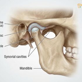
The stomatognathic system is a highly complex network comprising the teeth, the masticatory muscles, and the temporomandibular joints. For this system to function harmoniously, the upper and lower teeth must interlock seamlessly when the jaw closes. This ideal alignment allows the condyle—the rounded end of the lower jawbone—to rest comfortably within the glenoid fossa of the skull, cushioned by a cartilaginous articular disc. However, when an occlusal discrepancy exists, this delicate biomechanical balance is disrupted, initiating a cascade of pathological changes that culminate in temporomandibular joint disorder (TMD)[1].
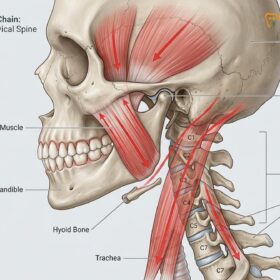
When the teeth do not fit together correctly, the brain receives proprioceptive feedback from the mechanoreceptors located in the periodontal ligaments surrounding the tooth roots. To avoid the discomfort of an uneven collision, the central nervous system automatically alters the chewing pattern. This subconscious adaptation forces the lateral pterygoid, masseter, and temporalis muscles to work asymmetrically and continuously, even during rest. Over time, this constant state of hyperactivity leads to muscle fatigue, lactic acid buildup, and severe myofascial pain.
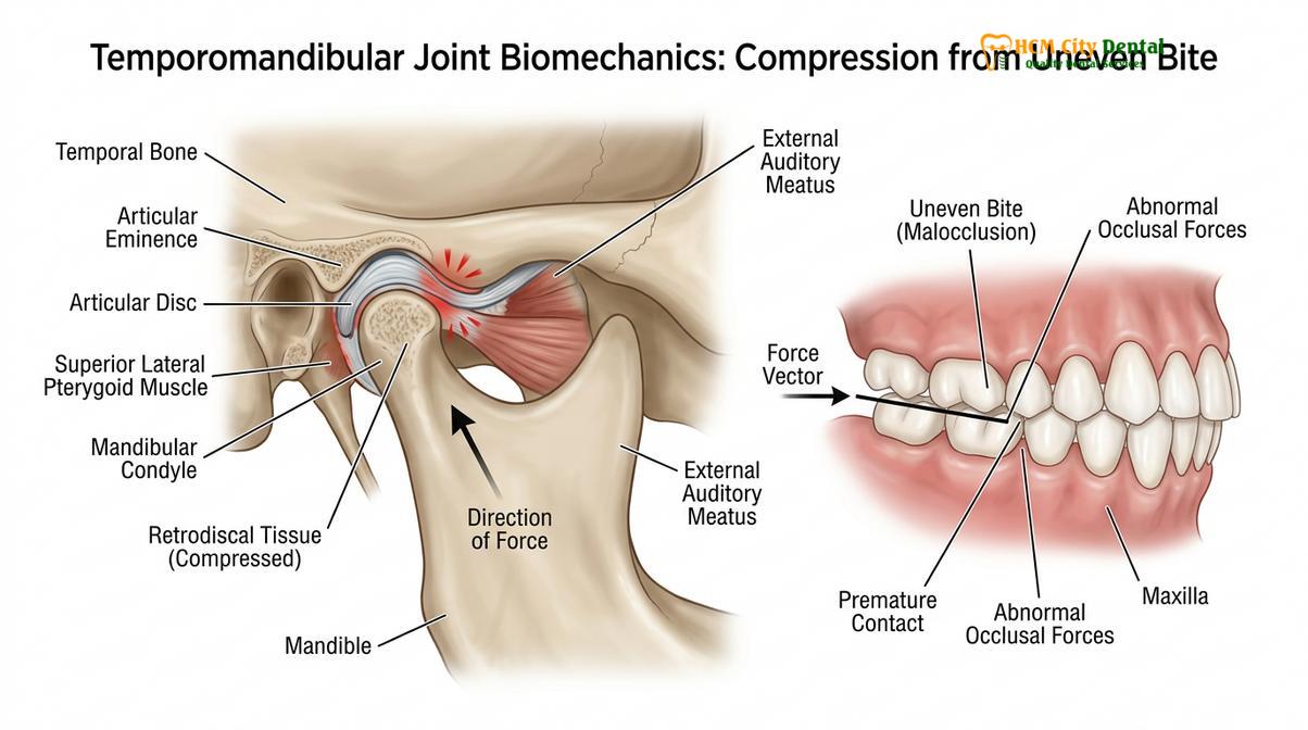
Furthermore, this muscular overcompensation physically alters the position of the jaw joint. As the muscles pull the mandible into an unnatural position to achieve maximum dental contact, the condyle is often forced backward and upward. This displacement compresses the highly vascularized and innervated retrodiscal tissue located behind the joint, triggering acute inflammation and pain. Dr. Nguyen Van Cuong, a leading specialist in restorative dentistry, frequently observes that patients often adapt to minor bite shifts for years, only seeking comprehensive temporomandibular joint therapy when the compensatory mechanisms finally fail and severe, radiating pain sets in.
“The temporomandibular joint is entirely at the mercy of the teeth and the muscles. If the bite is unstable, the muscles will fight to stabilize it, and the joint will ultimately bear the destructive physical burden of that continuous battle.”
Chronic inflammation within the joint capsule can eventually lead to the degradation of the synovial fluid, which normally lubricates the joint. Without adequate lubrication, the articular disc may become displaced, resulting in the characteristic clicking, popping, or grating sounds (crepitus) associated with advanced TMD. Addressing the root cause through precise bite analysis is therefore critical to halting this degenerative cycle.
What is Premature Contact and How Does It Strain the Joint?
Premature contact occurs when one or more teeth meet before the rest of the dental arch during closure, creating a fulcrum effect that destabilizes the jaw joint and triggers muscle spasms.
In a healthy, balanced occlusion, all posterior teeth should contact simultaneously and with equal intensity when the jaw closes into centric occlusion. This simultaneous contact distributes the immense forces of mastication evenly across the dental arch, protecting individual teeth and stabilizing the jaw joints. However, when a specific tooth or a high dental restoration strikes its opposing counterpart before the other teeth touch, it is clinically defined as having premature contact teeth.
The physics of a premature contact are highly destructive. Even a microscopic interference—measuring just a few microns—can act as a mechanical fulcrum. When the jaw closes and hits this high spot, the mandible is forced to slide or pivot off the interference to allow the rest of the teeth to come together. This micro-slide forces the condyle out of its physiologically stable position within the joint socket. The constant shifting strains the ligaments that hold the articular disc in place, gradually stretching them and leading to disc displacement[2].
Clinical Warning: Ignoring the sensation of a “high bite” after receiving a new filling or crown can lead to rapid onset TMJ inflammation. If your teeth do not feel like they fit together naturally after a dental procedure, prompt occlusal evaluation is strongly recommended.
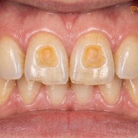
Premature contacts are particularly damaging during excursive movements—when the jaw slides side-to-side or forward during chewing or nighttime grinding (bruxism). In a healthy bite, the anterior teeth (canines and incisors) should guide these movements, smoothly disengaging the posterior teeth to protect them from lateral forces. If a posterior tooth interferes during these lateral movements (a working or non-working interference), it subjects the tooth to severe lateral stress, increasing the risk of enamel micro-fractures, abfraction lesions at the gumline, and periodontal ligament trauma.
The neurological response to premature contact teeth is equally significant. The brain perceives the high spot as a noxious stimulus. In an attempt to grind away the interference and level the bite, the central nervous system may trigger nocturnal bruxism. This involuntary clenching and grinding exponentially increases the load on the temporomandibular joint, exacerbating cartilage wear and muscle hypertrophy. Identifying and eliminating these interferences is a primary objective of advanced occlusal therapy.
Digital Occlusal Analysis vs. Traditional Articulating Paper
While traditional articulating paper only shows where teeth touch, digital occlusal analysis reveals the exact timing and force of those contacts, providing a dynamic, measurable map of your bite.
For decades, the standard method for evaluating a patient’s bite has been the articulating paper test. In this traditional approach, the dentist asks the patient to bite down and grind side-to-side on a piece of carbon-coated paper. The paper leaves colored ink marks on the occlusal surfaces of the teeth wherever contact occurs. The clinician then visually inspects these marks, assuming that larger or darker marks indicate areas of higher force.
However, extensive clinical research has demonstrated that the articulating paper test is fundamentally qualitative, not quantitative. The size and darkness of an ink mark are influenced by numerous variables, including the thickness of the paper, the presence of saliva, the morphology of the tooth, and the angle of contact. A broad, dark mark may simply represent a large surface area of light contact, while a tiny, faint mark could represent a highly destructive, high-force pinpoint interference. Relying solely on visual interpretation can lead to subjective and sometimes inaccurate occlusal adjustments.
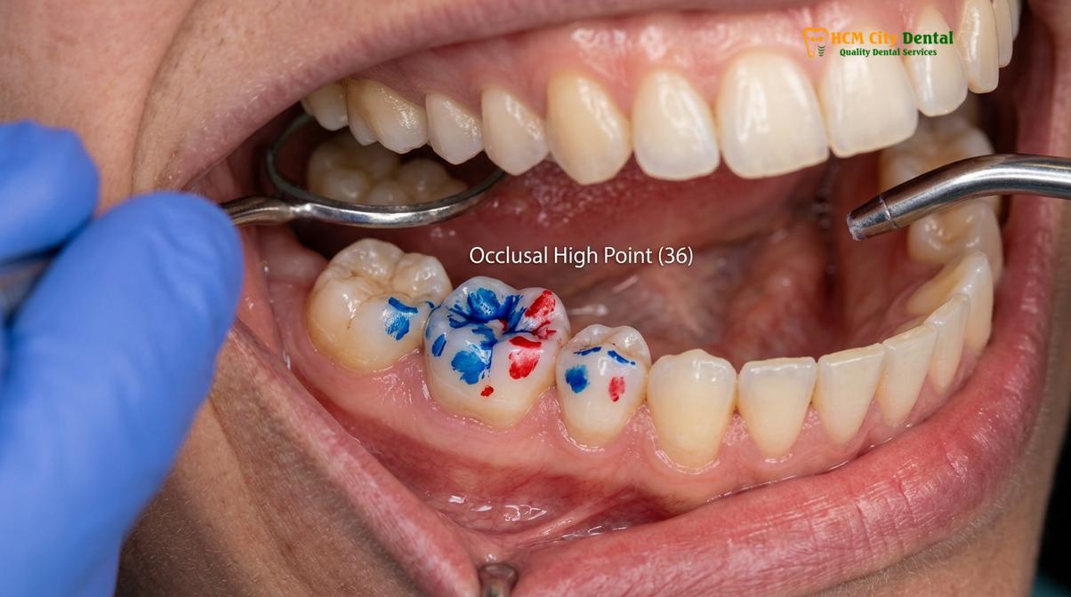
To overcome these limitations, modern specialized clinics employ computerized digital occlusal analysis. This advanced technology utilizes an ultra-thin, horseshoe-shaped electronic sensor connected to sophisticated software. As the patient bites down on the sensor, the system records the exact sequence, timing, and force of every tooth contact in real-time, typically capturing data at 100 frames per second[3].
| Diagnostic Feature | Traditional Articulating Paper | Digital Occlusal Analysis |
|---|---|---|
| Force Measurement | Cannot measure force; relies on visual estimation of ink marks. | Precisely quantifies relative force percentages for every tooth. |
| Timing of Contact | Cannot determine which teeth hit first or last. | Records the exact millisecond each tooth makes and breaks contact. |
| Data Type | Static, qualitative ink marks. | Dynamic, quantitative 3D digital video playback. |
| Clinical Accuracy | Subject to human interpretation and salivary interference. | Highly objective, data-driven, and reproducible. |
The digital interface displays a dynamic 3D bar graph, allowing the dentist to see exactly where the bite forces are concentrated. It reveals the “center of force” trajectory, showing how the balance of the bite shifts from the moment the first tooth touches until maximum intercuspation is achieved. This level of precision is invaluable for diagnosing complex TMD cases, as it eliminates the guesswork associated with traditional methods and allows for highly targeted, conservative treatments.
Computerized Bite Mapping Protocols at HCMC Dental
Our clinical workflow integrates 3D imaging with dynamic digital bite mapping to precisely diagnose occlusal discrepancies and formulate targeted, conservative TMJ treatment plans.
At HCMC Dental Clinic, the approach to diagnosing and managing temporomandibular joint disorders is rooted in comprehensive, data-driven analysis. The protocol for evaluating dental occlusion HCMC standards requires a multi-disciplinary perspective, combining structural imaging with dynamic functional assessment. This ensures that every aspect of the stomatognathic system is evaluated before any irreversible treatment is initiated.
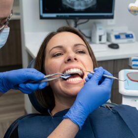
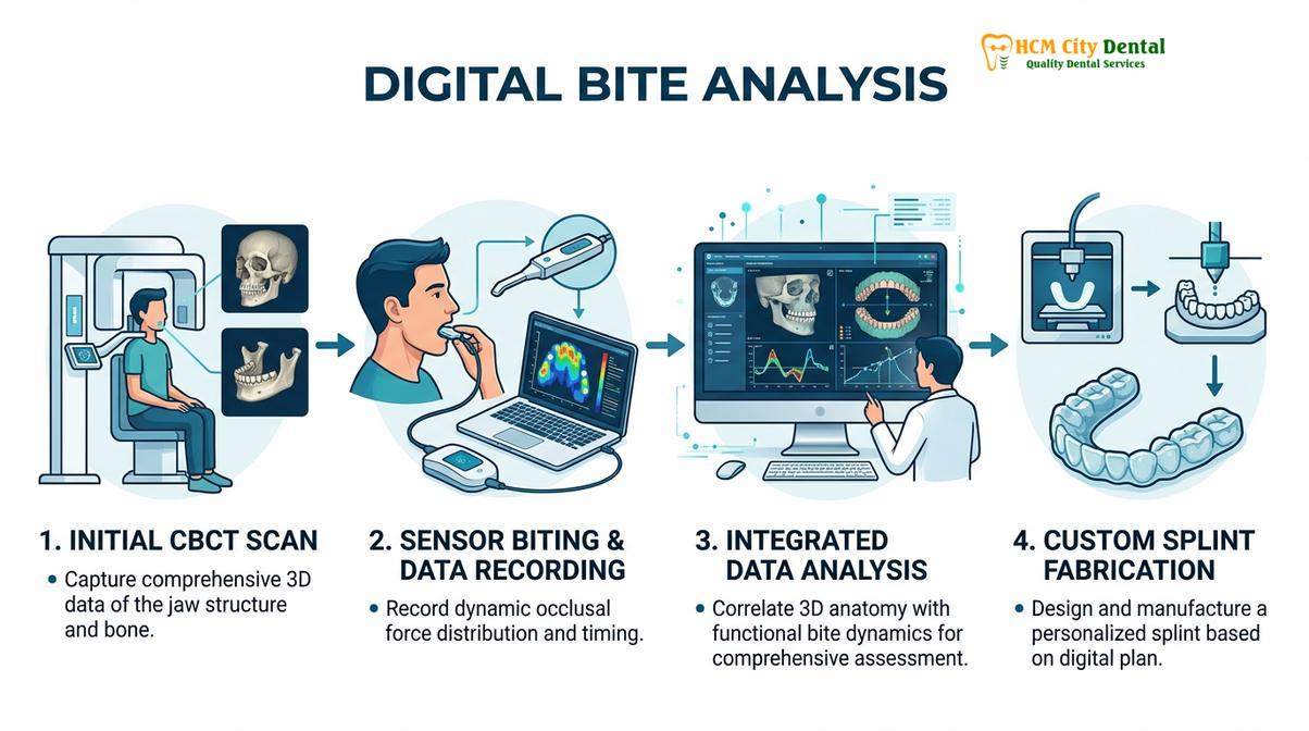
The clinical workflow begins with a thorough physical examination. The specialist palpates the muscles of mastication to identify trigger points and evaluates the range of motion of the mandible, noting any deviations, limitations, or joint sounds. Following the physical exam, a Cone Beam Computed Tomography (CBCT) scan is often utilized to visualize the bony architecture of the TMJ in three dimensions. This imaging is crucial for ruling out structural pathologies such as osteoarthritis, condylar resorption, or anatomical anomalies that may be contributing to the patient’s symptoms.
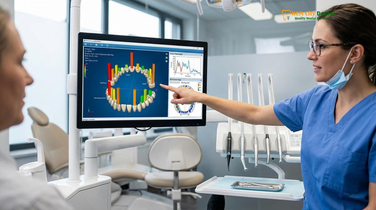
Once structural issues are assessed, the dynamic computerized bite mapping protocol is initiated. The patient is guided to bite naturally onto the digital sensor while the software records the occlusal dynamics. The clinician analyzes the data to identify premature contacts, force imbalances between the left and right sides of the dental arch, and prolonged disocclusion times during lateral movements. This data forms the blueprint for the treatment plan.
Clinical Case Example: A patient presented to HCMC Dental Clinic in Ho Chi Minh City with chronic morning headaches and severe masseter pain. Traditional exams showed no obvious decay. Digital bite mapping revealed a severe premature contact on a second molar, absorbing 40% of the total bite force. Following targeted micro-adjustment and the fabrication of a custom stabilization splint, the patient reported a complete cessation of headaches within two weeks.
Based on the digital findings, treatment is highly personalized. Dr. Cuong emphasizes a conservative, reversible approach as the first line of therapy. In many cases, the data is used to design and calibrate a custom precision nightguard or occlusal splint. Unlike over-the-counter mouthguards, a digitally calibrated splint provides an ideal, frictionless biting surface that allows the lower jaw to find its most relaxed physiological position, instantly decompressing the TMJ and reducing muscle hyperactivity[4].
If the occlusal discrepancies are minor and clearly identified by the digital software, a procedure known as occlusal equilibration (bite adjustment) may be performed. This involves selectively reshaping the microscopic enamel interferences to harmonize the bite. For more severe malocclusions, the bite mapping data may indicate the need for orthodontic realignment or the strategic placement of dental implants to replace missing teeth and restore posterior occlusal support.
When to See a Doctor for TMJ and Bite Issues
While occasional jaw fatigue or minor clicking without pain can be normal, certain symptoms indicate a progressive temporomandibular joint disorder that requires professional clinical evaluation. Delaying diagnosis can lead to chronic pain syndromes, irreversible damage to the articular disc, and severe attrition of the dental enamel.
You should seek a comprehensive occlusal and TMJ evaluation if you experience any of the following clinical red flags:
- Persistent Jaw Pain: A dull, aching pain in the jaw muscles or directly in front of the ear that worsens during chewing, speaking, or upon waking.
- Limited Range of Motion: Difficulty opening the mouth fully, or experiencing episodes where the jaw temporarily “locks” in an open or closed position.
- Sudden Changes in Bite: A distinct sensation that your upper and lower teeth no longer fit together correctly, or feeling that you are only biting down on one side.
- Painful Joint Sounds: Clicking, popping, or grating sounds in the jaw joint that are accompanied by sharp pain or a catching sensation.
- Chronic Unexplained Headaches: Frequent tension-type headaches, particularly in the temporal region, that do not respond to standard pain medication and are often worst in the morning.
Early intervention is paramount. A thorough diagnostic workup at a specialized facility in Ho Chi Minh City can identify the biomechanical root cause of your symptoms. By addressing occlusal imbalances early through conservative therapies like splints or minor adjustments, patients can achieve significant pain relief and avoid the need for complex, invasive surgical procedures in the future[5]. If you suspect your bite is contributing to facial pain, scheduling a consultation with an experienced restorative dentist is the critical first step toward recovery.
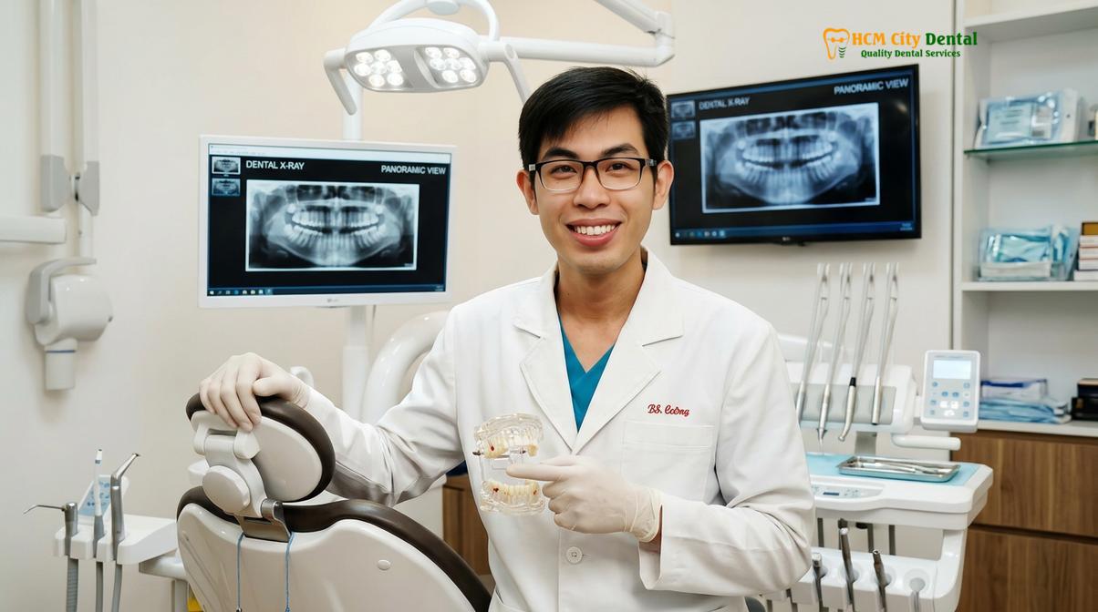
“Accurate diagnosis dictates successful treatment. By utilizing digital occlusal analysis, we transition from guessing where the bite is heavy to knowing exactly how the forces are distributed, allowing for highly predictable and conservative patient care.”
For a personalized assessment of your occlusal health and to explore advanced diagnostic options, you can learn more about our clinical team or reach out directly to schedule a comprehensive evaluation.
Frequently Asked Questions
What is occlusal analysis?
Occlusal analysis is a comprehensive clinical evaluation of how your upper and lower teeth meet and function together. By examining the contact points, forces, and timing of your bite, dental specialists can identify imbalances that contribute to temporomandibular joint disorders, muscle pain, and accelerated tooth wear. This process may involve physical examinations, traditional articulating paper, or advanced computerized digital mapping to create a precise functional profile of your stomatognathic system.
Can a bad bite cause teeth to fracture?
Yes, an uneven bite can concentrate excessive chewing forces onto a single tooth, significantly increasing the risk of micro-fractures and structural failure. Over time, this localized pressure weakens the enamel and dentin, often leading to cracked tooth syndrome or the failure of existing dental restorations. When forces are not evenly distributed across the dental arch, the overloaded teeth bear the brunt of the masticatory pressure, accelerating wear and potential breakage.
How does bite adjustment help with TMJ pain?
Bite adjustment helps with TMJ pain by eliminating premature tooth contacts that force the jaw into unnatural, strained positions. By reshaping specific enamel surfaces or utilizing orthotic splints, the procedure evenly distributes occlusal forces, allowing the masticatory muscles to relax and the jaw joint to decompress. This reduction in muscle hyperactivity and joint strain directly alleviates the chronic inflammation and myofascial pain associated with temporomandibular disorders.
Is computerized bite mapping painful?
Computerized bite mapping is a completely painless, non-invasive diagnostic procedure. Patients simply bite down on an ultra-thin, flexible electronic sensor while the software records the timing and force of their tooth contacts in real-time, requiring no anesthesia or physical alterations to the teeth. The process takes only a few minutes and provides the clinician with highly accurate, dynamic data without causing any discomfort to the patient.
How long does it take to correct an uneven bite?
The timeline for correcting an uneven bite varies from a single visit for minor occlusal adjustments to several months if orthodontic intervention or splint therapy is required. A personalized clinical assessment is necessary to determine the exact duration based on the severity of the occlusal discrepancy. Conservative treatments like custom nightguards can provide symptom relief within weeks, while comprehensive bite reconstruction requires a longer, phased approach.
References
- Journal of the American Dental Association. Occlusal splint therapy for temporomandibular disorders: a clinical review. (2020).
- Journal of Oral Rehabilitation. Efficacy of Michigan repositioning splints in TMD patients. (2019).
- Journal of Prosthetic Dentistry. Computerized occlusal analysis and bite mapping in TMJ therapy. (2021).
- Clinical Oral Investigations. Masseter Botox injections for chronic jaw pain and bruxism. (2018).
- International Journal of Oral and Maxillofacial Surgery. TMJ arthrocentesis and hyaluronic acid injections. (2022).