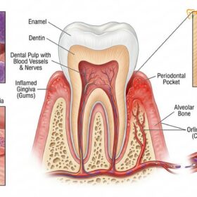
Treating peri-implantitis requires a multi-phased clinical approach to decontaminate the infected implant surface and halt progressive bone loss. Depending on disease severity, interventions range from mechanical debridement and antimicrobial therapy to advanced surgical bone regeneration, ensuring the long-term stability of the dental restoration.
Clinical Summary:
Peri-implantitis is a complex inflammatory condition characterized by the progressive destruction of the supporting bone around an osseointegrated dental implant. Unlike natural teeth, implants lack a protective periodontal ligament, making them highly susceptible to rapid bacterial invasion. Effective peri-implantitis treatment begins with accurate clinical diagnostics, including probing depths and radiographic assessment. Initial therapy focuses on non-surgical mechanical debridement using specialized instruments to disrupt the dysbiotic biofilm without damaging the titanium surface. However, due to the complex micro-topography of implant threads, surgical intervention is frequently required for advanced cases. Surgical protocols involve open flap debridement, rigorous chemical and mechanical surface decontamination, and often the application of regenerative bone grafts to restore lost osseous architecture. Long-term success relies heavily on customized professional maintenance and meticulous patient compliance.
Key Takeaways:
- Peri-implant diseases are categorized into reversible mucositis and irreversible peri-implantitis involving bone loss.
- Early detection through routine probing and radiographic monitoring is critical for conservative management.
- Non-surgical therapy aims to reduce bacterial load but is often insufficient for deep peri-implant pockets.
- Surgical access is required to thoroughly decontaminate exposed implant threads and facilitate bone regeneration.
- Strict adherence to a 3-to-6-month professional maintenance schedule is mandatory to prevent disease recurrence.
What is Peri-Implant Mucositis vs Peri-Implantitis?
Peri-implant mucositis is a reversible inflammatory lesion confined to the soft tissues, whereas peri-implantitis involves progressive, irreversible crestal bone loss around the integrated fixture.
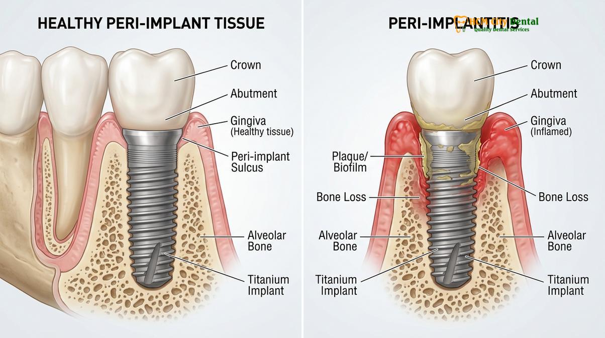
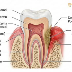
To understand the complexities of peri-implantitis treatment, it is essential to distinguish between the two primary classifications of peri-implant disease. While dental implants are highly successful restorative solutions, the biological seal surrounding a titanium fixture differs significantly from the anatomy of a natural tooth. Natural teeth are suspended in the alveolar bone by the periodontal ligament (PDL), which provides a robust vascular supply and a dense network of perpendicular collagen fibers that act as a physical barrier against bacterial ingress. In contrast, the connective tissue fibers around an implant run parallel to the fixture surface, creating a more fragile biological seal that is highly susceptible to bacterial penetration[1].
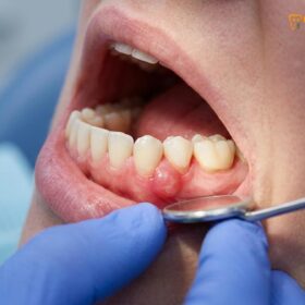
Peri-implant mucositis is the precursor to more severe disease. It is defined as an inflammatory lesion residing strictly within the peri-implant mucosa (the gums surrounding the implant). Clinically, it presents with redness, swelling, and bleeding on probing, but crucially, there is no loss of supporting alveolar bone. Because the inflammation is confined to the soft tissue, peri-implant mucositis is entirely reversible with prompt professional intervention and optimized home care. If left untreated, however, the dysbiotic biofilm matures and migrates apically along the implant body.
Peri-implantitis occurs when the inflammatory process extends beyond the soft tissue, triggering an aggressive host immune response that leads to the resorption of the surrounding crestal bone. This condition is characterized by deep peri-implant pockets, mucosal suppuration (pus), and radiographic evidence of marginal bone loss. Because the rough micro-topography of the implant surface—designed to enhance initial osseointegration—becomes exposed to the oral environment, it serves as an ideal harbor for pathogenic bacteria. Decontaminating these complex surfaces makes the treatment of peri-implantitis highly challenging and often necessitates surgical intervention to arrest disease progression[2].
Dr. Nguyen Van Cuong, a leading specialist in implantology, frequently emphasizes to his patients that recognizing early implant gum infection symptoms is the most critical factor in preserving the restoration. “The transition from mucositis to peri-implantitis can occur rapidly and often without significant pain,” notes Dr. Cuong. “Routine clinical monitoring allows us to intercept the disease while it is still reversible, sparing the patient from complex regenerative surgeries.”
Warning Signs: Bleeding Gums and Bone Loss Around Implants
The primary clinical indicators of peri-implant disease include profuse bleeding on probing, mucosal suppuration, increased pocket depths, and radiographic evidence of marginal bone resorption.
Diagnosing peri-implant diseases requires a meticulous clinical and radiographic evaluation. Because implants lack nerve endings, patients rarely experience pain during the early to moderate stages of infection. Consequently, the disease can progress silently until significant structural damage has occurred. Clinicians rely on a specific set of diagnostic parameters to evaluate the health of the peri-implant tissues and determine the appropriate course of therapy.
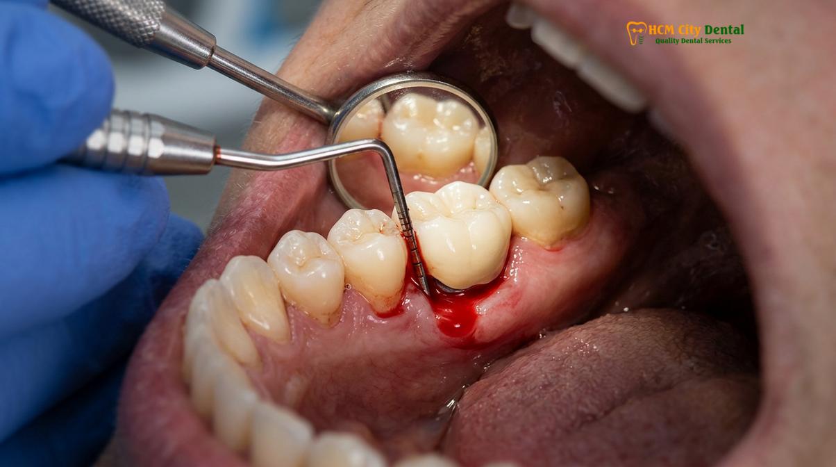
The most fundamental diagnostic tool is the periodontal probe. During a maintenance visit, the clinician will gently measure the depth of the sulcus (the crevice between the gum and the implant). Healthy peri-implant sulci typically measure between 2 to 4 millimeters. Probing depths of 5 millimeters or greater, especially when accompanied by bleeding on probing (BOP), are strong indicators of active inflammation and potential attachment loss. It is crucial that probing is performed with a controlled, light force (approximately 0.25 N) to avoid penetrating the fragile mucosal seal and causing false-positive readings.
Bleeding is a hallmark sign of mucosal ulceration and active infection. In healthy implants, the tissue should be firm, pink, and resilient, with no bleeding upon gentle manipulation. When a patient presents seeking a bleeding dental implant cure, the clinician must first determine if the bleeding is isolated to the soft tissue (mucositis) or if it is a symptom of deeper osseous destruction. In advanced cases, suppuration (the exudation of pus) may be observed when pressure is applied to the surrounding gums, indicating a severe, active bacterial infection that requires immediate antimicrobial and mechanical intervention.
“Accurate diagnosis of peri-implantitis relies on the identification of bleeding or suppuration on gentle probing, increased probing depths compared to baseline measurements, and the presence of progressive marginal bone loss documented via standardized radiographs.”
Radiographic assessment is the definitive method for confirming bone loss around implants. A baseline periapical radiograph should always be taken at the time of final prosthesis delivery to serve as a reference point. In subsequent visits, new radiographs are compared against the baseline to detect any crestal bone remodeling. According to current clinical guidelines, bone loss extending beyond the initial remodeling phase (typically >2mm from the expected marginal bone level) combined with clinical signs of inflammation confirms a diagnosis of peri-implantitis[3]. If a patient notices localized gingival swelling or a bad taste in their mouth, immediate radiographic evaluation is warranted to rule out progressive osseous destruction.
Key Causes of Implant Failure and Bacterial Infection
Implant infections are primarily driven by dysbiotic biofilm accumulation, exacerbated by risk factors such as smoking, uncontrolled diabetes, residual cement, and poor oral hygiene.
The etiology of peri-implantitis is multifactorial, involving a complex interplay between bacterial pathogens, host immune responses, and environmental risk factors. The primary initiating factor is the accumulation of a dysbiotic bacterial biofilm on the implant and restorative surfaces. The microbiome associated with peri-implantitis is highly diverse, predominantly consisting of Gram-negative anaerobic bacteria similar to those found in severe periodontitis, such as Porphyromonas gingivalis, Tannerella forsythia, and Treponema denticola. When this biofilm is not adequately disrupted through daily oral hygiene, it triggers a chronic inflammatory cascade that ultimately leads to tissue destruction.
Beyond bacterial plaque, several iatrogenic and systemic risk factors significantly increase a patient’s susceptibility to peri-implant diseases. One of the most common iatrogenic causes is excess residual dental cement. When an implant crown is cemented into place, excess cement can easily be extruded deep into the peri-implant sulcus. Because the titanium surface is highly retentive, this cement acts as a constant irritant and a highly porous scaffold for bacterial colonization. Careful delivery techniques and the use of custom abutments or screw-retained restorations are critical strategies to mitigate this risk.
Systemic health and lifestyle factors also play a profound role in implant longevity. Smoking is one of the most significant risk factors for peri-implantitis. The toxins in tobacco smoke impair peripheral blood flow, reduce the efficacy of the local immune response, and alter the composition of the oral microbiome, making smokers significantly more prone to rapid bone loss and delayed healing. Similarly, patients with uncontrolled diabetes mellitus exhibit an exaggerated inflammatory response and impaired tissue repair mechanisms, drastically increasing their risk of implant complications.
| Risk Factor | Mechanism of Action | Clinical Impact on Implants |
|---|---|---|
| Poor Oral Hygiene | Allows maturation of Gram-negative anaerobic biofilm. | Primary driver of mucositis and subsequent bone resorption. |
| Smoking / Tobacco Use | Causes vasoconstriction and impairs neutrophil function. | Accelerates bone loss; significantly reduces surgical success rates. |
| Residual Cement | Acts as a foreign body irritant and plaque-retentive surface. | Triggers rapid, localized acute inflammation and suppuration. |
| Uncontrolled Diabetes | Alters host immune response and impairs collagen synthesis. | Increases susceptibility to severe infection and delayed healing. |
| Lack of Keratinized Tissue | Reduces the physical barrier against mechanical trauma. | Leads to mucosal recession, discomfort during brushing, and plaque accumulation. |
Biomechanical factors, such as occlusal overload, can also contribute to implant failure. While bacterial infection is the primary cause of peri-implantitis, excessive biting forces—often due to bruxism (teeth grinding) or an improperly adjusted bite—can cause micro-fractures in the crestal bone, accelerating the rate of bone loss when inflammation is present. Comprehensive periodontal therapy programs must address both the biological and biomechanical aspects to ensure the long-term survival of restorative implant solutions.
Non-Surgical Treatments: Debridement and Antimicrobial Rinses
Initial therapy focuses on mechanical plaque removal using specialized titanium or carbon curettes, supplemented by localized antimicrobial agents to reduce bacterial load and soft tissue inflammation.
The management of peri-implant diseases follows a cumulative interceptive supportive therapy (CIST) protocol, which dictates that treatment should begin with the most conservative, non-surgical methods before progressing to surgical intervention. For patients diagnosed with peri-implant mucositis, non-surgical therapy is often sufficient to completely resolve the inflammation and restore tissue health. For those with early-stage peri-implantitis, non-surgical treatment serves as a critical first step to reduce the bacterial load and improve tissue quality prior to surgery.
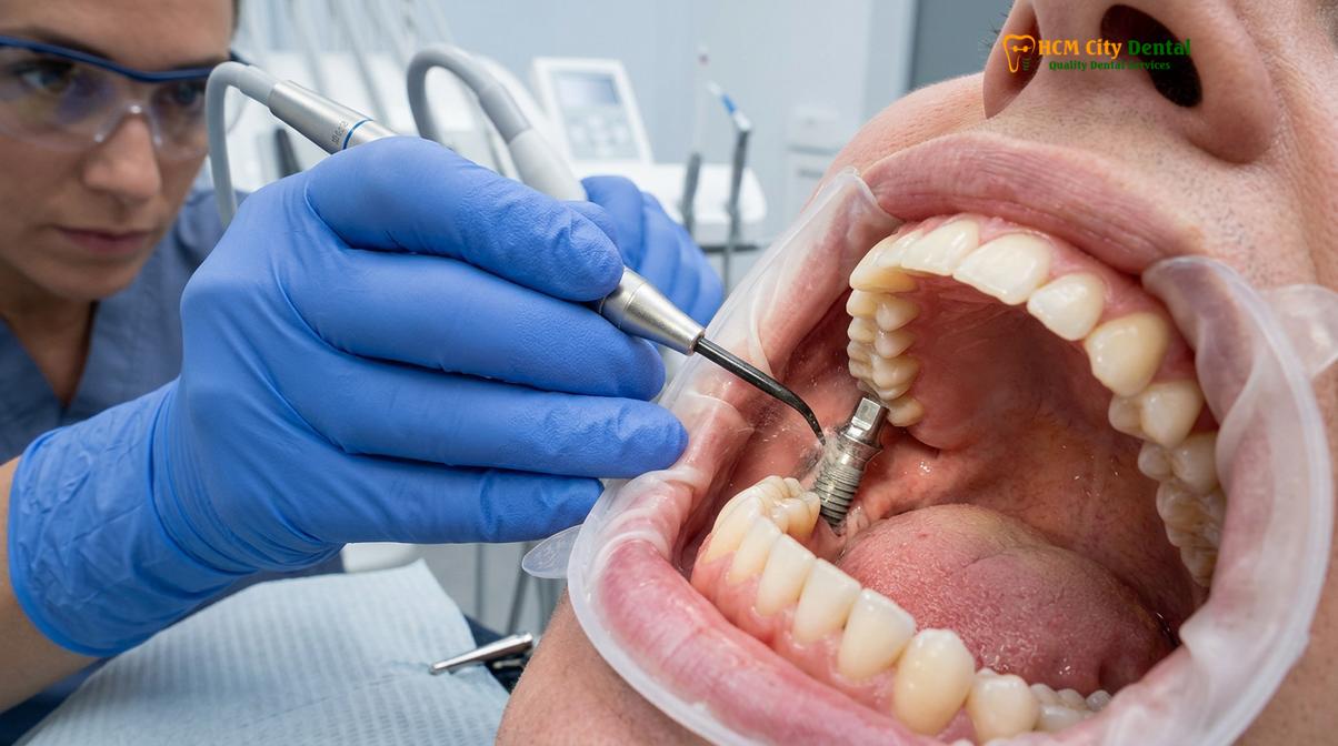
The cornerstone of non-surgical therapy is professional mechanical plaque removal (PMPR). However, cleaning a titanium implant requires specialized instruments. Traditional stainless steel scalers used on natural teeth are strictly contraindicated for implants, as they can scratch the delicate titanium surface. Scratches increase the surface roughness, creating microscopic niches that facilitate faster and more tenacious bacterial recolonization. Instead, clinicians utilize curettes made of carbon fiber, plastic, or specialized titanium alloys that are softer than the implant body. Ultrasonic scalers equipped with PEEK (polyether ether ketone) tips are also highly effective for safely dislodging calculus and biofilm without altering the implant topography[4].
Clinical Warning: Never use traditional stainless steel scaling instruments on dental implants. Altering the titanium surface topography through scratching significantly increases the risk of rapid bacterial recolonization and accelerates the progression of peri-implant disease.
In addition to hand and ultrasonic instrumentation, air polishing has emerged as a superior modality for implant decontamination. Using specialized subgingival nozzles, clinicians deliver a low-abrasive glycine or erythritol powder under air and water pressure. This technique is highly efficient at removing biofilm from the complex micro-threads of the implant without causing surface damage or soft tissue trauma. It is widely considered the gold standard for non-surgical subgingival scaling protocols around implants.
Adjunctive chemical therapies are frequently employed to enhance the efficacy of mechanical debridement. Localized irrigation with antimicrobial solutions, such as 0.12% chlorhexidine digluconate, hydrogen peroxide, or povidone-iodine, helps to flush out planktonic bacteria and disinfect the peri-implant pocket. In some cases, sustained-release antimicrobial chips or gels may be placed directly into the sulcus to provide prolonged bacterial suppression. However, it is important to note that systemic antibiotics are generally not recommended as a monotherapy for peri-implantitis, as the biofilm must be physically disrupted for any pharmacological agent to be effective.
While non-surgical therapy is highly effective for mucositis, clinical studies consistently show that it has limited efficacy in resolving true peri-implantitis. Once the probing depths exceed 5 millimeters and the bone loss exposes the rough implant threads, blind non-surgical instrumentation cannot adequately decontaminate the surface. In these scenarios, non-surgical therapy is utilized to reduce acute inflammation, paving the way for definitive surgical management.
Surgical Interventions: Bone Grafting and Laser Disinfection (Lapip)
Advanced peri-implantitis requires surgical access for thorough surface decontamination, often utilizing laser therapy and regenerative bone grafting to rebuild lost osseous support.
When non-surgical therapies fail to resolve deep pockets and arrest bone loss, surgical intervention becomes mandatory. The primary objective of surgical peri-implantitis treatment is to gain direct visual access to the contaminated implant surface, allowing for meticulous decontamination and the modification of the hard and soft tissue architecture to facilitate long-term maintenance. The surgical approach is dictated by the morphology of the bone defect—whether it is a horizontal pattern of bone loss or a crater-like (intrabony) defect.
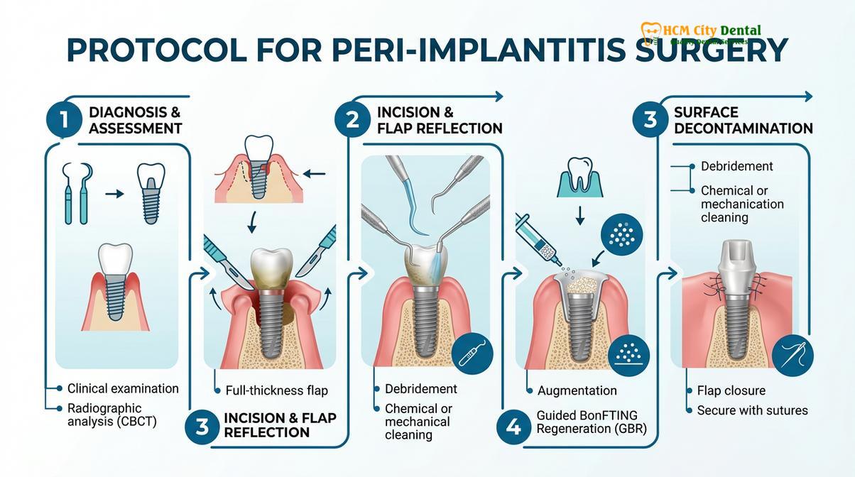
The procedure begins with an access flap debridement (AFD). The surgeon carefully raises a full-thickness mucoperiosteal flap to expose the underlying bone and the infected implant fixture. All inflamed granulation tissue is meticulously excised using surgical curettes. Once the defect is cleared, the critical phase of surface decontamination begins. Because the implant threads are highly porous, removing the embedded biofilm is exceptionally challenging. Clinicians employ a combination of mechanical and chemical methods. Titanium brushes rotated at low speeds are used to scrub the threads, followed by chemical detoxification using agents like sterile saline, citric acid, or EDTA to neutralize bacterial endotoxins[5].
In recent years, laser technology has revolutionized the decontamination process. The Laser-Assisted Peri-Implantitis Procedure (LAPIP) utilizes a specific wavelength (such as the Nd:YAG or Diode laser) that selectively targets and vaporizes diseased tissue and pigmented bacteria without overheating or damaging the titanium implant. This advanced laser decontamination not only sterilizes the surgical site but also stimulates the formation of a stable fibrin clot, which is essential for subsequent tissue regeneration. Patients seeking laser peri implantitis surgery saigon often benefit from reduced post-operative discomfort and faster healing times compared to traditional mechanical methods alone.
Clinical Case Example: A 55-year-old patient presented to HCMC Dental Clinic in Ho Chi Minh City with severe suppuration and 7mm probing depths around a lower molar implant. Dr. Nguyen Van Cuong performed an open flap debridement combined with laser decontamination. Following thorough sterilization of the titanium threads, a particulate xenograft was placed to regenerate the osseous defect. At the 12-month follow-up, probing depths were reduced to a healthy 3mm with complete resolution of bleeding and stable radiographic bone levels.
If the bone defect is crater-like (intrabony), the surgeon will attempt a reconstructive procedure. After decontamination, a bone graft material—often a deproteinized bovine bone mineral (DBBM) or an allograft—is packed into the defect to act as a scaffold for new bone growth. A resorbable collagen membrane is then placed over the graft to prevent fast-growing soft tissue cells from invading the space, allowing the slower-growing bone cells to regenerate. In cases where the bone loss is strictly horizontal and regeneration is not predictable, the surgeon may perform resective osseous flap procedures. This involves smoothing the irregular bone and performing “implantoplasty”—using specialized burs to polish away the exposed implant threads, creating a smooth surface that is less prone to plaque accumulation.
Step-by-Step Implant Care Guide for Long-Term Success
Sustaining implant health requires rigorous daily home care using interdental brushes and water flossers, combined with customized professional maintenance schedules every three to six months.
The successful treatment of peri-implantitis is not the end of the clinical journey; it marks the beginning of a lifelong commitment to rigorous maintenance. Because patients who have experienced peri-implant disease are at a significantly higher risk for recurrence, establishing a strict, customized care protocol is paramount. The longevity of the implant relies equally on the patient’s daily home care and the clinician’s professional oversight.
Daily oral hygiene must be meticulous. Standard tooth brushing alone is insufficient to clean the complex anatomy of an implant-supported restoration. Patients must utilize interdental brushes sized appropriately for the spaces between the implants and adjacent teeth. These brushes physically disrupt the biofilm at the critical junction where the crown meets the gum line. Additionally, oral irrigators (water flossers) are highly recommended. When used on a low-pressure setting, water flossers can effectively flush out unattached plaque and food debris from shallow peri-implant pockets without traumatizing the delicate mucosal seal.
“Long-term implant survival is directly correlated with patient compliance. A structured professional maintenance program, tailored to the individual’s risk profile, is the most effective strategy for preventing the recurrence of peri-implant infections.”
Professional maintenance is the second pillar of long-term success. Patients with a history of peri-implantitis should be placed on a strict recall schedule, typically every three to four months. During these visits, the clinician will perform a comprehensive evaluation, including gentle probing, assessment of bleeding indices, and periodic radiographic imaging. Professional mechanical plaque removal using air polishing or specialized implant scalers is performed to eliminate any biofilm that the patient missed. Furthermore, the clinician will assess the quality of the surrounding soft tissue. A thick band of keratinized gingiva is crucial for implant stability; if the tissue is thin or mobile, a soft tissue graft may be recommended to enhance the protective barrier.
When to See a Doctor (Clinical Considerations)
Early intervention is the key to saving a failing dental implant. Because peri-implant diseases often progress without significant pain, patients must be vigilant in monitoring their oral health. You should schedule an immediate clinical evaluation if you notice any of the following red flags:
- Bleeding during brushing or flossing: Healthy implants do not bleed. Any sign of blood is a primary indicator of active inflammation.
- Swelling or redness: Changes in the color or contour of the gums around the implant suggest an underlying infection.
- Pus or bad taste: Suppuration indicates a severe, active bacterial infection that is actively destroying bone.
- Gum recession: If the metallic threads of the implant become visible, the protective biological seal has been compromised.
- Implant mobility: This is a late-stage, critical warning sign. If an implant feels loose, it indicates catastrophic loss of osseointegration, and the fixture may need to be removed.
Do not wait for pain to develop. If you experience any of these symptoms, prompt evaluation at a specialized center like HCMC Dental Clinic is essential. Based on comprehensive clinical diagnostics, our specialists can implement targeted therapies to arrest the disease and protect your restorative investment.
Frequently Asked Questions
Can a dental implant get gum disease?
Yes, dental implants can develop a form of gum disease known as peri-implant disease. Because implants lack the protective periodontal ligament found around natural teeth, bacterial plaque can rapidly cause inflammation in the surrounding tissues, leading to mucositis or advanced peri-implantitis if left untreated. Regular monitoring is essential to prevent this progression.
Is peri-implantitis curable?
Peri-implantitis can be effectively managed and arrested, though complete regeneration of lost bone is not always predictable. With timely surgical intervention, surface decontamination, and strict maintenance protocols, clinicians can halt disease progression, resolve inflammation, and successfully preserve the implant’s function for the long term. Early detection significantly improves the prognosis.
What causes bone loss around a dental implant?
Bone loss around an implant is primarily caused by a dysbiotic bacterial biofilm triggering a severe host inflammatory response. Additional contributing factors include excess residual dental cement, biomechanical occlusal overload, smoking, uncontrolled systemic conditions like diabetes, and a lack of adequate keratinized tissue. Addressing all risk factors is crucial for stabilization.
How do dentists treat a bleeding implant?
Dentists treat a bleeding implant by first performing a thorough clinical assessment to determine the depth of infection. Initial therapy involves mechanical debridement using specialized non-scratching instruments, localized antimicrobial irrigation, and optimizing the patient’s home care routine to eliminate the bacterial biofilm causing the bleeding. If bone loss is present, surgical intervention may be required.
How do I prevent peri-implantitis?
Preventing peri-implantitis requires meticulous daily oral hygiene, including the use of interdental brushes and water flossers to clean under the prosthesis. Additionally, patients must adhere to a strict professional maintenance schedule, visiting their dentist every three to six months for specialized implant scaling and clinical monitoring to ensure the peri-implant tissues remain healthy.
References
- European Federation of Periodontology. Prevention and treatment of peri-implant diseases – The EFP S3 level clinical practice guideline. (2023).
- British Dental Journal. Surgical treatment of peri-implantitis and surface decontamination strategies. (2024).
- Clinical Oral Implants Research. Long-term outcome of surgical treatment of peri-implantitis. (2018).
- Journal of Clinical Periodontology. Efficacy of mechanical and physical approaches for implant surface decontamination. (2023).
- International Journal of Oral Implantology. Strategies for implant surface decontamination in peri-implantitis therapy. (2022).