Finding a loose tooth as an adult is never normal and often signals underlying structural damage to the jawbone or periodontal ligaments. Whether caused by advanced gum disease, sudden physical trauma, or chronic teeth grinding, immediate clinical evaluation is critical to prevent permanent tooth loss and restore oral stability. Understanding the biomechanics of tooth support is the first step in recognizing why this condition develops and how it can be effectively managed [1].
Clinical Summary:
Adult tooth mobility occurs when the supporting periodontium—comprising the alveolar bone, periodontal ligament, and cementum—is compromised. The primary etiology is untreated periodontitis, followed by occlusal trauma and systemic health conditions. Early intervention through scaling, root planing, occlusal adjustment, or dental splinting can often stabilize the dentition and promote tissue reattachment. Delaying treatment significantly increases the risk of irreversible bone resorption, severe localized infection, and the eventual necessity of surgical extraction. Comprehensive diagnostics, including periodontal probing and radiographic imaging, are essential for formulating an effective, individualized treatment plan.
Key Takeaways:
- Adult tooth mobility is a definitive sign of structural compromise and requires prompt clinical assessment.
- Advanced periodontitis is the leading cause, responsible for destroying the bone that anchors teeth in place.
- Non-surgical treatments, such as deep cleaning and bite adjustment, can often stabilize mild to moderate looseness.
- Dental splinting provides immediate mechanical support by bonding a mobile tooth to adjacent healthy teeth.
- Ignoring a loose tooth can lead to rapid bone loss, shifting of adjacent teeth, and complex restorative needs.
Why Adults Get Loose Teeth: Root Causes
Adult teeth become loose when the foundational structures, including the jawbone and periodontal ligaments, weaken or deteriorate due to chronic infection, physical trauma, or excessive bite forces.
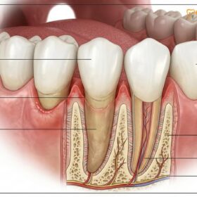
To understand why an adult tooth becomes mobile, it is essential to first understand the anatomy of the periodontium. The periodontium is the specialized system of tissues that surrounds and supports the teeth, keeping them firmly anchored in the maxillary (upper) and mandibular (lower) jawbones. This system consists of four main components: the gingiva (gums), the alveolar bone, the cementum (the outer layer of the tooth root), and the periodontal ligament (PDL). The PDL is a highly specialized connective tissue network that acts as a shock absorber, suspending the tooth within the bony socket and allowing for microscopic, natural movement during chewing [2].
When patients search for answers regarding why their teeth feel unstable, the clinical explanation almost always traces back to a disruption in this delicate supportive network. In a healthy state, the alveolar bone tightly hugs the tooth root, and the PDL fibers are taut and resilient. However, when these structures are subjected to chronic inflammation, physical injury, or systemic degradation, their integrity is compromised. The bone begins to resorb, and the ligament fibers detach, creating a widened space around the root. Without this rigid support, the tooth begins to exhibit noticeable mobility.
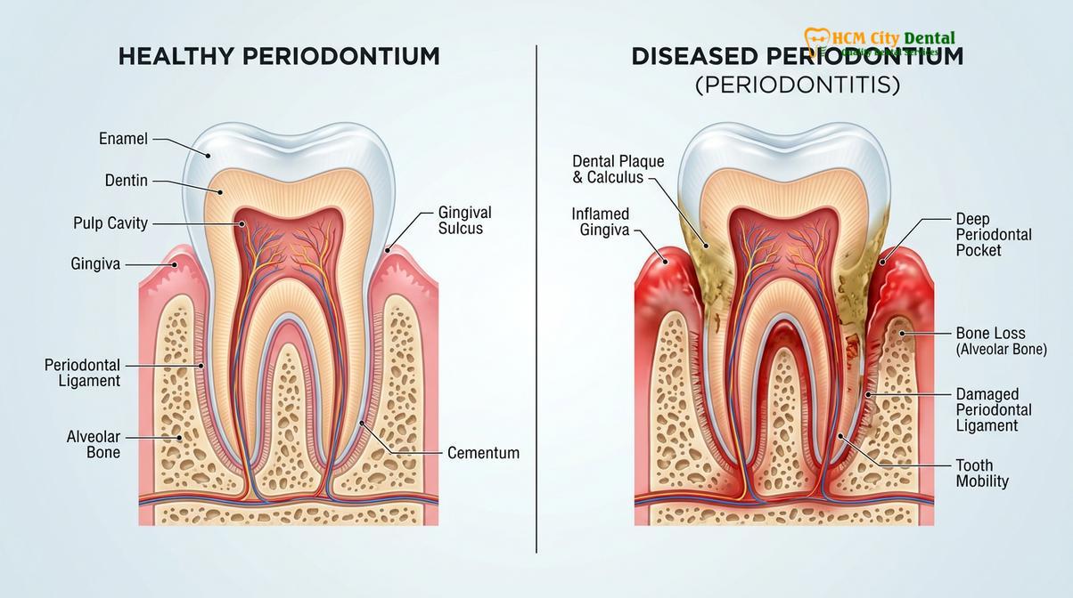
Beyond localized dental issues, systemic health conditions can profoundly impact the stability of the periodontium. Diabetes mellitus, particularly when poorly controlled, impairs the body’s immune response and restricts blood flow to the gum tissues, making diabetic patients significantly more susceptible to severe periodontal infections and subsequent tooth mobility. Osteoporosis, a condition characterized by decreased bone mineral density, can also affect the jawbone, reducing the structural foundation available to support the teeth.
Advanced Periodontitis: The Leading Cause of Tooth Mobility
Severe gum disease destroys the alveolar bone and connective tissues anchoring the tooth, making it the most common reason adults experience progressive tooth mobility and eventual tooth loss.
Periodontitis is a chronic, destructive inflammatory disease initiated by the accumulation of bacterial plaque and calculus (tartar) below the gumline. While gingivitis—the earliest stage of gum disease—only affects the superficial soft tissues and is entirely reversible, untreated gingivitis can progress into periodontitis. In this advanced stage, the bacterial biofilm harbors highly pathogenic, Gram-negative anaerobic bacteria. These bacteria release toxins that provoke a hyperactive immune response from the host’s body [4].
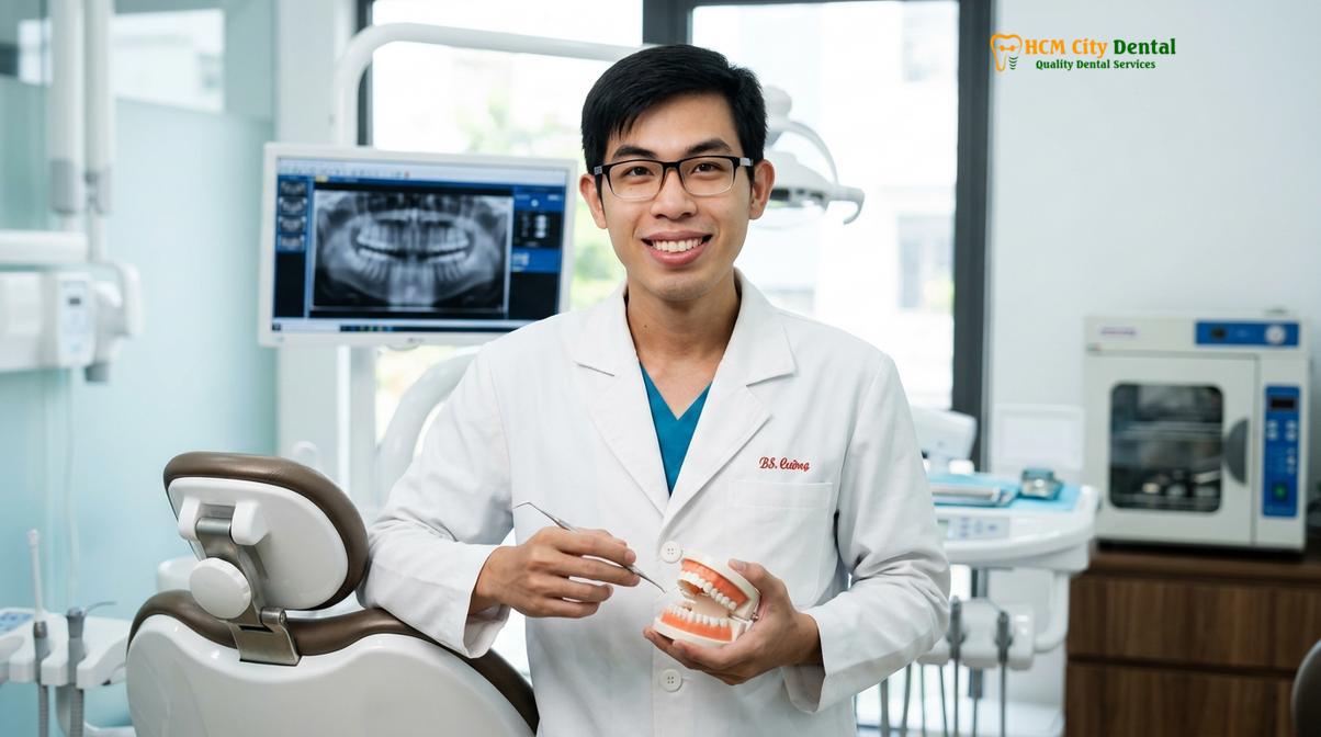
Dr. Nguyen Van Cuong, a leading specialist at HCMC Dental Clinic, frequently emphasizes to patients that bone loss is painless in its early stages. He notes that many adults are unaware of the ongoing destruction until a tooth begins to shift or wiggle during normal function. His clinical approach prioritizes rapid stabilization to preserve the natural dentition whenever possible, utilizing advanced diagnostics to map out the exact pattern of bone degradation before initiating comprehensive gum treatment.
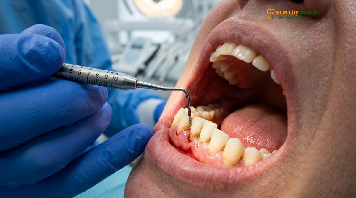
The host’s immune system, in an attempt to eradicate the bacterial invasion, releases inflammatory mediators that inadvertently destroy the host’s own tissues. The periodontal ligament fibers are severed, and osteoclasts (bone-resorbing cells) are activated, leading to the irreversible destruction of the alveolar bone. As the bone recedes, deep periodontal pockets form between the tooth and the gum, creating an even more favorable environment for anaerobic bacteria to thrive and multiply.
Symptoms That Accompany Adult Loose Teeth
A loose tooth rarely occurs in isolation; it is typically accompanied by a range of clinical symptoms that indicate the underlying cause, such as gum inflammation, recession, or localized discomfort.
Recognizing the secondary symptoms associated with tooth mobility is crucial for an accurate diagnosis. When periodontitis is the primary culprit, patients often notice that their gums bleed easily during brushing or flossing. The gingival tissues may appear swollen, red, or purplish, and there is frequently a persistent bad taste or halitosis (bad breath) caused by the volatile sulfur compounds produced by anaerobic bacteria in deep gum pockets.
“The insidious nature of periodontitis lies in its silent progression. By the time a patient notices significant tooth mobility, substantial and often irreversible alveolar bone loss has already occurred, shifting the clinical focus from prevention to complex structural salvage.”
Gum recession is another common accompanying symptom. As the underlying bone melts away, the gum tissue often follows, exposing the yellowish root surface of the tooth. This not only makes the tooth appear longer but also increases sensitivity to hot and cold temperatures. In cases where occlusal trauma is the cause, patients might not experience bleeding gums but may report a dull ache in the jaw, tension headaches upon waking, or a feeling that the tooth is “high” or hitting prematurely when they bite down.
Dental Trauma, Bruxism, and Occlusal Overload
Physical impacts from accidents or chronic grinding place excessive stress on the periodontal ligament, causing it to stretch and widen, which results in noticeable tooth wiggling.
While bacterial infection is the most common culprit, mechanical forces play a significant role in adult tooth mobility. The periodontal ligament is designed to withstand normal vertical chewing forces, but it is highly vulnerable to excessive, sustained, or lateral (side-to-side) pressures. When a tooth is subjected to forces that exceed the physiological adaptive capacity of the periodontium, a condition known as occlusal trauma occurs [3].
Primary occlusal trauma happens when excessive force is applied to a tooth with a perfectly healthy, intact periodontium. The most common cause of this is bruxism—the chronic, often subconscious clenching and grinding of teeth, particularly during sleep. The immense pressure generated by the masseter muscles during bruxism compresses the periodontal ligament, causing localized inflammation, widening of the PDL space, and subsequent tooth mobility. If the grinding habit is intercepted early, the tooth can often stabilize once the excessive force is removed.
Secondary occlusal trauma occurs when normal or excessive chewing forces are applied to a tooth that has already lost supporting bone due to periodontitis. In these cases, even the simple act of chewing soft food can exacerbate the looseness, creating a rapid downward spiral of tissue destruction.
When a Loose Tooth Becomes a Dental Emergency
A loose tooth transitions into a critical emergency when it is accompanied by severe pain, facial swelling, uncontrollable bleeding, or when it results from sudden physical trauma to the face or jaw.
Not all loose teeth require a midnight trip to the clinic, but certain symptoms indicate a critical situation that demands urgent attention. If a tooth becomes mobile after a sports injury, a fall, or a vehicular accident, it is classified as acute dental trauma. This can cause immediate luxation (displacement) or subluxation (loosening without displacement) of the tooth. These injuries require emergency stabilization to prevent necrosis of the dental pulp and permanent loss of the tooth.
Important Clinical Note: If your loose tooth is accompanied by a throbbing toothache, a swollen pocket of pus on the gums, or swelling extending into the cheek, jaw, or neck, it signifies an acute, spreading infection. Seek immediate medical or dental attention, as untreated facial infections can compromise systemic health and airway patency.
In scenarios involving acute trauma or severe abscesses, seeking Emergency Dental Care is vital. The clinical protocol involves draining the infection, prescribing appropriate antibiotics, and stabilizing the affected area. According to national guidelines on dental emergencies, rapid intervention within the first few hours of trauma significantly improves the prognosis for tooth survival [5].
How Dentists Diagnose and Measure Tooth Mobility
Clinicians utilize specialized instruments to apply gentle pressure to the tooth, grading its movement on a standardized scale while using radiographic imaging to assess the extent of underlying bone loss.
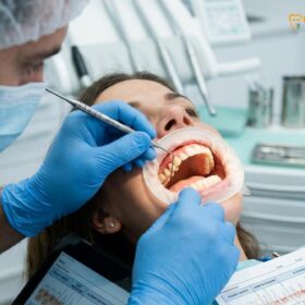
Accurate diagnosis is the cornerstone of effective treatment. When a patient presents with a loose tooth, the clinical examination begins with a thorough periodontal assessment. The dentist will use a calibrated periodontal probe to measure the depth of the sulcus (the space between the tooth and the gum). Healthy pockets typically measure between 1 to 3 millimeters. Depths of 4 millimeters or greater indicate attachment loss and the presence of periodontal disease.
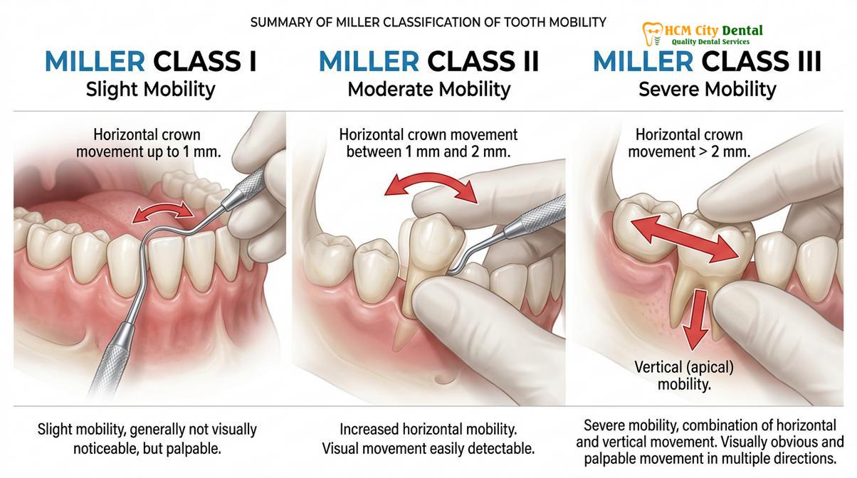
To quantify the degree of looseness, dentists universally employ the Miller Classification of Tooth Mobility. This standardized index allows clinicians to objectively grade the severity of the condition and monitor changes over time. The assessment is performed by placing the handles of two metallic dental instruments on either side of the tooth crown and applying alternating, gentle pressure.
| Classification | Clinical Description | Prognosis & Clinical Implication |
|---|---|---|
| Class I | Slight mobility; up to 1 mm of horizontal movement in a facial-lingual direction. | Highly favorable. Often resolves with non-surgical therapy and occlusal adjustment. |
| Class II | Moderate mobility; greater than 1 mm but less than 2 mm of horizontal movement. | Guarded to favorable. Requires targeted periodontal therapy and possible splinting. |
| Class III | Severe mobility; greater than 2 mm of horizontal movement, or any vertical depressibility into the socket. | Poor to hopeless. High risk of extraction unless advanced regenerative surgery is viable. |
Following the physical assessment, radiographic imaging is mandatory. Standard two-dimensional periapical and bitewing X-rays provide a clear view of the interproximal bone levels and the width of the periodontal ligament space. A widened PDL space on an X-ray is a classic hallmark of occlusal trauma. For complex cases, advanced 3D imaging is utilized to evaluate the exact three-dimensional architecture of the remaining bone, allowing for precise surgical planning.
Treatment Options to Save and Stabilize Loose Teeth
Depending on the severity and underlying cause, treatments range from deep cleanings and bite adjustments to fiber splinting, bone grafting, and surgical tissue regeneration to secure the tooth.
The approach to saving a loose tooth is highly dependent on the etiology, the Miller classification of mobility, and the amount of remaining healthy bone. Treatment is generally phased, beginning with the least invasive options to control active disease, followed by mechanical stabilization, and, if necessary, surgical regeneration.
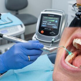
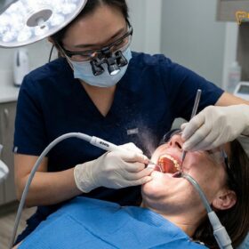
If the mobility is primarily caused by active periodontitis (Class I or mild Class II), the first line of defense is scaling and root planing (SRP). This meticulous deep cleaning procedure removes the bacterial biofilm, calculus, and endotoxins from the root surfaces deep beneath the gumline. By eliminating the source of inflammation, the gingival tissues can heal, shrink, and reattach more firmly to the clean root surface, often reducing tooth mobility significantly.
“Time is the most critical factor in dental traumatology. Splinting a traumatically loosened tooth within the first few hours dramatically increases the probability of periodontal ligament reattachment and long-term survival.”
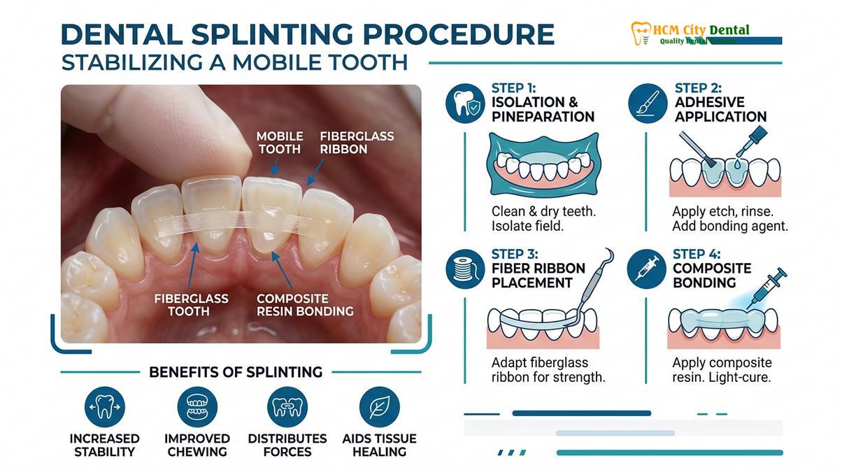
For teeth that remain mobile despite periodontal therapy, or those subjected to secondary occlusal trauma, dental splinting is a highly effective mechanical solution. Splinting involves joining the loose tooth to adjacent, stable teeth using a strong, thin fiberglass ribbon bonded with composite resin. This technique distributes chewing forces across multiple teeth, preventing the mobile tooth from being overloaded and allowing the periodontal ligament time to heal.
Clinical Case Study: Saving a Mobile Tooth in Ho Chi Minh City
A real-world clinical scenario demonstrates how prompt diagnosis and a combination of periodontal therapy and splinting can successfully rescue a severely compromised tooth.
Patient Profile: A 45-year-old patient visited HCMC Dental Clinic in Ho Chi Minh City complaining of a wiggling lower front tooth that made chewing uncomfortable.
Clinical Findings: Examination revealed Class II mobility on a central incisor, accompanied by localized bleeding and a 6mm periodontal pocket. Radiographs confirmed moderate horizontal bone loss but showed that the root apex was still firmly embedded in healthy bone.
Treatment Protocol: The clinical team initiated a comprehensive treatment plan starting with targeted scaling and root planing to eradicate the localized infection. Once the tissue inflammation subsided, a lingual fiberglass splint was applied to bond the mobile incisor to the adjacent stable canines.
Outcome: Within a few weeks, the patient reported complete resolution of discomfort during chewing, and follow-up probing showed a reduction in pocket depth to a healthy 3mm. This case highlights that with timely, appropriate intervention, even moderately loose teeth can be preserved and restored to full function.
Conclusion
Experiencing a loose tooth as an adult is a clear warning sign that the foundational structures of your smile are under distress. Whether the cause is silent periodontal disease, a sudden impact, or chronic grinding, ignoring the symptom will inevitably lead to further bone loss and eventual tooth extraction. Early diagnosis and targeted therapy are the keys to preserving your natural dentition. If you notice any abnormal movement in your teeth, do not wait for the condition to worsen. Schedule a comprehensive evaluation at HCMC Dental Clinic in Ho Chi Minh City to explore your stabilization options and protect your long-term oral health.
References
- Journal of Clinical Periodontology. Pathogenesis of periodontal disease and alveolar bone loss. (2021).
- International Endodontic Journal. Management of dental trauma and luxation injuries in adult patients. (2020).
- Journal of Prosthetic Dentistry. Biomechanics of occlusal trauma and dental splinting efficacy. (2019).
- American Academy of Periodontology. Clinical guidelines for scaling, root planing, and non-surgical therapy. (2022).
- Vietnam Odonto-Stomatology Association (VOSA). National guidelines on periodontal disease management and dental emergencies. (2023).