Black triangle teeth, clinically known as open gingival embrasures, are dark spaces that appear between teeth when the gum tissue (papilla) recedes or fails to fill the gap. These spaces can trap food, affect speech, and compromise smile aesthetics, requiring targeted dental interventions to restore proper contour.
Clinical Summary:
Open gingival embrasures, commonly referred to as black triangles, occur when the interdental papilla does not completely fill the space below the contact point of adjacent teeth. This condition is frequently caused by periodontal bone loss, orthodontic tooth movement, natural triangular tooth morphology, or age-related gingival recession. Beyond aesthetic concerns, these spaces act as plaque and food traps, potentially accelerating localized periodontal disease. Modern dental medicine offers several highly predictable treatment modalities to address this issue. Non-surgical approaches include injection-molded composite bonding and porcelain veneers, which alter the tooth shape to close the embrasure. Orthodontic solutions, such as Interproximal Reduction (IPR), reshape the teeth to allow closer approximation. While surgical papilla reconstruction via gum grafting remains clinically challenging due to limited blood supply, comprehensive periodontal management can halt further tissue loss. Proper diagnosis of the underlying etiology is essential for selecting the most appropriate restorative or regenerative workflow.
Key Takeaways:
- Black triangles are clinically known as open gingival embrasures, caused by a lack of interdental gum tissue.
- Periodontal bone loss and orthodontic alignment of crowded teeth are the most common triggers for these spaces.
- These gaps are not just cosmetic; they significantly increase the retention of plaque and food debris.
- Composite bonding using injection molding techniques offers a conservative, non-invasive method to close the spaces.
- Orthodontic Interproximal Reduction (IPR) can reshape teeth to eliminate gaps without restorative materials.
- Surgical reconstruction of the interdental papilla is highly complex and requires strict anatomical prerequisites.
What Are ‘Black Triangles’ (Gingival Embrasures)?
Black triangles are open spaces below the contact point of two adjacent teeth where the interdental papilla is missing, often exposing the darker oral cavity behind them.
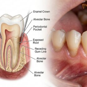
In a healthy, youthful dentition, the space between two adjacent teeth—from the point where they touch (the contact point) down to the underlying bone—is completely filled by a specialized triangle of pink gum tissue known as the interdental papilla. When this tissue is blunted, recedes, or is entirely lost, an empty space is left behind. Because the inside of the mouth is dark, this empty space appears as a distinct black triangle against the backdrop of white teeth. In clinical dental terminology, this condition is referred to as an open gingival embrasure.
The architecture of the interdental papilla is highly dependent on the underlying alveolar bone. The papilla does not stand up on its own; it is draped over the bone crest between the teeth. A landmark clinical study in periodontology established a critical metric known as Tarnow’s Rule. According to this principle, if the distance from the base of the tooth contact point to the crest of the underlying bone is 5 millimeters or less, the papilla will fill the space almost 100% of the time[1]. However, if that distance increases to 6 millimeters, the tissue only fills the space about 56% of the time. At 7 millimeters, the presence of a complete papilla drops to a mere 27%. This biological limitation explains why even minor bone loss can result in highly visible dark spaces.
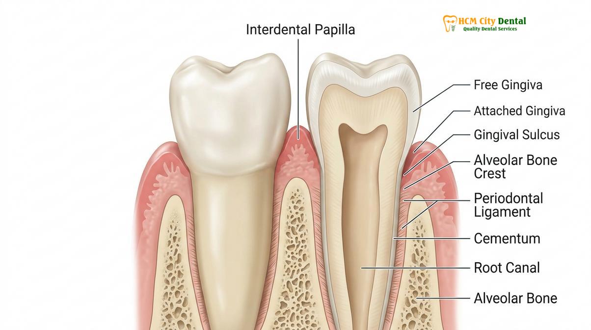
The biotype of the patient’s gingiva also plays a crucial role in the formation of these spaces. Patients with a thick, flat gingival biotype tend to have dense, fibrotic gum tissue that is highly resistant to recession. Conversely, patients with a thin, highly scalloped gingival biotype have delicate papillae that are highly susceptible to trauma, inflammation, and subsequent recession. Understanding this anatomical foundation is the first step in diagnosing and treating open embrasures effectively.
From a clinical perspective, Dr. Nguyen Van Cuong emphasizes that evaluating the underlying bone level via radiographic imaging is mandatory before promising any aesthetic closure of these spaces. If the bone has receded significantly, the treatment focus must shift from attempting to grow tissue to altering the shape of the teeth to meet the new, lower tissue level.
Main Causes of Gaps and Black Triangles Between Teeth
These dark spaces primarily result from periodontal bone loss, orthodontic tooth movement, natural triangular tooth anatomy, or age-related gum recession.
The development of open gingival embrasures is rarely due to a single factor; it is typically multifactorial, involving a combination of disease processes, anatomical predispositions, and sometimes even the unintended consequences of dental treatments. Understanding the etiology is critical for preventing further progression and selecting the correct restorative approach.
Periodontal Disease and Bone Loss: The most common pathological cause of black triangles is periodontitis. As plaque and calculus accumulate below the gumline, the body’s immune response triggers chronic inflammation. This inflammation activates osteoclasts, which are cells that break down the alveolar bone supporting the teeth. As the bone crest recedes apically (towards the root), the interdental papilla loses its structural foundation and follows the bone downward. This results in gaps in teeth gum recession, exposing the narrower root surfaces and creating large triangular voids[2].
Orthodontic Tooth Movement: It is a common clinical paradox that patients undergo orthodontic treatment to achieve a perfect smile, only to discover black triangles once their braces or clear aligners are removed. This occurs for two main reasons. First, when teeth are severely crowded and overlapping, the bone and gum tissue between them are often blunted or absent. However, because the teeth are overlapping, the missing tissue is hidden. Once the teeth are straightened and aligned side-by-side, the pre-existing lack of tissue is suddenly unmasked. Second, if the roots of the teeth are diverged (angled away from each other) during orthodontic movement, the contact point between the crowns moves higher up, increasing the distance to the bone crest and violating Tarnow’s 5mm rule.
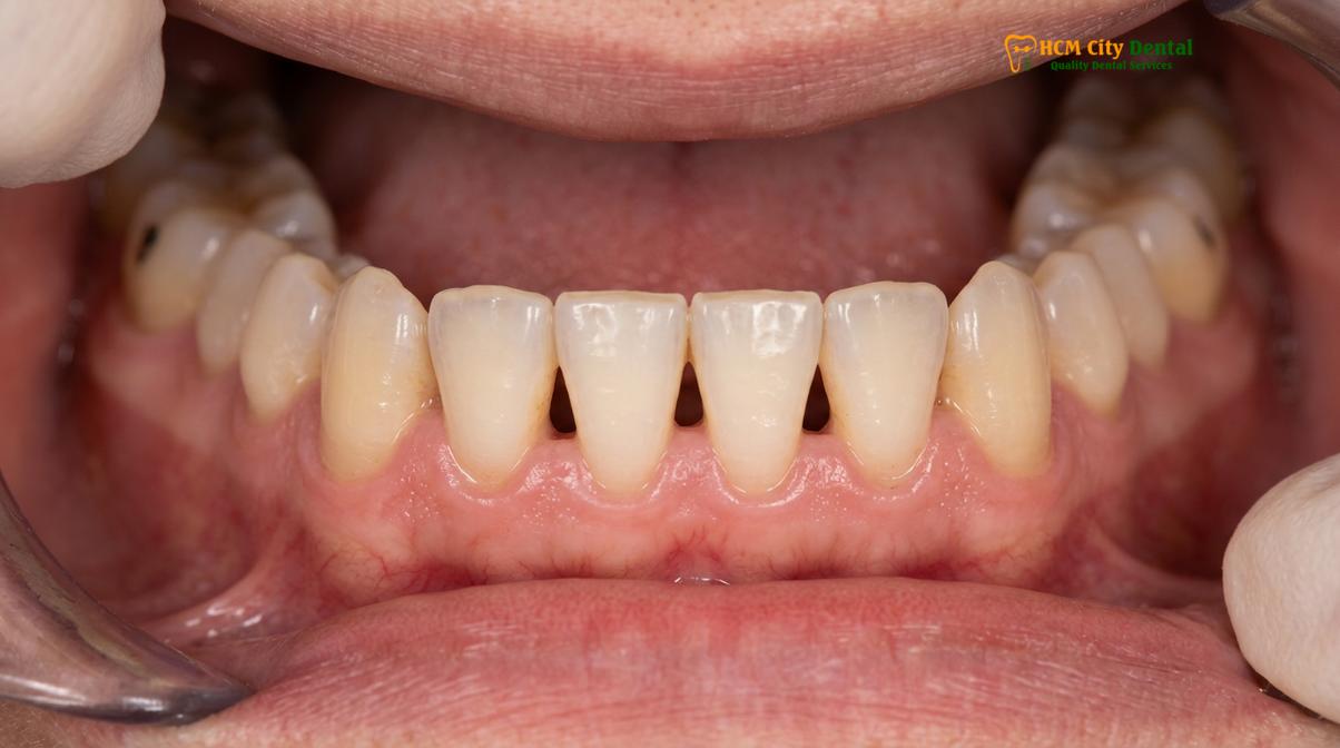
Tooth Morphology (Shape): The natural anatomical shape of a patient’s teeth heavily influences their susceptibility to open embrasures. Teeth generally fall into three shape categories: square, ovoid, and triangular. Square teeth have long, broad contact areas that extend deep toward the gumline, leaving very little space for a papilla; thus, they rarely develop black triangles. Triangular teeth, however, touch only at the very incisal (biting) edges and taper significantly toward the root. This creates a large, wide embrasure space that requires a massive, long papilla to fill it. Any slight recession in a patient with triangular teeth will immediately result in a highly visible dark space.
“The loss of the interdental papilla is one of the most frustrating aesthetic complications in dentistry. Because the papilla relies entirely on the underlying bone crest for support, any condition that causes bone resorption—whether inflammatory periodontitis or traumatic brushing—will inevitably lead to the collapse of the gingival architecture.”
Aggressive Oral Hygiene and Aging: While meticulous oral hygiene is essential, overly aggressive brushing with hard-bristled toothbrushes or improper flossing techniques can cause mechanical trauma to the delicate interdental tissue. Over time, this physical abrasion literally wears away the papilla. Furthermore, natural physiological aging is associated with a gradual, generalized recession of the periodontal tissues, making open embrasures more prevalent in older adult populations.
Why Gaps are More Than just a Cosmetic Issue
Beyond aesthetic concerns, open gingival embrasures create severe food traps, increase plaque retention, and can lead to phonetic issues like speech whistling.
While the aesthetic impact of black triangles is often the primary reason patients seek treatment, the clinical implications extend far beyond cosmetics. The interdental papilla serves a vital physiological function: it acts as a biological barrier that deflects food debris away from the interproximal spaces during mastication (chewing). When this barrier is lost, the resulting void becomes a significant functional liability.
Food Impaction and Plaque Retention: The most immediate consequence of an open embrasure is food impaction. Fibrous foods, seeds, and particulate matter easily become wedged in the dark spaces between the teeth. Because the space is wider at the gumline and narrower at the contact point, it acts like a funnel, trapping debris. This creates an ideal stagnation area for plaque biofilm. The accumulation of plaque in these hard-to-clean areas shifts the local oral microbiome toward anaerobic, disease-causing bacteria, which can accelerate localized gingivitis and further periodontal breakdown[3]. If you have black gaps between teeth, you are at a significantly higher risk for interproximal dental caries (cavities between the teeth) and localized bone loss.
Clinical Warning: Ignoring large open gingival embrasures can lead to a vicious cycle of periodontal destruction. The trapped food causes localized inflammation, which leads to further bone loss, which in turn makes the black triangle even larger and more prone to trapping food. Early intervention and meticulous interdental hygiene are critical to breaking this cycle.
Phonetic Impairment: The anterior teeth and the surrounding soft tissues play a crucial role in speech articulation. The interdental papillae help seal the spaces between the teeth, directing airflow properly when forming specific sounds. When large black triangles are present, air can escape through the gaps during speech. This often results in a noticeable whistling sound or a lisp, particularly when pronouncing sibilant sounds (like “s” and “z”) or fricative sounds (like “f” and “v”). For many patients, this phonetic impairment is just as distressing as the aesthetic compromise.
Saliva Ejection (Spitting): In severe cases where multiple large embrasures are present in the anterior maxilla (upper front teeth), patients may experience involuntary saliva ejection or “spraying” while speaking. The loss of the soft tissue seal allows saliva to be pushed through the gaps by the tongue and air pressure, causing significant social embarrassment and psychological distress.
Non-Surgical Treatments: Composite Bonding and Veneers
Dentists can close these spaces conservatively using injection-molded composite bonding or porcelain veneers to reshape the teeth and close the embrasure.
When the underlying bone loss is stable and the primary goal is to eliminate the dark spaces, restorative dentistry offers highly predictable, non-surgical solutions. Instead of attempting to grow the gum tissue back up to the contact point, the clinical strategy shifts to bringing the contact point down to meet the existing gum tissue. This is achieved by adding restorative material to the sides of the teeth, changing their shape from triangular to more square or rectangular.
Injection-Molded Composite Bonding (The Bioclear Method): One of the most significant advancements in modern restorative dentistry is the use of injection-molded composite resin to close black triangles. Traditional free-hand composite bonding often resulted in ledges or overhangs at the gumline, which trapped plaque and caused gingival inflammation. The modern approach utilizes specialized, anatomically curved clear mylar matrices that are slipped under the gumline.
The tooth is first meticulously cleaned using a micro-blaster with aluminum trihydroxide powder to remove all biofilm. The tooth is then etched and coated with a bonding agent. The clear matrices are placed, and a heated, highly flowable composite resin is injected into the mold, followed immediately by a paste composite. The heat allows the composite to flow seamlessly around the root and crown, creating a perfectly smooth, monolithic restoration with no plaque-retentive ledges. This technique is highly conservative, requiring zero removal of natural tooth structure, making it an ideal choice for patients looking to fix black triangles saigon without invasive procedures[4].
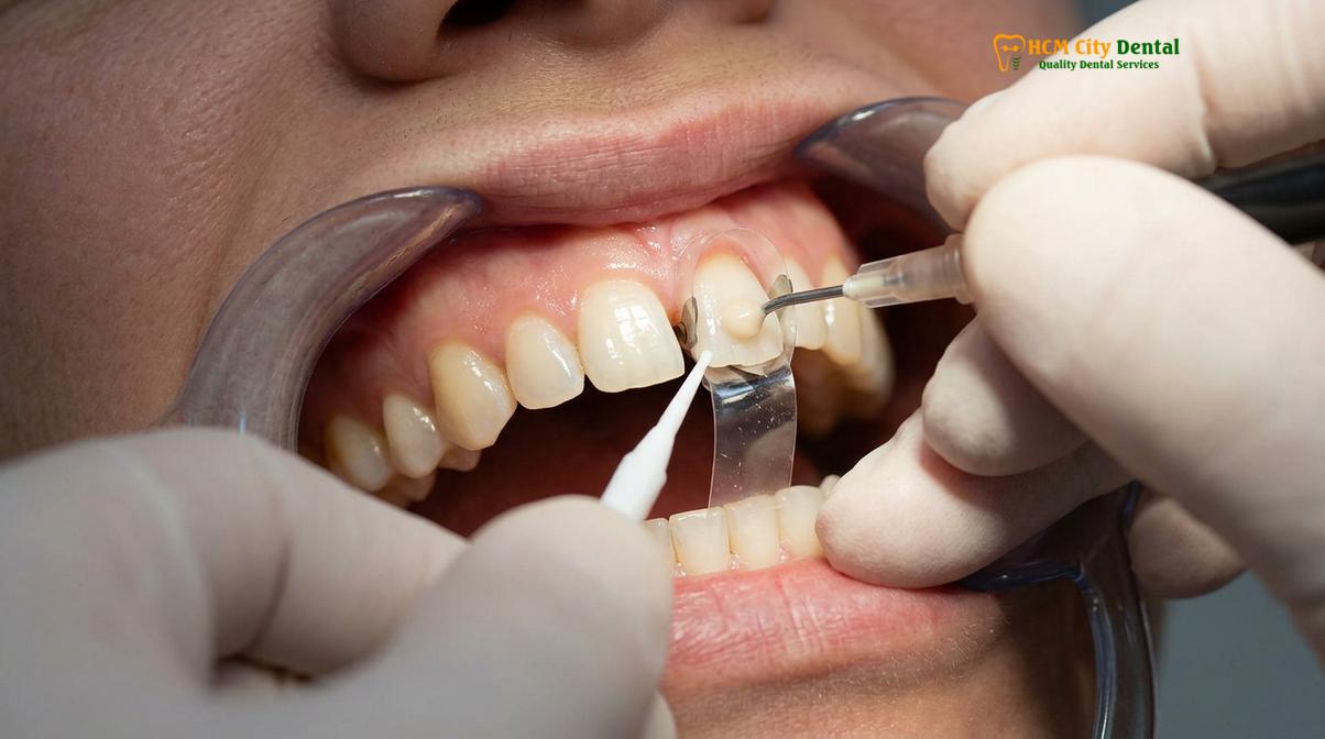
Clinical Case Example: A 45-year-old patient visited HCMC Dental Clinic in Ho Chi Minh City complaining of severe food impaction and aesthetic dissatisfaction due to large black triangles between her lower incisors following periodontal therapy. Because her bone levels were stable but reduced, surgery was contraindicated. The clinical team utilized injection-molded composite bonding to reshape the four lower incisors. The procedure was completed in a single visit without local anesthesia, successfully closing the embrasures, eliminating the food traps, and restoring a youthful contour to her smile.
Porcelain Veneers and Crowns: For patients who also wish to change the color, length, or overall facial alignment of their teeth, porcelain veneers offer a comprehensive solution. A veneer is a thin shell of custom-crafted ceramic that is bonded to the front and sides of the tooth. To close a black triangle, the dental ceramist designs the veneer with a modified emergence profile—meaning the porcelain bulges slightly at the sides near the gumline to physically fill the empty space.
While porcelain is highly durable, stain-resistant, and offers superior optical properties compared to composite resin, it typically requires the irreversible removal of a small amount of natural tooth enamel to accommodate the thickness of the ceramic. Therefore, veneers are usually recommended when the patient has multiple aesthetic concerns beyond just the open embrasures, such as severe discoloration, enamel wear, or minor misalignment.
Orthodontic Solutions: Interproximal Reduction (IPR)
Orthodontists use Interproximal Reduction (IPR) to gently reshape triangular teeth into rectangular forms, allowing them to be moved closer together to eliminate gaps.
When black triangles are caused by the natural triangular shape of the teeth or by diverging roots, orthodontic intervention can provide a biological solution that requires no restorative materials. The most effective orthodontic technique for managing open gingival embrasures is Interproximal Reduction (IPR), sometimes referred to as enamel stripping or reproximation.
The biomechanical principle behind IPR is straightforward. Triangular teeth touch only at their widest point near the biting edge. By using specialized diamond-coated strips or ultra-thin oscillating discs, the orthodontist carefully removes a microscopic amount of enamel (usually between 0.2mm to 0.5mm) from the sides of the teeth where they touch. This process flattens the contact point, effectively changing the tooth shape from a triangle to a rectangle.
Once the teeth have been reshaped, orthodontic forces (via traditional braces or clear aligners) are applied to push the teeth together, closing the newly created space. As the teeth move together, the contact point is physically lowered toward the gumline. By moving the contact point apically, the distance between the contact and the underlying bone crest is reduced. If this distance is brought back within Tarnow’s 5mm threshold, the interdental papilla will often naturally creep back up and fill the remaining space[5].
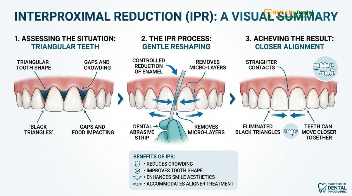
IPR is a highly safe and routine procedure when performed correctly. The amount of enamel removed is strictly calculated to ensure that the tooth remains structurally sound and resistant to decay. This approach is particularly advantageous because it provides a permanent solution using the patient’s natural tooth structure, avoiding the long-term maintenance associated with composite bonding or porcelain veneers. Patients undergoing comprehensive gum therapy and restorative planning often benefit from a multidisciplinary approach where orthodontics is used first to minimize the embrasure size, followed by minimal restorative touch-ups if necessary.
Surgical and Regenerative Options: Gum Grafting
While challenging, surgical gum grafting and microsurgical tissue regeneration can sometimes rebuild lost papilla, though case selection is highly critical for success.
Patients frequently ask if the missing gum tissue can simply be grafted back into place. While periodontal plastic surgery has achieved remarkable success in treating facial gum recession (recession on the front of the tooth), reconstructing the interdental papilla remains one of the most unpredictable and challenging procedures in all of dental microsurgery.
The primary limitation is blood supply. A standard gum graft placed on the front of a tooth receives a robust blood supply from the underlying periosteum and the surrounding lateral tissues. However, the interdental papilla is a tiny, isolated peak of tissue suspended between two avascular (bloodless) tooth roots. When a surgeon attempts to place a graft into this tiny space, the graft often fails to establish a sufficient blood supply and undergoes necrosis (dies) before it can integrate. Furthermore, because the underlying bone crest has usually resorbed, there is no physical “tent pole” to hold the new tissue up[6].
Despite these challenges, specific microsurgical techniques can be employed in highly selected cases where the bone loss is minimal and the tissue loss is primarily due to trauma rather than severe periodontitis.
| Treatment Modality | Mechanism of Action | Invasiveness | Predictability |
|---|---|---|---|
| Composite Injection Molding | Adds resin to tooth sides to close gap | Non-invasive (No drilling) | Highly Predictable |
| Porcelain Veneers | Custom ceramic shells alter tooth shape | Minimally invasive (Enamel prep) | Highly Predictable |
| Orthodontic IPR | Reshapes enamel, moves teeth together | Conservative (Micro-enamel removal) | Highly Predictable |
| Microsurgical Gum Grafting | Transplants tissue to rebuild papilla | Surgical (Donor site required) | Low to Moderate (Case dependent) |
Connective Tissue Grafting and Tunneling: In advanced gingival embrasures treatment, a periodontist may utilize a microsurgical tunneling technique. Instead of cutting the gums open, tiny instruments are used to gently separate the gum tissue from the bone, creating a small envelope or tunnel. A piece of connective tissue, usually harvested from the roof of the patient’s mouth, is then carefully threaded through this tunnel and suspended under the papilla to plump it up.
Dr. Nguyen Van Cuong notes that while surgical options exist, they must be approached with extreme caution and realistic patient expectations. In many cases, a combined approach is the most effective: utilizing orthodontics or restorative dentistry to close the majority of the space, and relying on meticulous periodontal maintenance to ensure the remaining tissue stays healthy and stable.
Daily Care Tips to Prevent Black Triangles from Growing
Preventing the expansion of black triangles requires meticulous interdental cleaning using correctly sized interproximal brushes and water flossers to halt further gum recession.
If you already have open gingival embrasures, your primary daily objective is to prevent them from getting larger. Because these spaces act as plaque traps, standard brushing is insufficient. The bristles of a regular toothbrush simply bridge over the gap, leaving the stagnant biofilm inside the triangle completely untouched.
Interdental Brushes: The gold standard for cleaning black triangles is the use of interdental brushes (often referred to as interproximal brushes). These look like tiny pipe cleaners designed to slide directly through the space between the teeth. It is critical to select the correct size. If the brush is too small, it will not effectively sweep the plaque off the adjacent root surfaces. If it is too large, forcing it through the space can cause mechanical trauma and actually worsen the gum recession. Your dental hygienist can measure your embrasures and prescribe the exact color-coded brush sizes needed for different areas of your mouth.
“Maintenance of the open embrasure is a lifelong commitment. The space must be kept immaculately clean to prevent the localized inflammatory response that leads to further alveolar bone resorption and subsequent enlargement of the black triangle.”
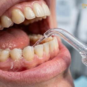
Water Flossers and Modified Brushing: Oral irrigators, or water flossers, are excellent adjuncts for flushing out loose food debris and disrupting the plaque biofilm deep within the embrasure space. They are particularly useful for patients who struggle with manual dexterity. When using a standard toothbrush, adopt the Modified Bass technique: angle the bristles at 45 degrees toward the gumline and use gentle, microscopic vibratory motions rather than aggressive back-and-forth scrubbing. Avoid using wooden toothpicks, as aggressive picking can splinter and severely traumatize the delicate interdental tissue.
When to See a Doctor
While small black triangles may simply be a cosmetic annoyance, sudden changes in your gingival architecture require professional evaluation. You should schedule a clinical examination if you notice the gaps between your teeth widening rapidly, if the gums between your teeth bleed easily when brushing or flossing, or if you experience chronic bad breath and localized pain, as these are hallmark signs of active periodontal disease. Furthermore, if food impaction becomes severe enough to cause constant discomfort, intervention is necessary to prevent interproximal decay. Patients in Ho Chi Minh City seeking comprehensive evaluation can consult with the periodontal and restorative teams at HCMC Dental Clinic to determine the underlying cause of tissue loss. A thorough radiographic assessment and periodontal probing will dictate whether the appropriate path forward is non-surgical management, orthodontic reshaping, or advanced general dentistry restorative care to protect the long-term health of your smile.
Frequently Asked Questions
Why do I have black triangles between my teeth?
Black triangles form when the interdental gum tissue (papilla) recedes or when bone loss occurs, leaving an open space. Common causes include periodontal disease, natural triangular tooth shape, aging, aggressive brushing, or the aftermath of orthodontic tooth alignment. Once the underlying bone support is lost, the gum tissue inevitably follows, exposing the dark space between the roots.
Can braces cause black triangles?
Yes, braces or clear aligners can reveal black triangles. When crowded, overlapping teeth are straightened, the hidden lack of gum tissue between them becomes visible. Additionally, moving roots apart can shift the contact point higher, creating an open embrasure. Orthodontists often anticipate this and may plan for interproximal reduction to mitigate the space.
How do dentists fix black triangles?
Dentists fix these spaces using several methods, including injection-molded composite bonding, porcelain veneers, orthodontic interproximal reduction (IPR) to reshape and close the teeth, or in specific cases, microsurgical gum grafting to rebuild the lost tissue. The choice of treatment depends heavily on the patient’s bone levels, tooth shape, and aesthetic goals.
Can gum grafting fix black triangles?
Gum grafting can sometimes improve black triangles, but it is clinically challenging. Because the interdental papilla lacks an underlying bone supply to support the graft, surgical reconstruction is unpredictable and usually reserved for specific, highly selected anatomical cases. Often, restorative or orthodontic approaches yield more predictable aesthetic results.
Do black triangles trap food?
Yes, open gingival embrasures act as significant food and plaque traps. Because the natural gum barrier is missing, debris easily accumulates in these spaces, increasing the risk of localized gum inflammation, bad breath, and further periodontal deterioration if not cleaned properly. Specialized interdental brushes are usually required to keep these areas clean.
References
- Journal of Periodontology. The effect of the distance from the contact point to the crest of bone on the presence or absence of the interproximal dental papilla. (1992).
- American Journal of Orthodontics and Dentofacial Orthopedics. Incidence and severity of open gingival embrasures after orthodontic treatment. (2010).
- Journal of Esthetic and Restorative Dentistry. Management of open gingival embrasures: a review of the literature. (2018).
- British Dental Journal. The use of injection molded composite for the management of black triangles. (2021).
- International Journal of Periodontics & Restorative Dentistry. Interproximal reduction (IPR) and its effects on interdental papilla contour. (2019).
- Clinical Advances in Periodontics. Microsurgical approaches to papilla reconstruction: limitations and outcomes. (2022).