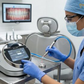
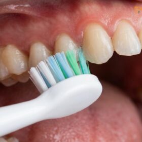
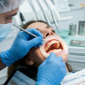
Gum flap surgery is a specialized periodontal procedure designed to treat advanced gum disease by temporarily separating the gum tissue from the teeth. This allows the periodontist to access deep periodontal pockets, thoroughly clean the tooth roots, and repair damaged underlying bone structure.
Clinical Summary:
Gum flap surgery, clinically referred to as osseous surgery, is a critical intervention for severe periodontitis when non-surgical treatments prove insufficient. By reflecting the gingival tissue, clinicians gain direct visual access to debride contaminated root surfaces, remove pathogenic biofilm, and recontour irregular alveolar bone. This surgical approach effectively reduces pocket depths, halts progressive bone loss, and creates a biologically healthy environment that patients can maintain through daily oral hygiene and professional care.
Key Takeaways:
- Indicated for deep periodontal pockets that do not respond to non-surgical scaling and root planing.
- Involves lifting the gum tissue to directly clean roots and reshape damaged alveolar bone.
- Routinely performed under local anesthesia to ensure patient comfort throughout the procedure.
- Initial soft tissue recovery generally takes one to two weeks, with sutures removed post-operatively.
- Offers a high success rate in preserving natural teeth when combined with strict long-term maintenance.
What is Gum Flap Surgery (Osseous Surgery)?
Gum flap surgery is a surgical procedure that folds back the gingival tissue to remove disease-causing bacteria, clean the tooth roots, and repair bone defects caused by advanced periodontitis.
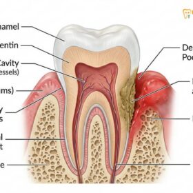
Periodontal disease is a progressive inflammatory condition initiated by dysbiotic bacterial plaque that accumulates below the gumline. In its early stages, known as gingivitis, the inflammation is confined to the soft tissues and is generally reversible. However, if left untreated, the bacterial infection penetrates deeper into the periodontium—the complex structure of tissues that support the teeth, including the gingiva, periodontal ligament, cementum, and alveolar bone. This advanced stage, characterized by the irreversible destruction of supporting bone, is where comprehensive gum treatment protocols become essential.
Gum flap surgery, frequently referred to in the dental community as osseous surgery, is the gold standard for managing moderate to severe cases of periodontitis. The primary objective of this procedure is to eliminate the anatomical spaces—known as periodontal pockets—where anaerobic bacteria thrive. In a healthy mouth, the gum tissue fits snugly around the teeth, with a natural sulcus depth of 1 to 3 millimeters. When periodontal disease advances, the tissue detaches from the tooth, creating pockets that measure 5 millimeters or deeper. These deep pockets are virtually impossible for a patient to clean at home and are highly resistant to standard professional cleanings.[1]
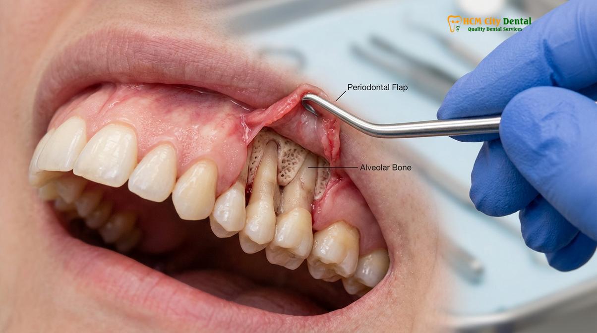
During the procedure, the periodontist makes precise incisions to gently separate the gums from the teeth, creating a “flap.” This temporary reflection of the soft tissue provides the surgeon with direct, unhindered visibility and access to the diseased root surfaces and the underlying alveolar bone. Once the area is exposed, the clinician can meticulously remove hardened calculus (tartar), inflammatory granulation tissue, and bacterial toxins. Furthermore, if the infection has caused the underlying bone to become pitted, cratered, or uneven, the surgeon will reshape the bone to restore a positive, healthy architecture. This specific application of the procedure is often necessary to address complex cases of osseous surgery periodontitis, ensuring that the gums can reattach firmly to a smooth, clean surface.
By reducing the depth of the periodontal pockets and eliminating the bacterial reservoir, gum flap surgery halts the progression of the disease. It transforms a highly diseased, unmaintainable environment into a healthy, shallow sulcus that the patient can effectively manage with daily brushing, flossing, and routine professional maintenance.
When is Surgical Periodontal Treatment Necessary?
Surgical intervention becomes necessary when deep periodontal pockets persist despite thorough non-surgical treatments, threatening the stability of the teeth and surrounding bone.
The decision to proceed with surgical periodontal therapy is based on a comprehensive clinical and radiographic evaluation. The initial phase of treating periodontitis almost always involves non-surgical therapy, primarily scaling and root planing (SRP). This deep cleaning procedure aims to remove plaque and calculus from the root surfaces beneath the gums. Following SRP, the patient undergoes a healing period of several weeks, after which the periodontist performs a re-evaluation to assess the tissue response.
If the re-evaluation reveals that the gums have healed, bleeding has ceased, and pocket depths have reduced to manageable levels (typically 4 millimeters or less), surgery may not be required. However, if deep pockets of 5 millimeters or greater persist, accompanied by bleeding on probing and radiographic evidence of ongoing bone loss, surgical intervention is strongly indicated. In these deep, tortuous pockets, non-surgical instruments simply cannot reach the base of the defect to remove all the calculus and bacteria. Leaving these deep pockets untreated allows the infection to smolder, inevitably leading to further bone destruction and eventual tooth loss.[2]
Clinical Warning: Ignoring persistent deep periodontal pockets can lead to severe consequences beyond tooth mobility and loss. Chronic periodontal infection serves as a constant source of systemic inflammation, which has been clinically linked to an increased risk of cardiovascular disease, poorly controlled diabetes, and other systemic health complications.
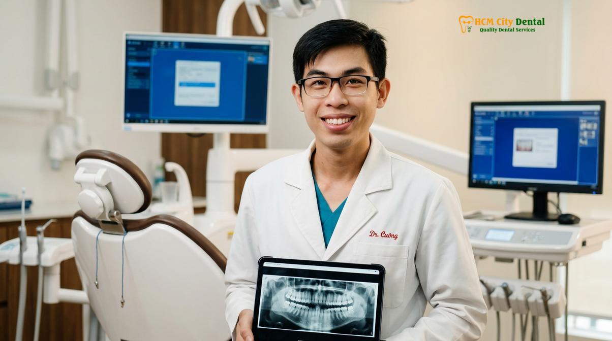
Dr. Nguyen Van Cuong, a leading specialist at HCMC Dental Clinic in Ho Chi Minh City, emphasizes that patient selection is critical for the success of periodontal flap surgery. Ideal candidates are those who demonstrate a strong commitment to improving their home oral hygiene and who have ceased habits that compromise healing, such as tobacco smoking. Smoking significantly impairs the microvascular blood supply to the gingival tissues, reducing the predictability of surgical outcomes and increasing the risk of disease recurrence.
Furthermore, surgical treatment is often necessary when the pattern of bone loss is irregular. Periodontal disease does not destroy bone evenly; it often creates vertical defects, craters, and trenches around the teeth. These anatomical deformities act as protected niches for bacteria. Without surgical access to recontour these defects, the overlying gum tissue will not adapt properly, and the deep pockets will remain. Therefore, the necessity of surgery is dictated not just by the depth of the pocket, but by the underlying morphology of the bone defect.
The Osseous Surgery Procedure Explained
The procedure involves administering local anesthesia, making precise incisions to create a tissue flap, debriding the roots, and suturing the gums back into a healthier position.
The execution of gum flap surgery requires a deep understanding of periodontal anatomy, microvascular blood supply, and precise surgical technique. The procedure is meticulously planned to maximize disease eradication while minimizing trauma to the delicate oral tissues. The workflow can be divided into several distinct clinical phases.
1. Anesthesia and Site Preparation: Patient comfort is paramount. The procedure begins with the administration of profound local anesthesia to completely numb the surgical site. Depending on the extent of the surgery and the patient’s anxiety levels, adjunctive sedation options, such as oral anxiolytics or intravenous conscious sedation, may be utilized. Once the area is anesthetized, the surgical field is isolated and prepared with antimicrobial rinses to reduce the bacterial load in the oral cavity.
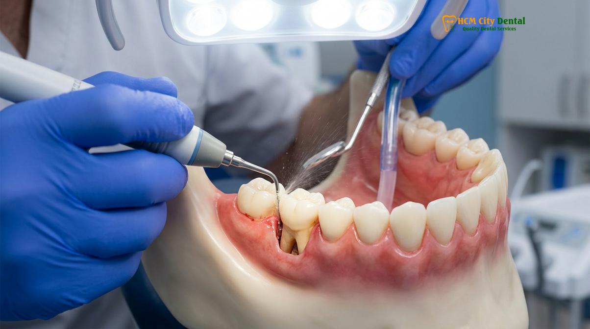
2. Incision Design and Flap Elevation: The surgeon uses a micro-scalpel to make highly precise incisions around the necks of the teeth. The design of the incision is critical; it must allow for adequate access to the underlying structures while preserving the blood supply to the tissue flap. A full-thickness mucoperiosteal flap is then carefully elevated using specialized instruments. This step separates the gingiva and the underlying periosteum from the bone, exposing the diseased root surfaces and the alveolar bone crest.
“The primary objective of flap elevation is to gain unhindered visual access to the root surface and the alveolar bone. This direct line of sight is what differentiates surgical therapy from non-surgical approaches, allowing for the complete eradication of subgingival calculus and the precise management of osseous defects.”
3. Debridement and Root Conditioning: With the roots fully exposed, the clinician utilizes a combination of ultrasonic scalers and hand curettes to meticulously remove all traces of calculus, bacterial biofilm, and infected granulation tissue. The root surfaces are planed until they are hard and smooth. In some advanced protocols, chemical root conditioning agents, such as citric acid or EDTA, may be applied to remove the smear layer and open the dentinal tubules, which can encourage the reattachment of healthy connective tissue fibers.[3]
4. Osseous Recontouring (if necessary): If the bone has been damaged by the infection, the surgeon will perform osseous recontouring (detailed in the next section) to restore a physiological architecture.
5. Flap Closure and Suturing: Once the roots are clean and the bone is properly shaped, the surgical site is thoroughly irrigated with sterile saline. The tissue flaps are then repositioned apically (lower down on the tooth root) to eliminate the pocket depth. The flaps are secured in place using fine surgical sutures. In some cases, a periodontal dressing—a soft, putty-like material—may be placed over the surgical site to protect the area, stabilize the tissues, and enhance patient comfort during the initial healing phase.
Pocket Reduction and Bone Contouring Steps
During this phase, the surgeon smooths irregular bone surfaces and reduces pocket depths to eliminate areas where harmful bacteria can hide and proliferate.
A critical component of many flap procedures is the management of the alveolar bone, which is why the procedure is frequently termed osseous surgery. When periodontitis destroys the bone supporting the teeth, it rarely does so in a flat, horizontal manner. Instead, it creates jagged edges, deep craters, and vertical defects. If the gum tissue is simply sutured back over these bony deformities, the deep pockets will immediately reform, as the tissue cannot adapt tightly to an irregular surface.
To achieve a successful surgical pocket reduction, the periodontist must reshape the bone to resemble its natural, healthy contours—a concept known as creating “positive architecture.” This involves two primary techniques: osteoplasty and ostectomy.
Osteoplasty refers to the reshaping of non-supporting bone. This involves thinning thick bony ledges or exostoses that prevent the gum tissue from lying flat against the tooth. Because osteoplasty does not remove bone that is directly attached to the tooth root, it does not compromise the structural support of the tooth.
Ostectomy, on the other hand, involves the careful removal of a small amount of supporting bone. This is often necessary to eliminate deep interdental craters or to level out vertical defects. While removing supporting bone may seem counterintuitive, it is a necessary compromise to establish a smooth, sloping bone profile. This physiological contour allows the gingival tissue to attach firmly to the tooth root, effectively eliminating the periodontal pocket and creating an environment that the patient can keep clean.
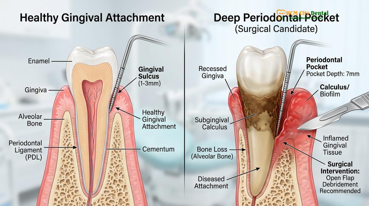
The morphology of the bone defect dictates the specific surgical approach. Periodontal bone defects are generally classified by the number of bony walls remaining around the tooth root.
| Defect Type | Clinical Description | Typical Surgical Approach |
|---|---|---|
| 1-Wall Defect | Only one bony wall remains; severe localized bone loss. | Primarily resective (ostectomy) to level the bone, or extraction if the tooth is deemed hopeless. |
| 2-Wall Defect (Crater) | Two walls remain (usually buccal and lingual); common between posterior teeth. | Osseous recontouring to ramp the bone and eliminate the crater, allowing tissue adaptation. |
| 3-Wall Defect | Three bony walls remain, creating a deep, narrow trench. | Highly amenable to regenerative procedures (bone grafting) rather than purely resective surgery. |
In cases where the bone defects are deep and narrow (such as 3-wall defects), the surgeon may opt for regenerative techniques rather than resective osseous surgery. This involves placing bone graft materials and barrier membranes into the defect to stimulate the body to regenerate the lost bone and periodontal ligament. The choice between resective and regenerative approaches is highly nuanced and depends on the specific anatomy of the defect, the tooth’s strategic value, and the patient’s overall health profile.[4]
Recovery After Gum Flap Surgery: Timeline and Care
Recovery involves managing mild discomfort and swelling for the first few days, followed by a gradual return to normal oral hygiene practices over several weeks.
The success of periodontal surgery relies heavily on the patient’s adherence to post-operative care instructions. While the surgical procedure removes the disease-causing factors, the actual healing and reattachment of the tissues occur in the weeks and months following the surgery. Understanding the timeline for gum surgery recovery helps patients manage their expectations and optimize their healing outcomes.
Immediate Post-Operative Phase (Days 1-3):Immediately following the procedure, it is normal to experience mild to moderate discomfort, swelling, and minor oozing from the surgical site. The periodontist will typically prescribe non-steroidal anti-inflammatory drugs (NSAIDs) to manage pain and reduce edema. Patients are instructed to apply an ice pack to the outside of the face in alternating 15-minute intervals during the first 24 hours to minimize swelling. Physical exertion should be strictly avoided, as elevated blood pressure can induce bleeding. During this time, mechanical brushing and flossing at the surgical site are prohibited to avoid disrupting the delicate blood clot and sutures.
Clinical Case Example: A 52-year-old patient presented to HCMC Dental Clinic in Ho Chi Minh City with advanced periodontitis and persistent 7mm pockets in the lower right quadrant. Dr. Nguyen Van Cuong performed osseous surgery to reduce the pockets and recontour the bone. By strictly following the prescribed soft-food diet and utilizing a chlorhexidine mouthrinse, the patient experienced minimal post-operative swelling. At the 6-month re-evaluation, the pocket depths had successfully reduced to a healthy, maintainable 3mm.
Early Healing Phase (Days 4-14):As the initial swelling subsides, the gingival tissues begin the process of epithelialization—forming a new surface layer. Discomfort usually diminishes significantly by the third or fourth day. Patients must maintain a soft-food diet, avoiding hard, crunchy, or spicy foods that could traumatize the healing gums. To maintain oral hygiene without mechanical brushing, a prescription antimicrobial mouthwash (such as 0.12% chlorhexidine gluconate) is used twice daily. Around the one-to-two-week mark, the patient will return to the clinic for a post-operative check-up, during which any non-resorbable sutures are gently removed.

Maturation Phase (Weeks 3-12):While the surface gums may appear healed within a few weeks, the underlying connective tissue and bone require several months to fully mature and remodel. During this phase, patients are gradually instructed to resume mechanical cleaning using an ultra-soft surgical toothbrush. The periodontist will provide specific techniques for cleaning the newly contoured areas, as the roots may be slightly more exposed, leading to temporary dentinal hypersensitivity. Desensitizing toothpastes containing potassium nitrate or stannous fluoride are highly effective in managing this transient sensitivity.
Long-term success dictates that the patient enters a strict periodontal maintenance program, typically requiring professional cleanings every three to four months. Without this ongoing professional care and meticulous home hygiene, the pathogenic bacteria will inevitably repopulate the sulcus, leading to disease recurrence.
Comparing Flap Surgery vs Non-Surgical Scaling (SRP)
While scaling and root planing treat early-stage gum disease non-invasively, flap surgery provides the direct access required to resolve advanced bone loss and deep pockets.
Understanding the distinction between non-surgical and surgical periodontal therapy is crucial for patients navigating their treatment options. Both modalities aim to eliminate bacterial biofilm and calculus from the root surfaces, but their applications, techniques, and capabilities differ significantly based on the severity of the disease.
Non-Surgical Scaling and Root Planing (SRP):SRP is the foundational treatment for all periodontal diseases. It is a “blind” procedure, meaning the clinician relies on tactile sensitivity rather than direct vision to detect and remove subgingival calculus. Using specialized curettes and ultrasonic instruments, the hygienist or dentist cleans the root surfaces beneath the gumline. SRP is highly effective for treating gingivitis and mild to moderate periodontitis, where pocket depths are generally between 4 and 5 millimeters. In these shallower pockets, the instruments can reach the base of the defect, and the tissues often respond well, shrinking tightly around the clean root.[5]
Periodontal Flap Surgery:When pocket depths exceed 5 millimeters, the efficacy of non-surgical SRP drops precipitously. Studies have shown that in pockets deeper than 6 millimeters, complete removal of calculus via blind scaling is rarely achieved. This is where flap surgery becomes indispensable. By reflecting the gum tissue, the surgeon gains direct, illuminated access to the entire root surface, including complex anatomical areas like root concavities and furcations (the areas where the roots of multi-rooted teeth divide).
Furthermore, SRP cannot alter the underlying bone architecture. If the disease has created bony craters or vertical defects, non-surgical therapy will leave these deformities intact, meaning the deep pockets will persist even if the roots are temporarily cleaned. Flap surgery allows for osseous recontouring, physically reshaping the bone to eliminate the anatomical niches where bacteria hide. In essence, while SRP manages the infection, flap surgery corrects the anatomical damage caused by the infection, creating a sustainable environment for long-term oral health.
When to See a Doctor
Periodontal disease is often described as a “silent” condition because it can progress significantly without causing severe pain. However, recognizing the clinical signs of advanced gum disease is critical for timely intervention. If you experience any of the following symptoms, it is imperative to seek a comprehensive evaluation from a dental professional.
“Early detection of periodontal attachment loss drastically improves the prognosis of the dentition. Patients should not wait for teeth to become loose or painful before seeking specialist care; persistent gingival bleeding is a definitive sign of active infection requiring clinical assessment.”
You should schedule a consultation for general dentistry and periodontal evaluation if you notice:
- Persistent Bleeding: Gums that bleed easily during normal brushing, flossing, or eating.
- Gingival Recession: Gums that appear to be pulling away from the teeth, making the teeth look longer than normal.
- Purulent Exudate: Pus visible between the teeth and gums when pressure is applied.
- Tooth Mobility: Teeth that feel loose or shift position when biting or chewing.
- Pathologic Migration: The development of new spaces between the teeth or a change in the way your upper and lower teeth fit together.
- Chronic Halitosis: Persistent bad breath or a bad taste in the mouth that does not improve with oral hygiene.
A thorough clinical examination, including periodontal probing and radiographic analysis, is necessary to determine the extent of the disease and whether surgical intervention is warranted. Only a qualified dental professional can provide an accurate diagnosis and formulate a personalized treatment plan based on your specific anatomical and biological needs.
Frequently Asked Questions
Why would someone need gum flap surgery?
A patient needs gum flap surgery when deep periodontal pockets cannot be effectively cleaned through non-surgical methods. This procedure allows the dentist to access the tooth roots directly, remove hardened bacteria, and halt progressive bone loss. It is typically recommended when pocket depths exceed 5 millimeters and show signs of active infection despite previous scaling and root planing.
How long does osseous surgery take to heal?
Initial healing of the gum tissue typically takes one to two weeks, at which point sutures are removed. However, complete maturation of the underlying bone and connective tissue can take several months of careful maintenance. During the first few days, patients may experience mild swelling and discomfort, which gradually subsides as the soft tissues reattach to the tooth roots.
Is gum flap surgery performed under sedation?
Gum flap surgery is routinely performed under local anesthesia to ensure the surgical site is completely numb. For patients with dental anxiety, oral sedatives or intravenous conscious sedation may be administered to enhance comfort. The choice of anesthesia depends on the complexity of the procedure, the duration of the surgery, and the patient’s individual medical history and comfort preferences.
What can I eat after periodontal surgery?
For the first few days after surgery, you should consume a soft-food diet such as yogurt, scrambled eggs, mashed potatoes, and lukewarm soups. Avoid hot, spicy, crunchy, or hard foods that could irritate the healing gums. It is also crucial to avoid using a straw, as the suction can dislodge the blood clot and disrupt the early stages of the healing process.
How much does osseous surgery cost?
The cost of osseous surgery varies based on the severity of the disease, the number of teeth involved, and whether bone grafting is required. A personalized clinical consultation is necessary to provide an accurate treatment estimate. Factors such as the need for advanced regenerative materials, the complexity of the bone recontouring, and the specific quadrant being treated will all influence the final clinical fee.
References
- Journal of Periodontology. Clinical efficacy of periodontal flap surgery in advanced periodontitis management. (2021).
- International Journal of Periodontics & Restorative Dentistry. Osseous resective surgery: biological rationale and clinical outcomes. (2020).
- Clinical Oral Investigations. Healing dynamics of the periodontium following surgical pocket reduction. (2019).
- Journal of Clinical Periodontology. Comparative analysis of surgical versus non-surgical periodontal therapy. (2022).
- American Academy of Periodontology. Guidelines for the management of severe periodontal diseases. (2018).