A bone graft for gum disease is a specialized surgical procedure designed to rebuild jawbone tissue destroyed by severe periodontitis. By placing grafting material into areas of bone loss, dentists create a scaffold that stimulates natural bone regeneration, stabilizing loose teeth and restoring a healthy foundation.
Clinical Summary:
Advanced periodontal disease leads to the irreversible destruction of the alveolar bone that anchors teeth in the jaw. To combat this, a periodontal bone graft is utilized as a regenerative therapy. This procedure involves cleaning the infected root surfaces and placing a biocompatible grafting material—often protected by a barrier membrane—into the bony defect. The graft acts as a biological scaffold, facilitating osteoconduction and osteoinduction, which encourages the patient’s own body to generate new bone cells. Over several months, this process restores structural integrity, saves teeth that would otherwise be lost to mobility, and creates a robust foundation for future restorative work such as dental implants. Success relies heavily on precise surgical execution and strict patient compliance with oral hygiene protocols.
Key Takeaways:
- Bone grafting is a targeted surgical intervention to reverse bone loss caused by severe periodontitis.
- The procedure utilizes various materials, including human, animal, or synthetic bone, to act as a regenerative scaffold.
- Guided Tissue Regeneration (GTR) often accompanies grafting to prevent soft tissue from interfering with bone growth.
- Complete biological healing and integration of the graft material typically spans four to nine months.
- Successful grafting stabilizes highly mobile teeth and ensures sufficient bone volume for successful implant placement.
How Advanced Periodontitis Causes Bone Loss
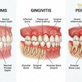
Advanced periodontitis triggers a chronic inflammatory response where the body’s immune system, while fighting bacterial infection, inadvertently destroys the supporting alveolar bone. This progressive bone loss compromises tooth stability and requires clinical intervention to halt.
To understand the necessity of regenerative surgery, one must first examine the pathophysiology of periodontal disease. The oral cavity is a complex microbiome. When oral hygiene is inadequate, pathogenic bacteria accumulate along the gingival margin, forming a sticky biofilm known as dental plaque. Over time, minerals from saliva calcify this plaque into hard calculus (tartar), which cannot be removed by standard brushing. This calculus harbors virulent bacteria that release toxins directly into the delicate gum tissue.
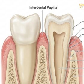
In response to these bacterial toxins, the body’s immune system mounts a vigorous defense. However, this chronic inflammatory response becomes a double-edged sword. The immune cells release inflammatory mediators, such as cytokines and interleukins, which inadvertently stimulate osteoclasts—the specialized cells responsible for breaking down bone tissue[1]. As the osteoclast activity outpaces the bone-building activity of osteoblasts, the alveolar bone that forms the socket around the tooth begins to resorb and melt away.
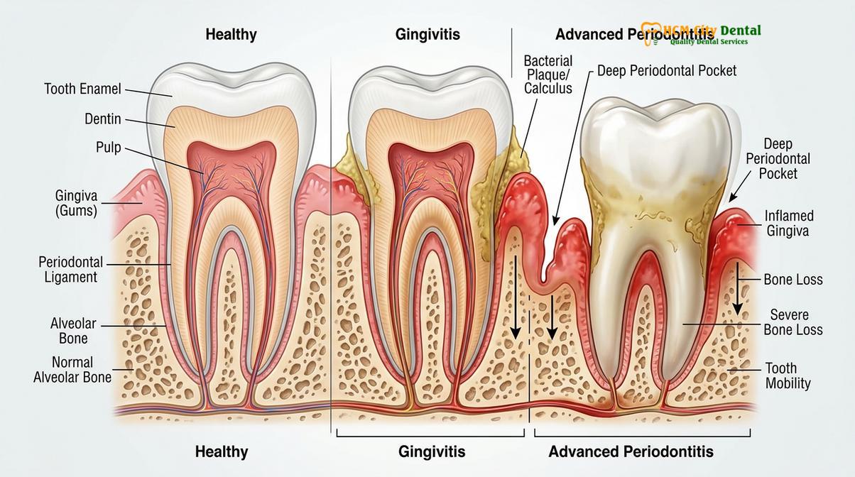
As the bone recedes, the periodontal ligaments that attach the tooth root to the bone are also destroyed. This creates deep periodontal pockets between the gums and the teeth, providing an even deeper, oxygen-deprived environment for anaerobic bacteria to thrive. The cycle of infection and bone destruction accelerates, leading to significant attachment loss. Without comprehensive gum treatment to eradicate the infection and regenerative procedures to rebuild the lost architecture, the affected teeth will eventually become highly mobile and exfoliate or require extraction.
What is a Periodontal Bone Graft?
A periodontal bone graft is a regenerative surgery that places bone-like material into defects caused by gum disease. It acts as a biological scaffold, encouraging your body to grow new bone cells and restore structural integrity.
When the alveolar bone has been significantly compromised, simply cleaning the infection is not enough to restore the tooth’s original stability. The lost bone does not naturally grow back on its own because fast-growing gum tissue tends to fill the empty space before the slower-growing bone cells have a chance to regenerate. This is where a periodontal bone graft becomes a critical clinical intervention.
The primary objective of this procedure is to achieve true periodontal regeneration—meaning the formation of new alveolar bone, new cementum (the calcified layer covering the tooth root), and new periodontal ligaments. The grafting material placed into the bony defect serves three fundamental biological functions:
- Osteoconduction: The graft material acts as a physical matrix or scaffold. It provides a three-dimensional framework upon which the patient’s own bone-forming cells (osteoblasts) can migrate, attach, and begin depositing new bone matrix.
- Osteoinduction: Certain grafting materials contain bone morphogenetic proteins (BMPs) and other growth factors that actively stimulate undifferentiated stem cells in the surrounding tissue to transform into active osteoblasts.
- Osteogenesis: In cases where autogenous bone (the patient’s own bone) is used, living bone cells are transplanted directly into the site, actively contributing to new bone formation immediately upon placement.
“The primary goal of a periodontal bone graft is not just to fill a void, but to signal the body’s natural regenerative mechanisms to rebuild the lost alveolar crest, effectively turning back the clock on periodontal destruction.”
By utilizing these biological principles, dentists can successfully reverse the damage inflicted by periodontitis, transforming a compromised, weakened tooth into a stable, functional component of the dental arch.
Types of Bone Grafting Materials Used in Dentistry
Dentists utilize four main types of grafting materials: autografts (your own bone), allografts (human donor bone), xenografts (animal-derived bone), and alloplasts (synthetic materials). The choice depends on the defect size and clinical goals.
The selection of the appropriate grafting material is a critical decision in periodontal surgery. The choice is dictated by the morphology of the bone defect, the patient’s medical history, and the specific goals of the regeneration. Modern dentistry offers a highly refined selection of biomaterials, each processed under strict medical protocols to ensure safety, sterility, and efficacy[2].
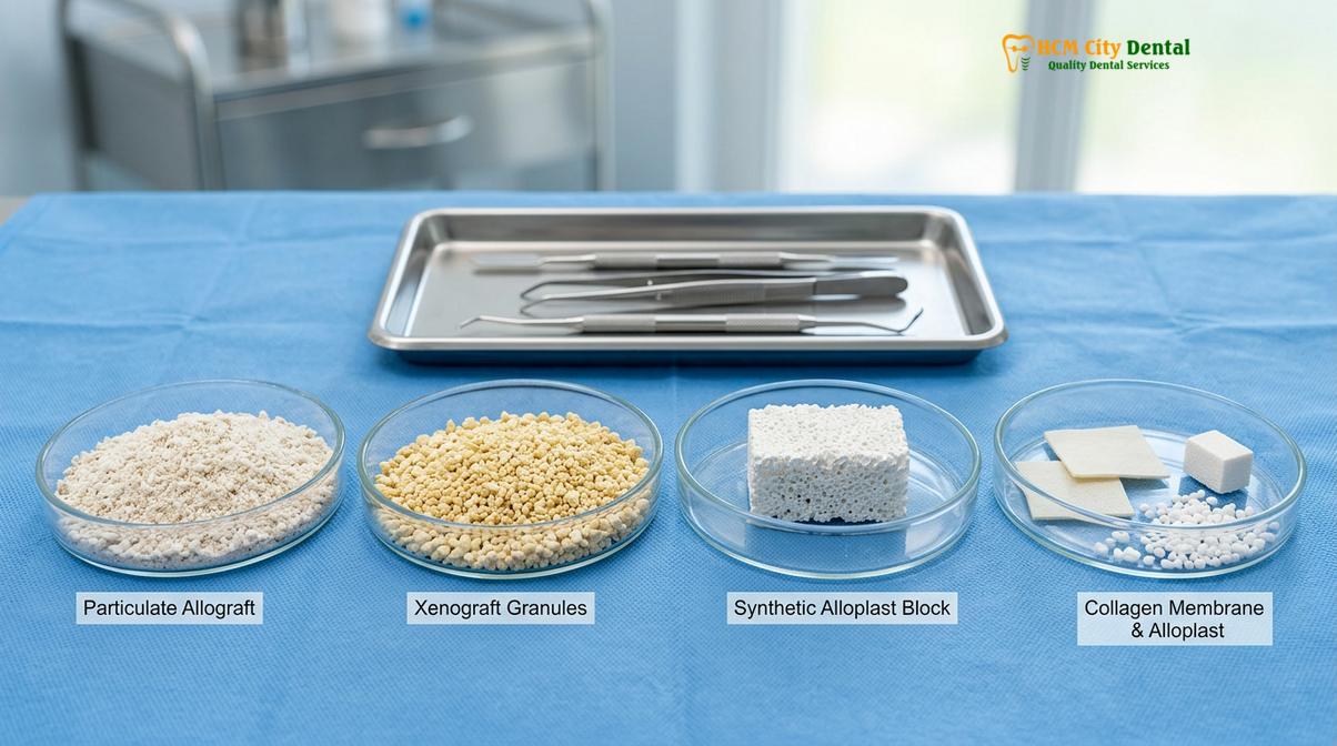
The four primary categories of bone grafting materials include:
| Graft Type | Source Material | Mechanism of Action | Clinical Advantages |
|---|---|---|---|
| Autograft | The patient’s own bone (often harvested from the jaw, chin, or tuberosity). | Osteogenic, Osteoinductive, Osteoconductive. | Considered the “gold standard” as it contains living cells and carries zero risk of disease transmission or rejection. |
| Allograft | Human donor bone obtained from a certified, rigorously screened tissue bank. | Osteoinductive and Osteoconductive. | Eliminates the need for a second surgical site to harvest bone. Highly effective for periodontal defects. |
| Xenograft | Animal-derived bone, most commonly bovine (cow) or porcine (pig), sterilized and processed. | Strictly Osteoconductive. | Provides an excellent, long-lasting scaffold that maintains space well while natural bone slowly replaces it. |
| Alloplast | Synthetic, laboratory-made materials such as hydroxyapatite or calcium phosphate. | Strictly Osteoconductive. | Completely sterile, readily available in unlimited quantities, and poses no risk of biological cross-contamination. |
In many complex periodontal cases, clinicians may choose to mix different types of grafts—for example, combining an autograft with a xenograft. This hybrid approach leverages the rapid healing and living cells of the patient’s own bone while utilizing the slow-resorbing nature of the animal bone to maintain the physical volume of the scaffold over the long term.
The Bone Grafting Procedure Step-by-Step
The dental bone grafting procedure involves administering local anesthesia, accessing the bone defect through a gum flap, thoroughly cleaning the infected root, placing the graft material with a protective membrane, and suturing the gums closed.
Executing a dental bone grafting procedure requires meticulous surgical precision and a deep understanding of periodontal anatomy. The process begins long before the actual surgery, starting with a comprehensive clinical evaluation. High-resolution 3D Cone Beam Computed Tomography (CBCT) scans are often utilized to map the exact dimensions, depth, and topography of the bone defect. This allows the surgical team to select the precise volume and type of grafting material required.
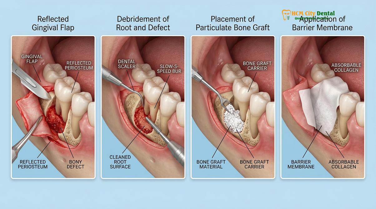
On the day of the surgery, the procedure follows a strict, sterile protocol to ensure optimal outcomes and minimize the risk of post-operative complications[3]:
- Anesthesia and Comfort Management: The targeted area is profoundly numbed using advanced local anesthetics. For patients with dental anxiety, conscious sedation options may be administered to ensure a completely relaxed and comfortable experience throughout the surgery.
- Flap Reflection (Access): The periodontist makes precise micro-incisions along the gum line. The gingival tissue is then gently lifted and folded back (creating a “flap”). This crucial step provides the surgeon with direct, unobstructed visual access to the diseased tooth root and the underlying degraded jawbone.
- Debridement and Root Conditioning: All inflammatory granulation tissue, bacterial plaque, and deep calculus are meticulously removed from the bone defect and the root surface using ultrasonic scalers and specialized hand instruments. The root surface may then be treated with chemical conditioning agents (like EDTA or citric acid) to detoxify the area and open the dentinal tubules, which encourages new tissue attachment.
- Graft Placement: The selected bone grafting material, often mixed with sterile saline or the patient’s own Platelet-Rich Fibrin (PRF) to accelerate healing, is carefully packed into the bony defect. The material is condensed to eliminate air pockets and ensure intimate contact with the surrounding natural bone.
- Barrier Membrane Application: To practice Guided Tissue Regeneration (GTR), a thin, biocompatible barrier membrane is placed over the graft material. Because gum tissue grows much faster than bone tissue, this membrane acts as a physical barricade, preventing the soft gum cells from invading the space reserved for the slower-growing bone cells.
- Suturing and Closure: Finally, the gingival flap is carefully repositioned over the membrane and graft, and secured with fine surgical sutures. These sutures hold the tissue tightly in place to facilitate primary wound closure and protect the surgical site during the initial healing phase.
Important Clinical Warning: Smoking and poor oral hygiene drastically reduce the success rate of bone grafts by constricting blood vessels and introducing bacteria. Patients must strictly adhere to post-operative care instructions, including the complete cessation of tobacco use, to prevent graft failure and severe infection.
Healing and Bone Regeneration Timeline
Initial soft tissue healing takes one to two weeks, but complete jawbone regeneration requires several months. The graft material slowly integrates with your natural bone, forming a solid foundation over a period of four to nine months.
Patience is a vital component of periodontal regenerative therapy. While the surgical procedure itself is completed in a single visit, the biological process of jawbone regeneration gum disease treatment is a complex, multi-phased journey that occurs on a cellular level over many months.
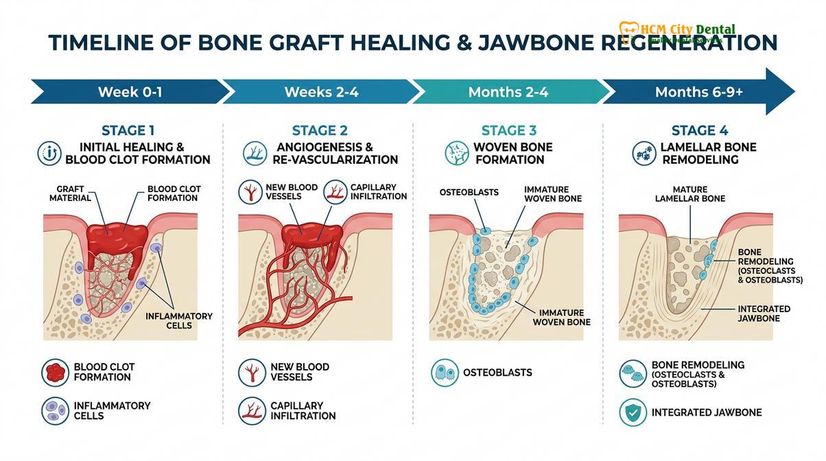
The healing timeline can be broadly categorized into distinct biological phases[4]:
- Phase 1: Hemostasis and Clot Formation (Days 1-3): Immediately following surgery, a blood clot forms within the surgical site. This clot is crucial; it acts as the initial biological matrix, rich in platelets and growth factors, which signals the start of the healing cascade.
- Phase 2: Granulation and Angiogenesis (Weeks 1-3): The blood clot is gradually replaced by granulation tissue. During this phase, angiogenesis occurs—new blood vessels sprout and grow into the graft site. A robust blood supply is absolutely essential, as it delivers the oxygen, nutrients, and immune cells required for bone formation. The surface gums will typically heal and close over during this period.
- Phase 3: Woven Bone Formation (Months 1-3): Osteoblasts begin to migrate along the scaffold provided by the graft material. They start depositing a primitive, unorganized form of bone known as woven bone. The graft particles are slowly surrounded and incorporated into this new cellular matrix.
- Phase 4: Lamellar Bone Remodeling (Months 4-9+): The primitive woven bone is gradually broken down and replaced by highly organized, mature, load-bearing lamellar bone. The original grafting material is slowly resorbed by the body and entirely replaced by the patient’s own natural, dense jawbone.
Clinical Case Example: A 52-year-old patient presented at HCMC Dental Clinic in Ho Chi Minh City with severe tooth mobility in the lower anterior region due to advanced periodontitis. Dr. Nguyen Van Cuong performed a comprehensive debridement followed by guided tissue regeneration using a particulate xenograft and a resorbable collagen membrane. After six months of monitored healing, radiographic imaging confirmed significant bone fill, successfully stabilizing the teeth and saving them from planned tooth extraction.
How Bone Grafts Save Loose Teeth and Support Implants
By regenerating lost alveolar bone, grafts provide essential biomechanical support to stabilize loose teeth. Furthermore, they ensure sufficient bone volume and density are present to securely anchor dental implants if tooth replacement is necessary.
The ultimate goal of periodontal therapy is the preservation of the natural dentition. When gum disease erodes the alveolar bone, the crown-to-root ratio becomes highly unfavorable. The tooth loses its mechanical anchorage, leading to mobility, shifting, and pain during mastication. A bone graft for loose teeth directly addresses this structural deficit. By regenerating the bone height and width around the compromised root, the procedure re-establishes the necessary biomechanical support, firming up the tooth within its socket and significantly extending its lifespan.
However, in cases where periodontal disease has progressed to the point where a tooth is deemed unsalvageable and must be extracted, bone grafting takes on a different, yet equally vital, role: implant site development. When a tooth is removed, the surrounding bone naturally begins to atrophy and shrink. If severe periodontitis was the cause of the extraction, the bone deficit is often already profound.
“Without adequate bone volume, even the most advanced restorative treatments will fail. Bone grafting is the critical bridge between treating severe periodontal disease and achieving long-term success with implantology.”
To successfully place dental implants, a surgeon requires a foundation of bone that is sufficiently wide, tall, and dense to completely encase the titanium post. If an implant is placed into inadequate bone, it lacks primary stability and is at a high risk of failure. Bone grafting procedures, such as socket preservation immediately following extraction or ridge augmentation prior to implant surgery, rebuild the necessary three-dimensional architecture[5]. This ensures that when the implant is eventually placed, it integrates flawlessly, providing a permanent, aesthetic, and fully functional replacement for the lost tooth.
When to See a Doctor
Periodontal bone loss is often a “silent” disease in its early stages, progressing without significant pain until the damage is severe. It is crucial to seek professional evaluation if you notice any warning signs of advanced gum disease. Symptoms that warrant immediate clinical attention include gums that bleed easily during brushing, persistent bad breath (halitosis), gums that are pulling away from the teeth (recession), the appearance of elongated teeth, or any noticeable shifting or looseness of your teeth.
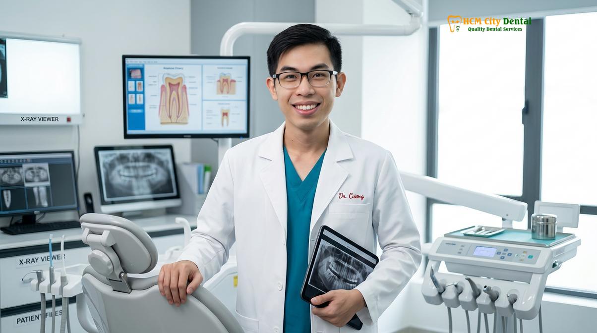
If you are experiencing these symptoms, early intervention is paramount. We highly recommend scheduling a comprehensive periodontal evaluation. At HCMC Dental Clinic in Ho Chi Minh City, our team, led by experienced specialists like Dr. Nguyen Van Cuong, utilizes advanced diagnostic imaging to assess the exact extent of bone loss. Based on these precise clinical diagnostics, we can formulate a personalized regenerative treatment plan to halt the disease progression and restore your oral health. Whether you require advanced surgical intervention or routine general dentistry maintenance, timely care is the key to preserving your natural smile.
Frequently Asked Questions
Can bone loss from gum disease be reversed?
Yes, bone loss from gum disease can be partially or fully reversed through regenerative surgical procedures. By utilizing bone grafts and barrier membranes, dentists can stimulate the body to grow new alveolar bone and periodontal ligaments in areas damaged by severe infection. The extent of reversal depends heavily on the specific anatomy of the bone defect; deep, narrow defects generally respond better to regeneration than broad, horizontal bone loss.
Is bone grafting for gum disease painful?
Bone grafting for gum disease is not typically painful during the procedure, as profound local anesthesia is administered. Post-operative discomfort is generally mild to moderate, easily managed with prescribed analgesics, and usually subsides within the first few days of healing. Patients may experience some swelling and minor bruising around the surgical site, which is a normal part of the body’s initial inflammatory healing response.
How long does a bone graft take to heal?
Initial soft tissue healing over a bone graft takes about one to two weeks. However, the complete biological process of bone regeneration, where the graft material integrates and transforms into solid natural jawbone, typically requires four to nine months. During this extended period, the graft acts as a scaffold while your body’s osteoblasts slowly deposit new, mature bone cells to fill the defect.
Do I need a bone graft before getting implants?
You will need a bone graft before getting implants if periodontal disease has destroyed the necessary bone volume. Implants require a dense, wide, and tall bone foundation for primary stability and long-term success, which grafting procedures reliably restore. If a tooth is extracted due to severe infection, a socket preservation graft is often placed immediately to prevent the natural shrinkage of the jawbone while awaiting implant placement.
What is the success rate of dental bone grafting?
The success rate of dental bone grafting is exceptionally high, often exceeding ninety percent in healthy individuals. Success depends heavily on meticulous surgical technique, the patient’s overall health, strict adherence to post-operative care, and the complete cessation of smoking. Conditions such as uncontrolled diabetes or poor daily oral hygiene can significantly compromise blood flow and introduce bacteria, leading to graft failure.
References
- Journal of Periodontology. Pathogenesis of periodontal disease and alveolar bone loss mechanisms. (2021).
- International Journal of Oral and Maxillofacial Implants. Biomaterials and scaffolds for periodontal regeneration. (2020).
- Clinical Oral Investigations. Surgical protocols in guided tissue regeneration and bone grafting. (2019).
- Journal of Clinical Periodontology. Histological healing and osteogenesis following bone grafting procedures. (2022).
- Journal of the American Dental Association. Ridge preservation, periodontal therapy, and implant site development. (2023).
For more information on preserving your oral health or to discuss regenerative treatment options, please contact our team to schedule a consultation with our experienced dental specialists.