Digital scan veneers in Vietnam combine state-of-the-art intraoral scanning with premium ceramic materials to deliver flawless smile makeovers. By eliminating traditional molds, patients experience maximum comfort, micron-level precision, and an express turnaround at a fraction of Western dental costs.
Clinical Summary:
The integration of digital dentistry has revolutionized the fabrication of Porcelain Veneers. Utilizing advanced intraoral scanners such as the iTero Element 5D and Medit i700, clinicians can capture highly accurate 3D topographies of the dentition without the discomfort of traditional impression materials. This digital workflow facilitates precise marginal adaptation, respects the biological width, and allows for meticulous gingival zenith calibration. Coupled with a direct lab partnership in Ho Chi Minh City, the CAD/CAM milling process ensures an express turnaround. Patients benefit from certified materials like Ivoclar E.max and Cercon HT Zirconia, ensuring optimal flexural strength and lifelike aesthetics. Furthermore, the integration of Digital Smile Design (DSD) empowers patients to visualize their outcomes in real-time, making the cosmetic journey predictable, minimally invasive, and highly efficient for international dental tourists.
Key Takeaways:
- Digital Precision: Intraoral scanners capture sub-micron 3D data, ensuring a flawless marginal fit and helping to prevent post-bonding microleakage.
- Patient Comfort: Eliminates the gag reflex associated with traditional goopy silicone impressions.
- Premium Materials: Exclusive use of certified German and Liechtenstein ceramics (E.max and Zirconia HT) for superior durability and aesthetics.
- Rapid Turnaround: Direct lab partnerships enable complete smile makeovers within an express timeframe.
- Cost Efficiency: Transparent pricing with significant savings, including a pre-arrival discount for WhatsApp bookings.
The Evolution of Dental Impressions: Goodbye Goopy Trays
Traditional silicone impressions are uncomfortable and prone to distortion, whereas digital scans offer a goopy tray alternative that ensures precise, gag-free veneer fabrication.
For decades, the standard protocol for capturing the anatomical structure of a patient’s teeth involved the use of alginate or polyvinyl siloxane (PVS) impression materials. While these materials have served the dental profession well, they present several clinical and patient-experience challenges. The process requires the patient to bite down on a bulky tray filled with a viscous, slow-setting putty for several minutes. This often triggers a severe gag reflex, causing significant anxiety and discomfort. Furthermore, traditional impressions are highly technique-sensitive; factors such as saliva contamination, blood, or improper mixing ratios can lead to dimensional instability and voids in the final mold.
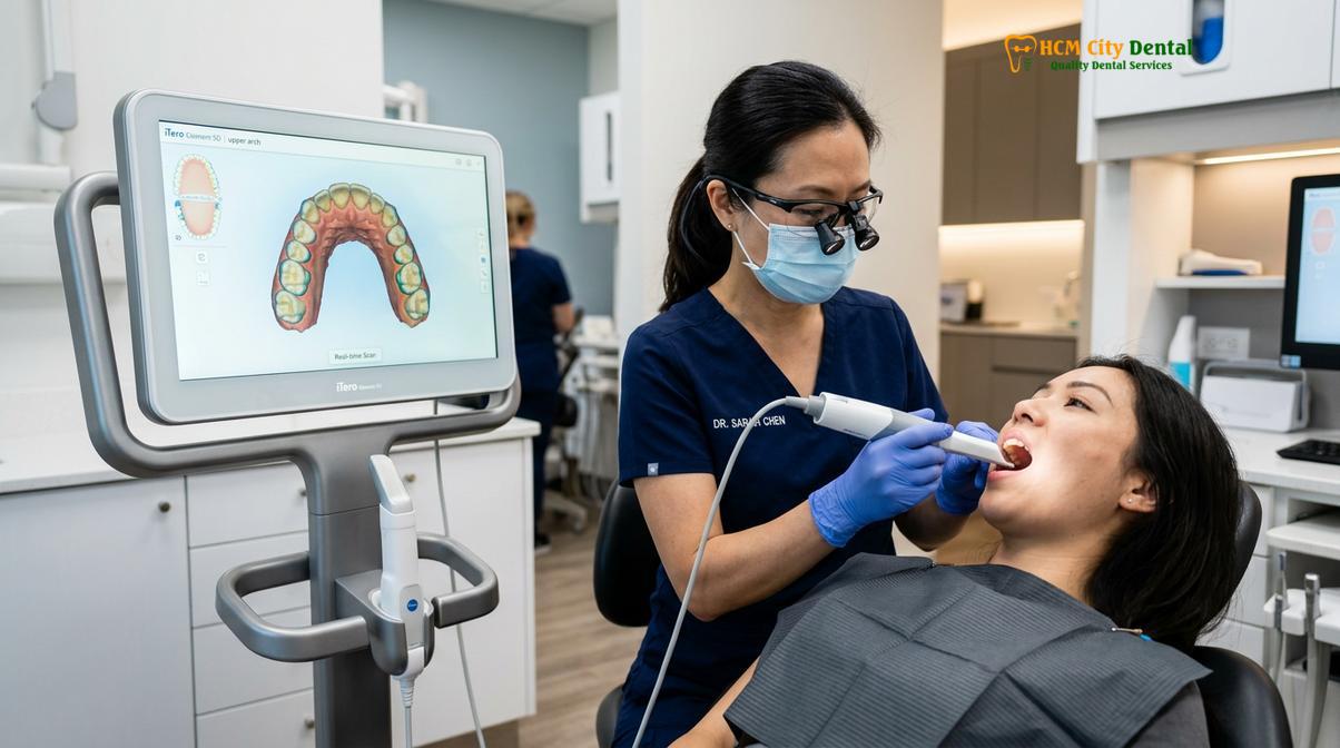
From a clinical perspective, any distortion in the physical impression transfers directly to the stone model poured by the dental laboratory. This cascading effect of minor inaccuracies can result in porcelain restorations that require extensive chairside adjustments, compromising the structural integrity of the ceramic. The advent of the intraoral digital scanner has fundamentally eliminated these variables. By utilizing optical scanning technology, clinicians can capture the exact geometry of the prepared teeth in a matter of minutes, completely bypassing the physical limitations of PVS materials.
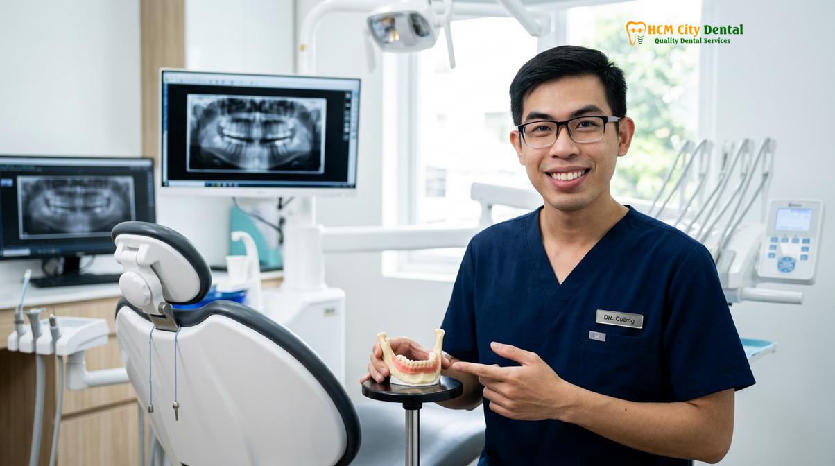
Dr. Nguyen Van Cuong, a leading prosthodontist at HCMC Dental Clinic, emphasizes that the transition to digital impressions is fundamentally about clinical excellence. As detailed in his insights on Dental Crown vs Veneer: Which Is Right For You? | Dr. Cuong, a digital scan provides a distortion-free foundation that allows our ceramists to design restorations with absolute confidence. This technological evolution ensures that the subsequent CAD/CAM (Computer-Aided Design and Computer-Aided Manufacturing) processes are based on flawless data, leading to superior clinical outcomes.
iTero Element 5D and Medit i700: Digital Intraoral Scanning
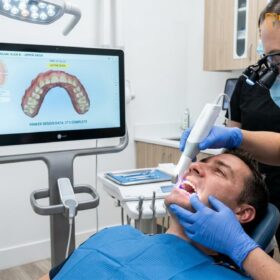
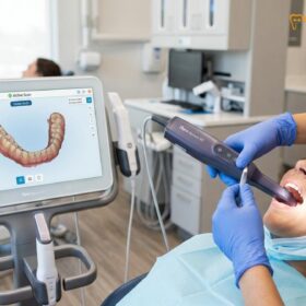
Utilizing the iTero Element 5D and Medit i700 intraoral digital scanner allows clinicians to capture highly detailed 3D topographies of prepared teeth for flawless veneer design.
The cornerstone of modern cosmetic dentistry lies in the hardware used to capture the oral environment. At the forefront of this technology are the iTero Element 5D and the Medit i700 intraoral scanners. These devices utilize advanced confocal microscopy and Near-Infrared Imaging (NIRI) technology to capture thousands of frames per second, stitching them together to create a high-definition, full-color 3D model of the patient’s dental arches. This level of detail is critical when planning how many veneers you need and executing minimal-prep or prep-less veneer protocols.
“The precision of intraoral scanning allows for the fabrication of ultra-thin ceramic restorations that seamlessly integrate with the natural tooth structure, preserving the maximum amount of healthy enamel while achieving optimal aesthetic results.”
When preparing teeth for veneers, the goal is maximum enamel preservation. Traditional crowns require aggressive reduction, but modern veneer preparations are incredibly conservative. A standard minimal-prep veneer requires only 0.3mm to 0.7mm of enamel reduction, while prep-less options like Lumineers may require as little as 0.2mm. Capturing the exact finish line (the margin where the prepared tooth meets the unprepared tooth) at these microscopic depths is incredibly challenging with traditional putty. The iTero and Medit scanners excel in this regard, clearly delineating the preparation margins even in subgingival areas, provided proper tissue management and retraction techniques are employed [1].
Furthermore, these scanners play a vital role in analyzing the biological width and facilitating gingival zenith calibration. The biological width is the natural distance between the base of the gingival sulcus and the alveolar bone crest. Violating this space with a poorly fitting restoration leads to chronic inflammation and bone loss. Digital scans allow the clinician and the lab technician to precisely map the gingival contours, ensuring that the final veneer respects the periodontal tissues and promotes long-term gingival health [2].
Accuracy and Margin Fit: Preventing Post-Bonding Cavities
Achieving margin fit precision through digital scanning helps prevent microleakage, ensuring the long-term biological integration of the veneer and protecting the underlying enamel from decay.
The longevity of any dental restoration is heavily dependent on the quality of its marginal fit. The margin is the critical junction where the porcelain veneer meets the natural tooth structure. If this junction is not perfectly sealed, it creates a microscopic gap. Over time, oral fluids, bacteria, and plaque can infiltrate this gap—a process known as microleakage. Microleakage is the primary cause of secondary caries (decay under the veneer), sensitivity, and eventual failure of the restoration. Achieving margin fit precision is therefore a non-negotiable clinical objective.
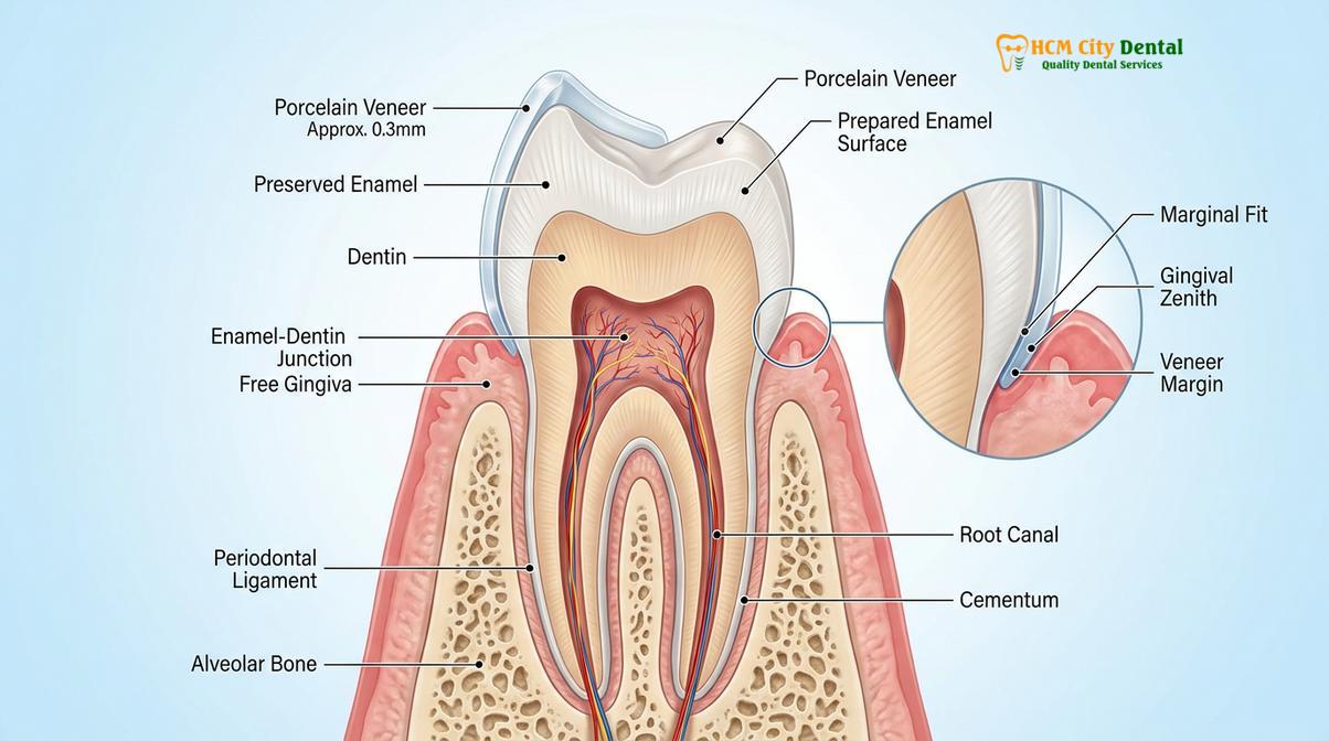
Digital scanning significantly mitigates the risk of microleakage. Because the 3D dataset is accurate to the sub-micron level, the CAD software can design a veneer with a virtually seamless transition from porcelain to enamel. When the restoration is milled, the internal adaptation is so precise that the space required for the luting cement is perfectly uniform, typically calibrated to around 50-100 microns. This uniform cement space ensures that when the veneer is bonded, the dual-cure resin cement flows evenly, creating a robust, hermetic seal [3].
The cementation protocol itself is a highly meticulous procedure that benefits from this accuracy. The process involves isolating the teeth (often with a rubber dam), etching the enamel with phosphoric acid, and treating the intaglio (internal) surface of the ceramic with hydrofluoric acid followed by a silane coupling agent. A bonding resin is then applied, and the veneer is seated and light-cured. If the margin fit is poor, excess cement can pool at the gingival margin, leading to periodontal irritation. The precision afforded by digital scans ensures that the margins are flush, making the removal of excess cement straightforward and preventing iatrogenic gum disease.
Speed: Sending 3D Scans Instantly to the Ceramic Lab via Cloud
Cloud-based transmission of digital impressions to our direct lab partnership enables an express turnaround, perfect for international patients seeking efficient smile makeovers.
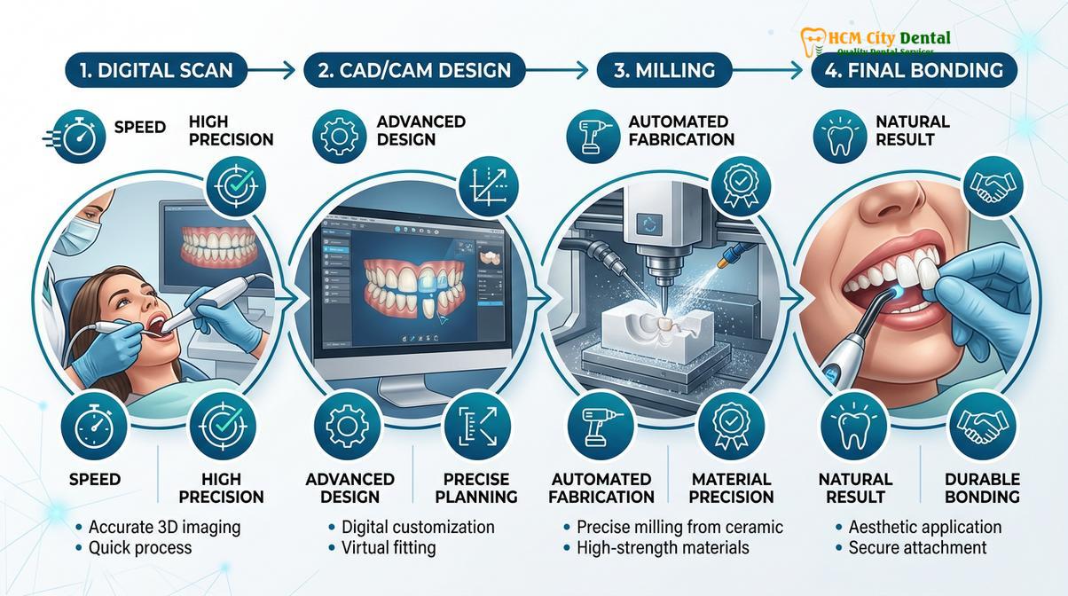
One of the most transformative aspects of the digital workflow is the speed of data transmission. In the past, physical impressions had to be disinfected, packaged, and couriered to a dental laboratory, a process that could take days, especially if the lab was off-site. Today, the moment the intraoral scan is completed, the encrypted 3D stereolithography (STL) or Polygon File Format (PLY) files are uploaded to a secure cloud server and instantly accessed by the master ceramists.
At HCMC Dental Clinic, our direct lab partnership with a leading facility in Ho Chi Minh City allows for unprecedented collaboration between the prosthodontist and the laboratory technicians. Because the lab is integrated into our digital ecosystem, we can offer an express turnaround for complete smile makeovers. This rapid timeline is specifically tailored for patients engaging in dental tourism, allowing them to complete their treatment within a standard vacation window without compromising on the meticulous craftsmanship required for premium ceramics.
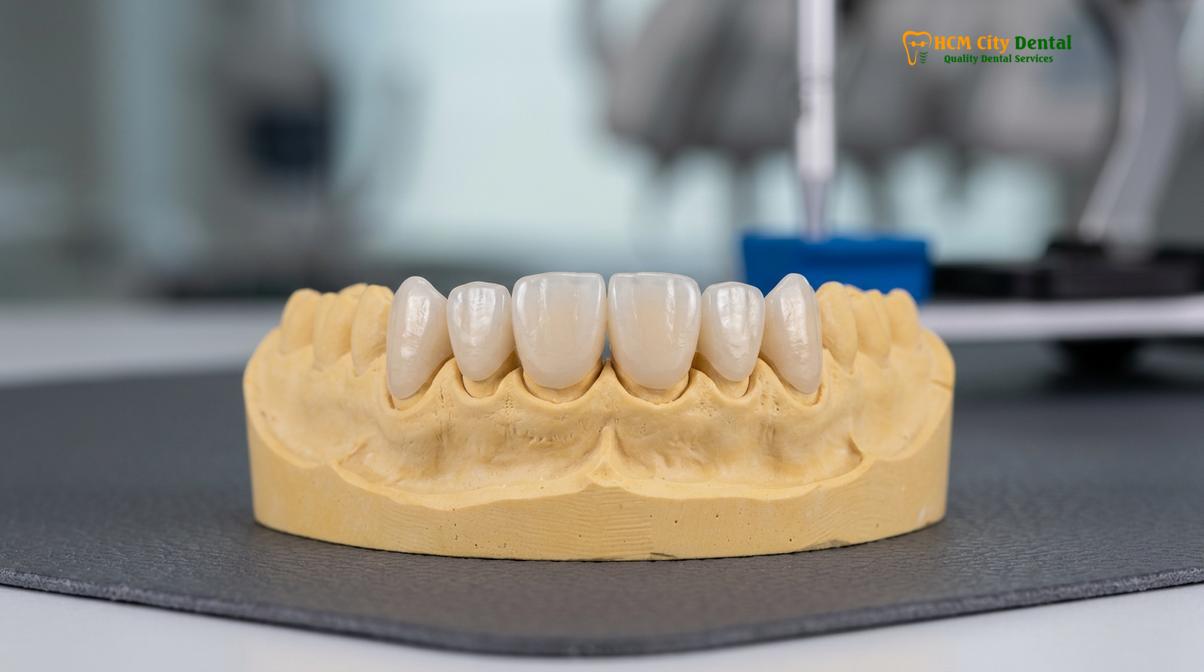
Once the lab receives the digital file, the CAD/CAM milling process begins. The software proposes a design based on the patient’s specific anatomical parameters and the clinician’s prescription. The technician refines this design, paying close attention to the contact points, occlusal scheme, and surface texture. The data is then sent to a 5-axis milling machine, which sculpts the veneer from a solid block of high-strength ceramic. Following milling, the veneer undergoes a crystallization firing process in a ceramic furnace, followed by hand-characterization and glazing to achieve a lifelike appearance. This seamless digital relay ensures that patients receive their final restorations rapidly, minimizing the time spent wearing temporary veneers.
Patient Comfort: Real-Time Visualization of Your Teeth on Screen
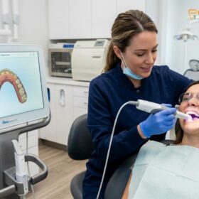
A real-time 3D teeth scan allows patients to instantly visualize their current dentition and co-design their future smile using Digital Smile Design (DSD) software.
The integration of digital scanning technology profoundly enhances patient communication and comfort. Beyond eliminating the physical discomfort of impression trays, the scanner provides an immediate, high-definition visual representation of the patient’s oral cavity on a chairside monitor. This real-time 3D teeth scan serves as a powerful educational tool, allowing the dentist to point out areas of enamel wear, micro-fractures, or alignment issues that necessitate cosmetic intervention.
Clinical Case Study: Digital Workflow for Diastema Closure
A 34-year-old patient traveled to HCMC Dental Clinic in Ho Chi Minh City seeking to close a prominent midline diastema (gap between the front teeth) and address mild tetracycline staining. Using the Medit i700, a digital scan was captured in under two minutes. Through DSD, the patient previewed the proposed closure and selected a natural BL1 translucent shade. The direct lab partnership facilitated the milling of 8 E.max veneers. The restorations were bonded on the fourth day of her trip, achieving a flawless, gap-free smile with zero need for physical impressions.
This visual data is the foundation of Digital Smile Design (DSD). DSD is a conceptual protocol that utilizes digital technology to analyze the patient’s facial and dental proportions. By integrating the 3D intraoral scan with dynamic video recordings and high-resolution facial photography, the clinician can design a smile that is in perfect harmony with the patient’s facial midlines, interpupillary lines, and lip dynamics.
“Digital Smile Design empowers the patient to transition from a passive recipient of dental care to an active co-author of their own aesthetic transformation, ensuring predictable and highly personalized outcomes.”
During the DSD phase, critical aesthetic decisions are made, such as custom shading and translucency matching. For patients desiring a bright, Hollywood-style aesthetic, a VITA BL1 shade might be selected, while others may prefer a more subtle, natural B1 or A1 shade. The software allows the patient to preview these variations. Once the digital design is approved, a 3D-printed mock-up can be created and temporarily placed over the patient’s un-prepared teeth, allowing them to “test drive” their new smile before committing to the final Porcelain vs Composite Veneers treatment plan.
Clinical Workflow & Material Selection (E.max vs. Zirconia)
Selecting authentic, certified materials like Ivoclar E.max or Cercon HT Zirconia ensures optimal flexural strength and lifelike aesthetics tailored to your specific bite dynamics.
The success of a veneer case relies heavily on the selection of appropriate restorative materials. At our clinic, we strictly utilize authentic, material-certified ceramics from industry-leading manufacturers in Germany and Liechtenstein. The two primary materials utilized in our digital workflow are Lithium Disilicate (E.max by Ivoclar) and High-Translucency Zirconia (Cercon HT by Dentsply Sirona). The choice between these materials is dictated by the patient’s functional requirements and aesthetic goals.
E.max (Lithium Disilicate) is widely considered the gold standard for anterior (front teeth) cosmetics. It offers outstanding lifelike aesthetics, exceptional translucency matching, and natural opalescence. With a flexural strength of approximately 400-500 MPa, it is highly durable when bonded correctly to enamel. Conversely, Zirconia HT is utilized for patients with heavier bite forces, signs of mild bruxism, or when masking dark underlying tooth structure (such as severe tetracycline stains). Zirconia boasts a flexural strength exceeding 1000 MPa, making it incredibly robust, though it is slightly more opaque than E.max. Understanding the nuances between these materials is crucial when discussing Porcelain Veneers vs Porcelain Crowns: Which Is Right for You? | Dr. Cuong.
In cases where the gingival zenith requires minor contouring for optimal symmetry, clinicians may utilize diode laser gingival sculpting prior to the final digital scan, ensuring a harmonious pink-and-white aesthetic balance [4]. To provide transparency for our international patients, we offer a highly competitive pricing structure. By utilizing our direct lab partnership, we eliminate middleman costs, passing the savings directly to the patient.
| Treatment / Material | Standard Walk-in Price | WhatsApp Discount (-40%) |
|---|---|---|
| Composite Veneer (Chair-side) | ~$80 (2.0M VND) | From $48 (1.2M VND) |
| E.max Lithium Disilicate Veneer | ~$350 (8.5M VND) | From $210 (5.1M VND) |
| Cercon HT Zirconia Veneer | ~$300 (7.5M VND) | From $180 (4.5M VND) |
When to Consult a Prosthodontist & Important Considerations
Understanding the clinical prerequisites and maintenance requirements is essential before embarking on a digital veneer transformation.
While digital precision ensures a perfect fit, patients who suffer from sleep bruxism (involuntary teeth grinding) exert immense occlusal forces that can fracture even the strongest ceramics. It is a strict clinical requirement for bruxers to wear a custom-fabricated nightguard during sleep to protect their investment and prevent failure of the porcelain restorations. If you experience jaw pain, frequent headaches upon waking, or notice flattened wear facets on your natural teeth, it is imperative to consult with a prosthodontist before proceeding with any cosmetic enhancements.
According to the clinical guidelines set forth by the Vietnam Odonto-Stomatology Association (VOSA), the adoption of digital impressions is highly recommended for minimally invasive aesthetic restorations to ensure both patient safety and marginal integrity [5]. Patients exploring an Alternative for Veneers: 3 Secrets for a Perfect Smile | Dr. Cuong should also seek a comprehensive digital evaluation to determine if orthodontic alignment or professional whitening might achieve their aesthetic goals without the need for ceramic restorations.
Proper maintenance is equally critical for the longevity of your new smile. Although porcelain does not decay, the underlying natural tooth structure remains susceptible to cavities, particularly at the margin line. Maintaining a rigorous oral hygiene routine, including daily flossing and the use of a non-abrasive fluoride toothpaste, is essential. Regular professional cleanings and examinations every six months will allow your dentist to monitor the integrity of the margins and the health of the surrounding gingival tissues, ensuring your porcelain veneers last for many years.
References
- Journal of Esthetic and Restorative Dentistry. Translucency and mechanical properties of lithium disilicate. (2021).
- International Journal of Prosthodontics. Enamel preservation, biological width, and bond strength in veneer restorations. (2020).
- The Journal of Prosthetic Dentistry. Flexural strength and marginal adaptation of CAD/CAM porcelain restorations. (2019).
- Clinical Oral Investigations. Diode laser gingival sculpting parameters for aesthetic dentistry. (2022).
- Vietnam Odonto-Stomatology Association (VOSA). Guidelines on digital impressions and minimally invasive aesthetic restorations. (2023).
Transform Your Smile at HCMC Dental Clinic
If you are ready to experience the precision and comfort of digital dentistry, our team is here to guide you. Contact HCMC Dental Clinic in Ho Chi Minh City today to schedule your comprehensive 3D digital scan and discover how our advanced veneer workflow can safely and beautifully revitalize your smile.