Extracting failing teeth before implant denture preparation is a critical clinical step that removes active infection and creates a healthy foundation for prosthetics. This sequence ensures optimal bone volume, proper soft tissue healing, and a highly stable environment for long-lasting, functional implant-supported restorations.
Clinical Summary:
The transition from a failing natural dentition to an implant-supported denture requires meticulous surgical and prosthodontic planning. The process of extraction before implant denture preparation focuses heavily on atraumatic tooth removal to preserve the delicate alveolar bone architecture. Following extraction, clinicians must actively manage the socket through ridge preservation techniques or immediate implant placement, depending on the patient’s bone density and the presence of localized infection. Proper management of both the hard and soft tissues ensures that the final prosthesis has a stable, functional, and highly aesthetic foundation. Dr. Nguyen Van Cuong emphasizes that preserving the alveolar ridge during this initial phase is absolutely paramount for the long-term biomechanical success of the implant placement preparation and the overall longevity of the final restoration.
Key Takeaways:
- Atraumatic extractions minimize trauma to the buccal plate, preserving critical bone volume for future implants.
- Placing a bone graft immediately after extraction significantly reduces natural dimensional bone shrinkage.
- Immediate implant placement can streamline treatment timelines when the socket walls are fully intact and infection-free.
- Thick, keratinized soft tissue is essential to create a durable and comfortable denture fitting foundation.
- A carefully phased prosthodontic treatment sequence allows for predictable osseointegration and superior aesthetic outcomes.
The Prosthodontic Sequence: Extracting to Replace Teeth
The prosthodontic sequence begins with the strategic, atraumatic extraction of non-restorable teeth to eliminate active oral infections and establish a pristine surgical site for future implant placement.
The journey toward a fully restored smile using implant-supported dentures often begins with the difficult but necessary decision to remove failing natural teeth. Teeth that have been severely compromised by advanced periodontal disease, extensive structural decay, or catastrophic root fractures can no longer serve as reliable abutments for traditional prosthetics. In these scenarios, retaining the diseased teeth poses a significant risk to the surrounding healthy bone and systemic health. The initial phase of the prosthodontic treatment sequence involves a comprehensive diagnostic evaluation to determine exactly which teeth must be sacrificed and which, if any, can be salvaged. This diagnostic phase relies heavily on three-dimensional Cone Beam Computed Tomography (CBCT) imaging, which provides clinicians with an exact topographical map of the patient’s maxillofacial structures, revealing the precise extent of bone loss and the proximity of vital anatomical landmarks such as the inferior alveolar nerve and the maxillary sinuses.
When evaluating when a tooth restoration fails beyond the point of repair, extraction becomes the definitive treatment to halt the progression of localized osteomyelitis (bone infection) and periodontitis. However, in the context of implant dentistry, an extraction is not merely the removal of a tooth; it is the first critical step in site development. Traditional extraction techniques, which often involve heavy luxation and the aggressive use of forceps, can easily fracture the delicate buccal bone plate—the thin wall of bone on the cheek side of the tooth. Loss of this buccal plate severely compromises the site, making future implant placement highly unpredictable without extensive and costly reconstructive grafting.
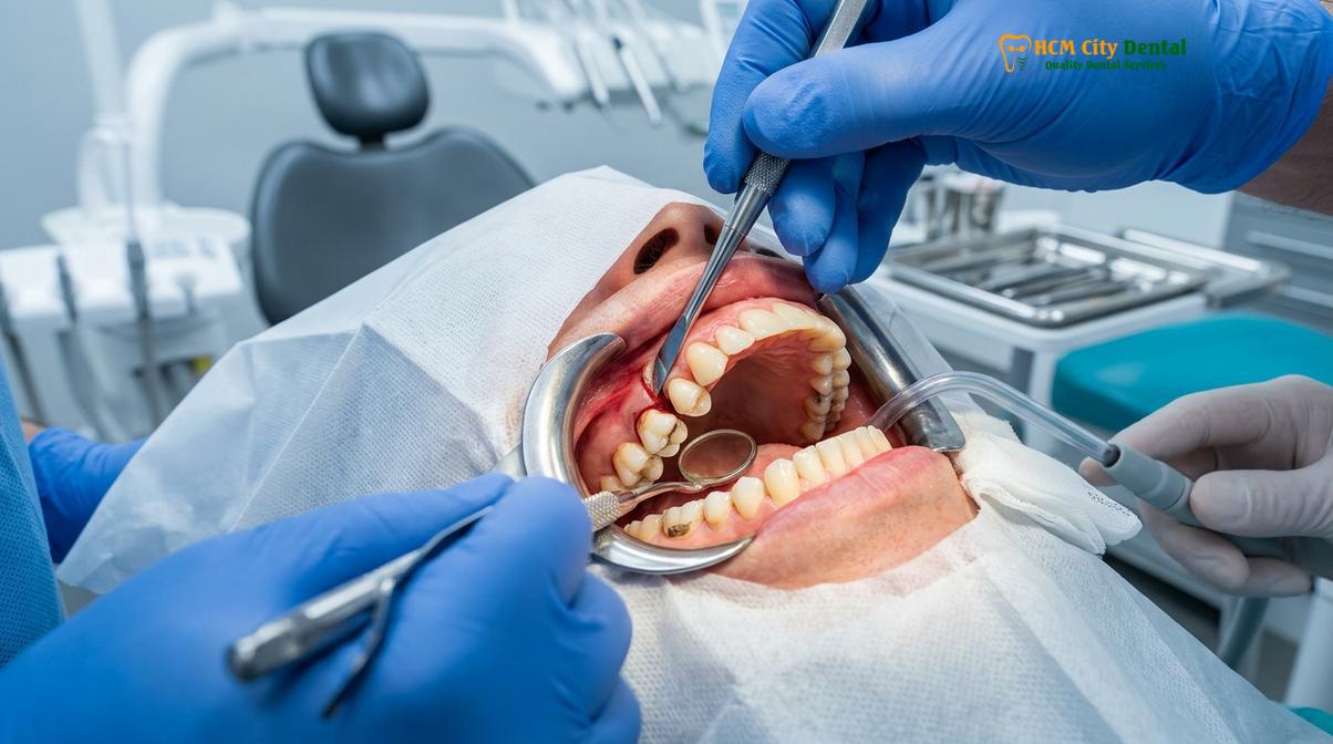
To prevent this structural damage, modern implantologists employ atraumatic extraction protocols. This technique utilizes specialized micro-surgical instruments, such as periotomes and piezosurgery devices, to gently sever the periodontal ligament fibers that attach the tooth root to the surrounding bone. By carefully separating the tooth from its socket without expanding the bone walls, the clinician preserves the maximum amount of native bone. In cases involving multi-rooted molars, the tooth is often sectioned into multiple pieces using a high-speed surgical handpiece, allowing each root to be elevated individually along its natural path of withdrawal. This meticulous approach minimizes surgical trauma, reduces post-operative inflammation, and maintains the critical three-dimensional architecture of the alveolar ridge.
“The success of an implant-supported prosthesis is directly proportional to the quality of the surgical site development; atraumatic extraction is the cornerstone of preserving the vital bundle bone necessary for predictable osseointegration.”
Following the removal of the tooth, the extraction socket undergoes a complex biological healing process. Immediately after extraction, the socket fills with a blood clot, which serves as a biological matrix for the migration of fibroblasts and osteoblasts. Over the next several weeks, this clot is replaced by highly vascularized granulation tissue, which eventually mineralizes into woven bone and, later, mature lamellar bone. However, this healing process is accompanied by an unavoidable physiological phenomenon: the resorption of the bundle bone. The bundle bone is a tooth-dependent structure that relies on the mechanical stimulation of the periodontal ligament to maintain its volume. Once the tooth is removed, the bundle bone rapidly resorbs, leading to significant dimensional changes in the alveolar ridge [1]. Understanding and mitigating this resorption is the primary objective of the subsequent phases of the implant placement preparation.
Ridge Preservation: Why Placing a Bone Graft Prevents Bone Shrinkage
Alveolar ridge preservation involves placing a specialized bone graft into the extraction socket immediately after tooth removal to counteract the natural dimensional collapse of the jawbone.
The physiological response to tooth loss is characterized by rapid and progressive bone remodeling. Clinical studies have demonstrated that without intervention, the alveolar ridge can lose up to 50% of its horizontal width and a significant portion of its vertical height within the first six months following an extraction [2]. This dimensional collapse presents a severe challenge for future implant placement. Dental implants require a minimum volume of dense, healthy bone to achieve primary mechanical stability and to support the immense biomechanical forces generated during chewing. If the ridge is allowed to resorb, the resulting bone volume may be insufficient to house an implant of adequate length and diameter, necessitating complex, secondary block grafting procedures that increase treatment time, cost, and patient morbidity.
To intercept this resorptive process, clinicians routinely perform alveolar ridge preservation at the time of extraction. This procedure involves meticulously debriding the empty socket to remove any residual granulation tissue or cystic remnants, ensuring a sterile and highly vascularized bony housing. Once the socket is prepared, an extraction bone graft is carefully packed into the void. The graft material serves as a three-dimensional osteoconductive scaffold, maintaining the physical space of the socket and providing a framework upon which the patient’s native bone cells (osteoblasts) can migrate, proliferate, and deposit new bone matrix.
Clinical Warning: Smoking and poor oral hygiene significantly impair local blood flow and can severely compromise the integration of a bone graft. Patients must adhere strictly to post-operative care instructions to prevent graft failure and ensure a stable foundation for future implants.
Various types of bone graft materials are utilized in ridge preservation, each selected based on the specific clinical requirements of the site. Allografts, which are derived from human donor tissue, are highly favored for their excellent remodeling capabilities and their ability to be completely replaced by the patient’s own bone over time. Xenografts, typically derived from bovine (cow) or porcine (pig) sources, resorb much more slowly and are excellent space maintainers, making them ideal for preserving the external contour of the ridge. In some cases, synthetic alloplasts made from calcium phosphate or hydroxyapatite are used. Regardless of the material chosen, the graft is typically covered with a barrier membrane. This membrane, often made of cross-linked porcine collagen, serves a critical function: it prevents the rapidly growing soft tissue (epithelium) from migrating down into the socket and interfering with the slower-growing bone cells.
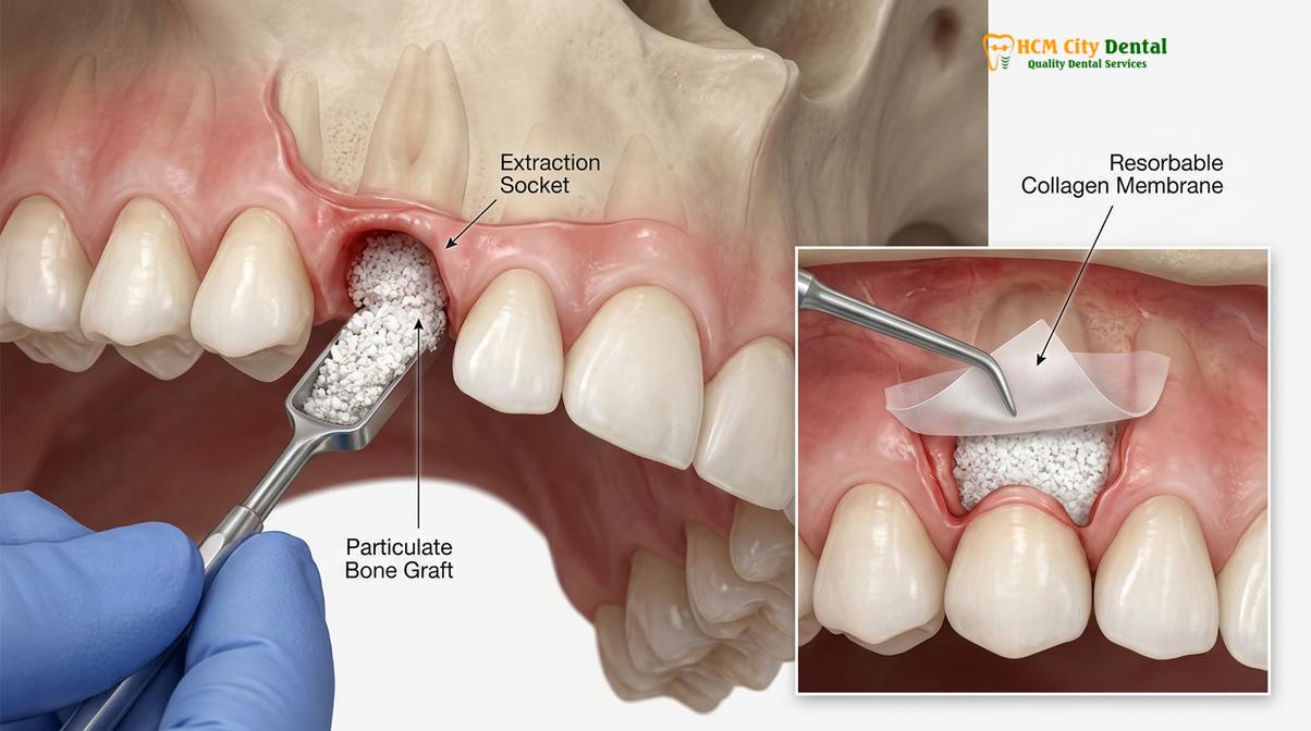
By implementing these ridge preservation techniques, the clinician effectively establishes a robust and dimensionally stable denture fitting foundation. When the patient returns for implant placement three to six months later, the surgeon is presented with a wide, dense ridge that facilitates optimal three-dimensional implant positioning. This precise positioning is crucial not only for the biomechanical distribution of occlusal forces but also for the aesthetic emergence profile of the final prosthesis. Whether the patient is transitioning to a removable overdenture or a fixed hybrid prosthesis, the quality of the underlying bone dictates the long-term success of the entire restorative endeavor.
Immediate Implants: Placing the Post in the Fresh Socket
Immediate implant placement involves inserting the titanium post directly into the jawbone during the same surgical visit as the tooth extraction, significantly streamlining the overall treatment timeline.
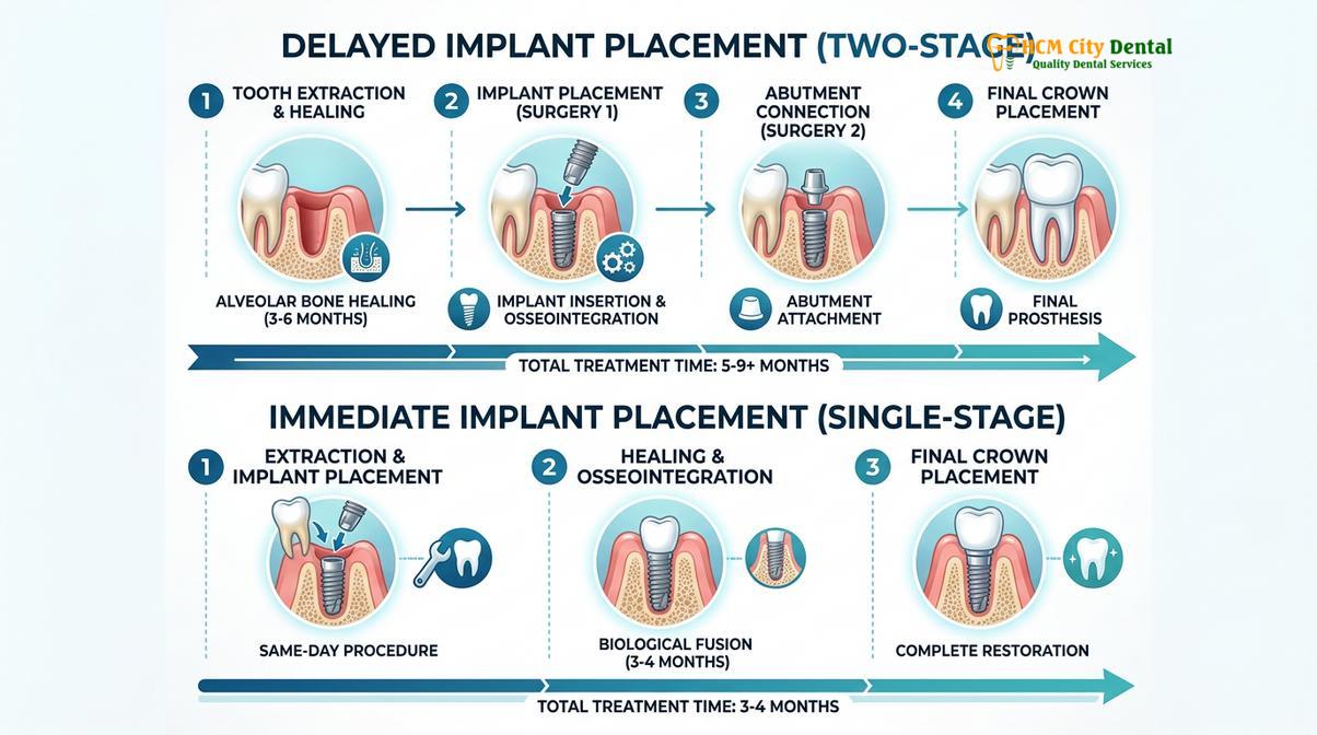
While delayed implant placement following ridge preservation is the traditional and highly predictable gold standard, advancements in implant macro-geometry and surgical protocols have made immediate implant placement a viable and increasingly popular option for select patients. Immediate placement involves extracting the failing tooth and immediately drilling the osteotomy (the receptor site for the implant) into the apical bone beyond the extraction socket. The titanium implant is then threaded into place during the exact same surgical appointment. This approach offers several distinct advantages: it reduces the total number of surgical interventions, shortens the overall treatment timeline, and helps to preserve the natural scalloped architecture of the surrounding gingival tissues.
However, immediate implant placement is a highly technique-sensitive procedure that requires strict adherence to specific clinical criteria. Not every extraction site is suitable for immediate placement. The primary prerequisite is the presence of intact socket walls, particularly the delicate buccal plate. If the buccal bone has been destroyed by chronic infection or fractured during extraction, immediate placement is generally contraindicated, as the implant will lack the necessary bony housing to integrate successfully. Furthermore, the site must be completely free of acute purulent infection. While chronic, asymptomatic periapical lesions can often be thoroughly debrided to allow for immediate placement, active, pus-producing infections significantly increase the risk of early implant failure.
Achieving primary stability is the most critical biomechanical requirement for immediate implants. Primary stability refers to the mechanical friction between the implant threads and the surrounding bone at the time of insertion. Because the upper portion of the extraction socket is wider than the implant, the implant must engage the dense native bone at the apex (bottom) and the palatal/lingual walls of the socket to achieve a high insertion torque (typically greater than 35 Ncm) [3]. If primary stability is not achieved, the micro-movements of the implant during the healing phase will lead to fibrous encapsulation rather than true osseointegration, resulting in implant failure.
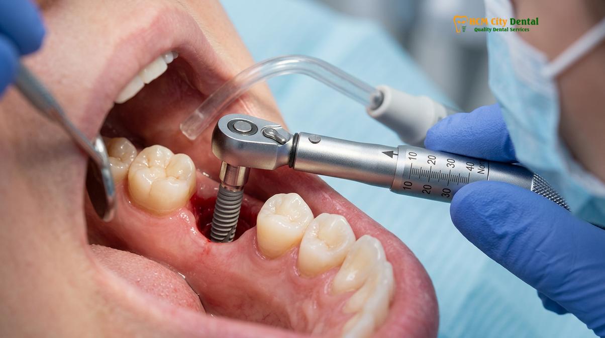
When an implant is placed immediately into a fresh socket, there is almost always a void between the surface of the implant and the inner walls of the socket. This void is clinically referred to as the “jumping distance.” If this gap is larger than 2 millimeters, it must be grafted with particulate bone material to ensure that the bone fully bridges the gap and integrates with the implant surface. This simultaneous grafting procedure is a critical component of the implant placement preparation, ensuring that the final restoration is supported by a thick, healthy wall of bone.
| Clinical Parameter | Immediate Placement | Delayed Placement (Post-Grafting) |
|---|---|---|
| Treatment Timeline | Shorter (3-4 months total) | Longer (6-9 months total) |
| Number of Surgeries | One primary surgical phase | Two distinct surgical phases |
| Bone Quality Requirement | Requires excellent native apical bone | Allows for regeneration of deficient bone |
| Infection Status | Contraindicated in acute infection | Ideal for clearing severe infections first |
| Aesthetic Predictability | Excellent for preserving natural gum contours | May require secondary soft tissue grafting |
The decision between immediate and delayed placement is made on a case-by-case basis, heavily relying on 3D imaging and the surgeon’s intraoperative assessment. For patients undergoing full-arch rehabilitation, a combination of both techniques may be employed, utilizing immediate placement in healthy sites and delayed placement in areas requiring extensive reconstruction. This tailored approach ensures that the foundation for Dental Implants is optimized for long-term durability.
Preparing the Soft Tissues: Ensuring Healthy Gums for Dentures
Proper soft tissue management following extraction ensures the development of a thick, keratinized gingival barrier that protects the underlying implants and provides a comfortable, resilient seat for the final denture.
While much of the focus in implant dentistry is rightfully placed on the preservation and regeneration of the jawbone, the management of the overlying soft tissues is equally critical to the long-term success of the prosthesis. The gingiva (gum tissue) serves as the primary biological seal, protecting the underlying bone and the implant surface from the hostile, bacteria-rich environment of the oral cavity. In the context of an implant-supported denture, the soft tissue must not only provide this protective seal but also withstand the frictional forces and compressive loads exerted by the prosthetic base.
The ideal soft tissue profile around a dental implant consists of a thick band of attached, keratinized gingiva. Keratinized tissue is dense, fibrous, and firmly bound to the underlying periosteum, making it highly resistant to mechanical trauma and bacterial infiltration. Conversely, non-keratinized alveolar mucosa is thin, mobile, and highly susceptible to inflammation and recession. Clinical studies have consistently shown that a lack of adequate keratinized tissue around implants is associated with higher rates of plaque accumulation, peri-implant mucositis, and marginal bone loss [4]. Therefore, establishing a robust soft tissue profile is a fundamental component of the denture fitting foundation.
“The peri-implant soft tissue seal is the first line of defense against biological complications; without a thick band of keratinized gingiva, even the most perfectly integrated implant remains vulnerable to peri-implantitis.”
Soft tissue management begins at the time of extraction. Atraumatic surgical techniques and the careful handling of the gingival flaps minimize scarring and preserve the natural blood supply to the tissues. During the healing phase, clinicians may utilize custom healing abutments or provisional restorations to actively sculpt the emergence profile of the gums. These temporary components guide the healing tissues, creating a natural-looking scalloped contour that mimics the appearance of a healthy natural tooth emerging from the gums.
In cases where the patient presents with a naturally thin gingival biotype or where significant tissue loss has occurred due to previous infections, soft tissue grafting may be necessary. Procedures such as connective tissue grafts (CTG) or free gingival grafts (FGG) involve harvesting a small piece of dense tissue, typically from the patient’s palate, and transplanting it to the implant site. This thickens the tissue biotype, enhancing both the aesthetic outcome and the biological stability of the site. When advanced restorative materials are eventually used to fabricate the final denture, they must interface seamlessly with this optimized soft tissue to prevent food impaction and ensure patient comfort.
Planning Your Complete Smile Reconstruction in Saigon
Comprehensive smile reconstruction requires a highly coordinated multidisciplinary approach, combining advanced 3D digital imaging with expert surgical execution to transition patients smoothly from extractions to final implant dentures.
Transitioning from a state of terminal dentition to a fully restored, implant-supported smile is a profound life change that requires meticulous planning and expert execution. At HCMC Dental Clinic in Ho Chi Minh City, the approach to full-arch rehabilitation is deeply rooted in digital dentistry and evidence-based surgical protocols. The process begins with a comprehensive consultation, where the clinical team evaluates the patient’s chief complaints, medical history, and aesthetic goals. This is followed by the acquisition of high-resolution digital data, including intraoral optical scans and CBCT imaging. These digital files are merged in specialized implant planning software, allowing the surgeon to perform a virtual surgery before ever touching the patient.
Dr. Nguyen Van Cuong, a highly respected clinician in the field of implantology, emphasizes that this digital workflow is the key to predictable outcomes. By planning the exact position, angle, and depth of every implant in a 3D virtual environment, the surgical team can fabricate highly precise, 3D-printed surgical guides. These guides fit securely over the patient’s remaining teeth or bone, directing the surgical drills with sub-millimeter accuracy. This level of precision ensures that the implants are placed in the optimal biomechanical positions to support the final denture, while safely avoiding critical anatomical structures [5].
Clinical Case Overview: A 62-year-old patient visited HCMC Dental Clinic in Ho Chi Minh City presenting with severe generalized periodontitis and multiple failing teeth. The comprehensive treatment plan involved the atraumatic extraction of the remaining upper dentition, simultaneous alveolar ridge preservation, and the placement of six maxillary implants using a fully guided digital workflow. Following a four-month osseointegration period, the patient was successfully restored with a fixed, implant-supported hybrid denture, dramatically improving both masticatory function and facial aesthetics.
For patients requiring extensive work, the ability to consolidate treatments is a significant advantage. The clinical team often coordinates the surgical phase to address all extractions, bone grafting, and implant placements in a single, well-planned visit. This approach, similar to the efficiency seen when consolidating multiple dental treatments, minimizes the number of times the patient must undergo anesthesia and significantly reduces the overall recovery burden. Furthermore, the integration of an in-house dental laboratory allows for the rapid fabrication of provisional immediate dentures, ensuring that patients never leave the clinic without a functional and aesthetically pleasing smile.
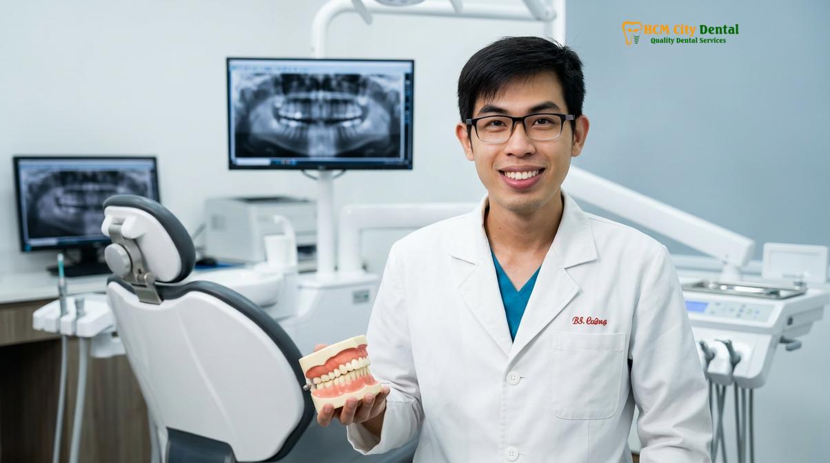
The role of a dedicated Saigon implant specialist extends beyond the surgery itself; it encompasses the meticulous design of the final prosthesis. Whether the patient opts for a removable overdenture retained by locator attachments or a premium fixed zirconia hybrid prosthesis, the final restoration is custom-milled to harmonize with the patient’s unique facial features and functional dynamics. By adhering strictly to the principles of extraction before implant denture preparation, Dr. Cuong and the team at HCMC Dental Clinic ensure that every smile reconstruction is built upon a foundation of uncompromising clinical excellence.
When to See a Doctor for Implant Evaluation
Determining the exact moment when a tooth can no longer be saved and must be replaced with an implant requires a professional clinical diagnosis. You should schedule a comprehensive evaluation with a dental specialist if you experience any of the following symptoms:
- Severe Tooth Mobility: Teeth that are noticeably loose or shift during chewing often indicate advanced periodontal bone loss that cannot be reversed.
- Chronic Pain or Recurrent Abscesses: Persistent swelling, pain, or the presence of a pimple-like fistula on the gums suggests a deep-seated infection that may be destroying the surrounding jawbone.
- Extensive Structural Damage: Teeth that have fractured below the gumline or have decay so extensive that managing severe dental decay with traditional crowns is no longer biomechanically viable.
- Failing Dental Bridges or Dentures: If your current prosthetics are loose, uncomfortable, or causing damage to the supporting abutment teeth, an implant-supported solution may be required.
It is important to note that while implant dentistry offers highly predictable and successful outcomes, it is not a universal solution for every patient. Systemic health conditions, such as uncontrolled diabetes or a history of bisphosphonate therapy, can affect bone healing and must be carefully evaluated. A personalized consultation, complete with 3D radiographic imaging, is essential to determine your specific candidacy and to develop a safe, effective treatment plan tailored to your unique anatomical needs. For comprehensive care, explore our comprehensive general dentistry services to begin your journey toward optimal oral health.
Frequently Asked Questions
How long do I wait after extraction to get a dental implant?
You typically wait three to six months after a standard tooth extraction before placing a dental implant. This healing period allows the alveolar bone to regenerate and consolidate, providing a dense, stable foundation for the titanium post. However, in cases where the extraction socket is completely free of infection and the surrounding bone architecture is fully intact, an immediate implant may be placed during the exact same surgical visit. Your clinician will evaluate your bone density using 3D CBCT imaging to determine the safest and most predictable timeline for your specific anatomical conditions.
Why is a bone graft needed if I am getting a denture?
A bone graft is required to prevent the natural shrinkage of the jawbone that occurs immediately after a tooth is extracted. When teeth are removed, the alveolar ridge rapidly loses both width and height due to the lack of mechanical stimulation from the tooth roots. By placing an extraction bone graft into the empty socket, clinicians preserve the necessary bone volume required to securely anchor dental implants. This preserved bone mass ensures that the final implant-supported denture has a stable, retentive, and highly functional foundation for long-term chewing efficiency.
Can I wear a temporary denture right after tooth extraction?
Yes, you can usually wear an immediate temporary denture directly after your teeth are extracted. This provisional prosthesis is fabricated prior to your surgery and inserted immediately, ensuring you never leave the clinic without teeth. While it provides immediate aesthetic restoration and basic phonetic function, it is primarily designed to protect the surgical sites and extraction bone grafts during the initial healing phase. Because your gums and bone will shrink as they heal, this temporary denture will require periodic relining to maintain a proper fit until your final implant denture is ready.
Is the extraction process painful when preparing for implants?
The extraction process is performed under profound local anesthesia, ensuring you do not feel sharp pain during the surgical procedure. Modern atraumatic extraction techniques focus on gently severing the periodontal ligament rather than applying heavy force, which significantly minimizes tissue trauma. While you will feel pressure and vibration during the removal, the procedure is highly controlled. Post-operative discomfort is typically manageable with prescribed anti-inflammatory medications and analgesics, and most patients report that the recovery is much smoother than anticipated when proper surgical protocols are followed.
How many implants are required to support a full denture?
A full lower denture typically requires a minimum of two to four implants, while an upper denture usually requires four to six implants for optimal stability. The upper jaw consists of softer, less dense bone, necessitating a greater number of implants to distribute the chewing forces evenly. The exact number depends on whether you are receiving a removable overdenture that snaps onto locators or a fixed hybrid denture that is permanently screwed into place. A comprehensive clinical evaluation and 3D imaging will dictate the precise biomechanical requirements for your smile reconstruction.
References
- Journal of Periodontology. Alveolar ridge preservation following tooth extraction: A systematic review. (2021).
- Clinical Oral Implants Research. Dimensional changes of the alveolar ridge post-extraction. (2020).
- International Journal of Oral and Maxillofacial Implants. Primary stability and osseointegration in immediate implant placement. (2019).
- Journal of Prosthetic Dentistry. Soft tissue thickness and peri-implant marginal bone loss. (2022).
- Clinical Implant Dentistry and Related Research. Digital workflows in full-arch implant rehabilitation. (2023).
For pricing, booking, and a free clinical assessment, visit our Tooth Extraction service page at HCMC Dental Clinic in Ho Chi Minh City.