When a dental filling fails, the threshold between replacing it with another filling versus placing a dental crown depends entirely on the remaining structural tooth integrity. If the defect compromises more than half the tooth’s width or involves load-bearing cusps, a crown is often clinically required to help prevent catastrophic fracture and preserve the underlying root.
Clinical Summary:
The decision to transition from a filling to a crown is governed by strict biomechanical principles, specifically the volume of healthy dentin and enamel remaining after decay removal. While composite resins are excellent for conservative restorations, they lack the tensile strength to bind weakened tooth walls together under heavy occlusal forces. Exceeding established isthmus width parameters or repeatedly replacing massive fillings drastically increases the risk of vertical root fractures. A dental crown provides full-coverage circumferential protection, distributing chewing forces evenly and preserving the underlying tooth structure for long-term functional stability.
Key Takeaways:
- A failed restoration must be evaluated based on remaining wall thickness, not just the size of the recurrent cavity.
- The isthmus width rule dictates that if a cavity preparation exceeds one-third to one-half of the intercuspal distance, full coverage is typically indicated.
- An endodontically treated posterior tooth becomes brittle and almost universally requires a crown to help prevent splitting.
- Over-filling a severely compromised tooth creates a wedge effect, significantly increasing the risk of unrestorable cusp fractures.
- Timely intervention with a full-coverage restoration can often save a structurally compromised tooth from extraction following a catastrophic failure.
The Limits of Composite Resin: When Cavities Outgrow Fillings
Composite resin is ideal for small to moderate cavities, but it cannot structurally reinforce a tooth once the decay or previous filling exceeds fifty percent of the tooth’s total volume.
Modern dentistry relies heavily on composite resin for direct restorations due to its excellent aesthetic properties and ability to bond micromechanically to tooth structure. However, the physical limitations of this material become apparent when dealing with extensive tooth loss. To understand the filling vs crown threshold when a filling fails, one must first understand the biomechanics of how teeth bear the immense forces of mastication. A natural, healthy tooth is a highly engineered structure where the enamel acts as a rigid outer shell and the underlying dentin provides flexible support. When a cavity is small, a filling simply plugs the hole, allowing the surrounding intact enamel to continue bearing the brunt of the chewing load.
The clinical paradigm shifts dramatically when a large restoration fails. A failed composite filling often masks secondary decay that has silently hollowed out the dentin beneath the visible surface, leading to a significant reduction in the longevity of the tooth if not properly addressed[1]. By the time the old filling is removed along with the newly decayed tissue, the remaining tooth walls are frequently left thin, undermined, and structurally compromised. In these scenarios, placing another, even larger filling is a biomechanical misstep.
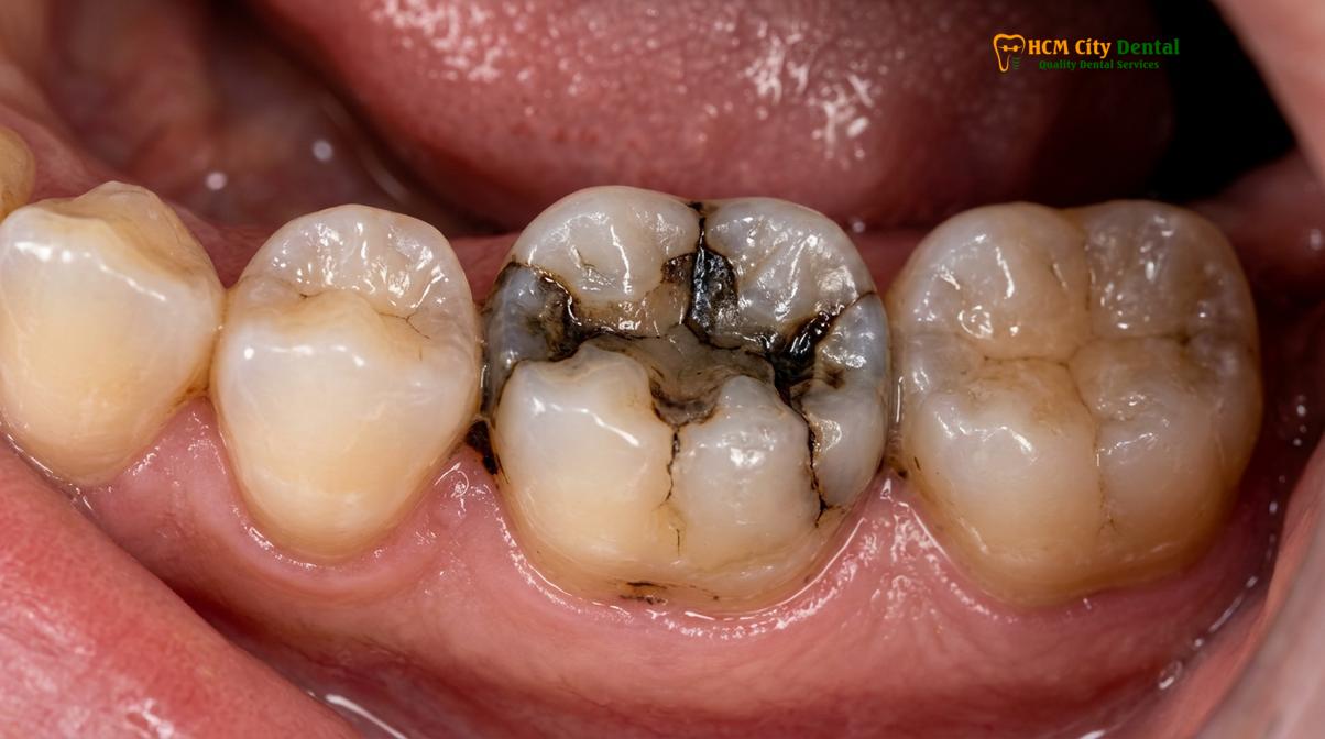
Composite resin has a high compressive strength but relatively low tensile and shear strength. It is not designed to hold a broken tooth together. When a patient bites down, the forces are transmitted through the filling to the remaining walls. If those walls are too thin, they will flex. This continuous microscopic flexing—known as cyclic fatigue—eventually breaks the adhesive bond between the tooth and the filling, leading to marginal leakage, recurrent decay, and ultimately, structural failure.
The transition from a filling to a crown is a necessity dictated by physics. A crown changes the force distribution entirely. Instead of resting inside the tooth and pushing outward against the walls, a crown encases the outside of the tooth, holding the walls together. This circumferential bracing effect, known as a ferrule, is a highly reliable way to restore a tooth that has lost its inherent structural integrity due to massive decay or repeated filling failures.
The Isthmus Width Rule: Evaluating Tooth Wall Thickness
The isthmus width rule is a fundamental biomechanical guideline stating that if a cavity spans more than half the distance between the cusps, the tooth walls are generally too thin to support a filling.
In clinical dentistry, the decision to place a crown over a filling is heavily influenced by a geometric measurement known as the isthmus width. The isthmus is the narrowest part of a cavity preparation, typically located between the buccal (cheek side) and lingual (tongue side) cusps of a molar or premolar. The isthmus width rule serves as a critical diagnostic threshold. When the width of the cavity preparation is less than one-third of the total intercuspal distance, the tooth retains enough bulk to safely support a direct filling. The remaining walls are thick enough to resist flexing under occlusal loads.
However, when a filling fails and requires replacement, the new cavity preparation is inevitably larger than the original. If the resulting defect expands the isthmus to greater than one-third, but less than one-half of the intercuspal distance, the tooth enters a gray area where advanced restorative techniques such as ceramic inlays or onlays may be considered. Once the isthmus width exceeds one-half of the intercuspal distance, the biomechanical verdict is clear: the remaining walls are too weak to survive long-term functional loading without full-coverage protection[2].
“The preservation of tooth structure is our highest priority, but we must differentiate between preserving healthy enamel and leaving behind fragile, undermined walls. When the isthmus width rule is violated, placing a filling is no longer conservative; it is a gamble that often ends in a non-restorable fracture.”
To illustrate the clinical decision-making process, dentists evaluate several parameters of the remaining tooth structure before recommending a crown. The table below outlines the general thresholds used to determine the appropriate restoration:
| Clinical Parameter | Direct Filling Indicated | Inlay / Onlay Indicated | Dental Crown Indicated |
|---|---|---|---|
| Isthmus Width | < 1/3 of intercuspal distance | 1/3 to 1/2 of intercuspal distance | > 1/2 of intercuspal distance |
| Remaining Wall Thickness | > 2.0 mm of solid dentin/enamel | 1.5 mm to 2.0 mm | < 1.5 mm (undermined walls) |
| Cusp Involvement | No cusps involved | 1 to 2 cusps compromised | Multiple cusps missing or undermined |
| Endodontic Status | Vital pulp | Vital pulp | Root canal treated (Posterior) |
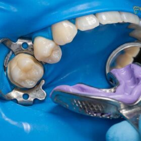
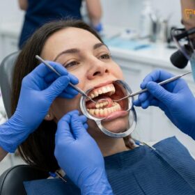
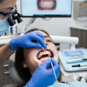
Evaluating these parameters requires precise diagnostics. During a comprehensive examination, a dentist will use high-magnification loupes, transillumination, and tactile exploration to assess the integrity of the remaining walls. If the walls are less than 1.5 millimeters thick, they are considered structurally compromised. Attempting to pack a large composite filling into a tooth with such thin walls can lead to severe complications, as the walls will inevitably fracture under the immense pressure of the human bite.
Why Root Canal Treated Teeth Almost Always Require Crowns
Endodontic therapy removes the tooth’s internal blood supply, rendering the remaining dentin brittle and highly susceptible to splitting under normal chewing forces without full-coverage protection.
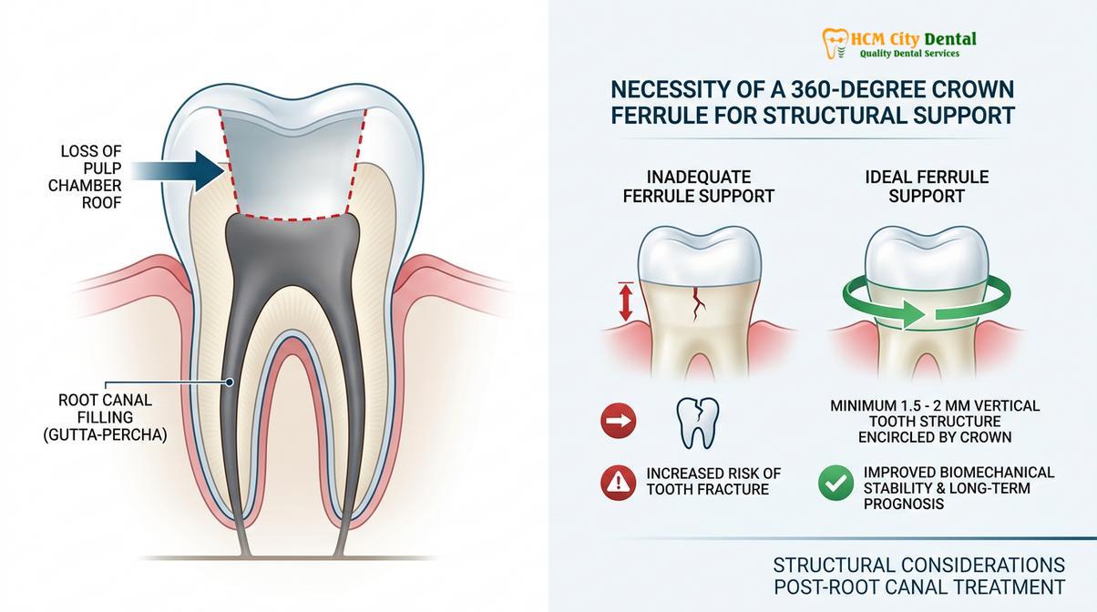
One of the most absolute thresholds in restorative dentistry involves teeth that have undergone endodontic therapy. A root canal treated tooth is fundamentally different from a vital tooth in both its physical properties and its biomechanical behavior. To perform a root canal, the dentist must drill an access hole through the biting surface of the tooth, deep into the pulp chamber. This process inherently destroys the “roof” of the pulp chamber, which is a critical structural component that ties the buccal and lingual cusps together.
Furthermore, the removal of the pulp tissue eliminates the tooth’s internal blood supply and hydration mechanism. Over time, the dentin in a root canal treated tooth loses its moisture content, becoming significantly more brittle and less resilient to stress, which drastically lowers its survival rate if only restored with a direct filling[3]. Additionally, the tooth loses its proprioception—the internal nerve feedback that tells your brain how hard you are biting. Without this feedback, patients subconsciously exert more force on endodontically treated teeth, increasing the risk of mechanical failure.
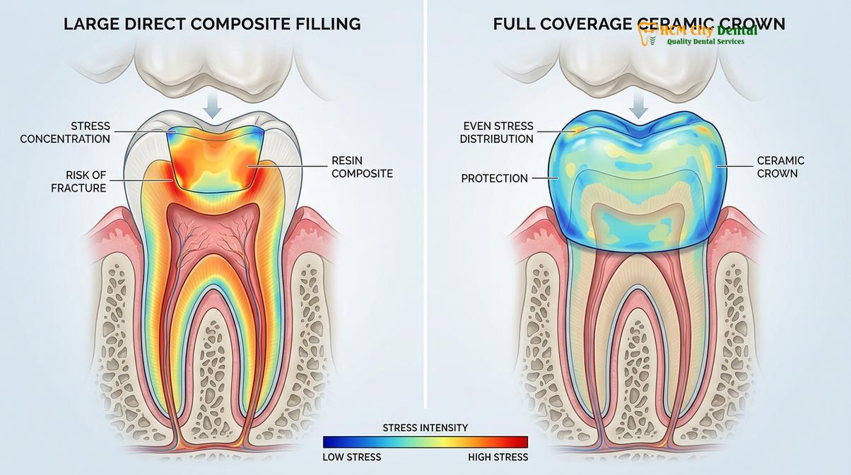
For posterior teeth (molars and premolars), which bear the brunt of masticatory forces, the combination of structural loss, dentin brittleness, and increased occlusal loading makes a direct filling an inadequate long-term solution. If a large filling is placed in a root-canaled molar, the wedge effect of chewing will almost certainly cause the freestanding cusps to fracture. Often, these fractures propagate vertically down the root, rendering the tooth completely unrestorable and necessitating extraction.
The standard of care dictates that posterior root canal treated teeth receive full-coverage crowns to provide a protective ferrule. The ferrule is a band of solid tooth structure, ideally at least 1.5 to 2 millimeters high, that the crown grips around the base of the tooth. This bracing effect neutralizes the outward forces that would otherwise split the tooth.
The Danger of Over-Filling: Cusp Fractures and Tooth Loss
Placing a massive filling inside a hollowed-out tooth acts like a wedge during chewing, forcing the weakened enamel walls apart and often leading to unrestorable vertical fractures.
A common misconception among patients is that a filling is always the more conservative and preferable option compared to a crown. While preserving natural tooth structure is the ultimate goal of modern dentistry, placing an excessively large filling in a severely compromised tooth is a false economy. This practice, often referred to as “over-filling,” ignores the fundamental principles of cusp fracture prevention and places the entire tooth at grave risk.
To understand the danger, one must visualize the mechanics of a large intracoronal restoration. When a cavity consumes more than half the width of the tooth, the resulting filling is essentially a large block of composite resin sitting between thin walls of natural enamel. When the opposing teeth bite down on this filling, the force is not directed straight down the long axis of the root. Instead, the filling acts as a wedge, translating downward force into lateral pressure that pushes outward against the remaining cusps[4].
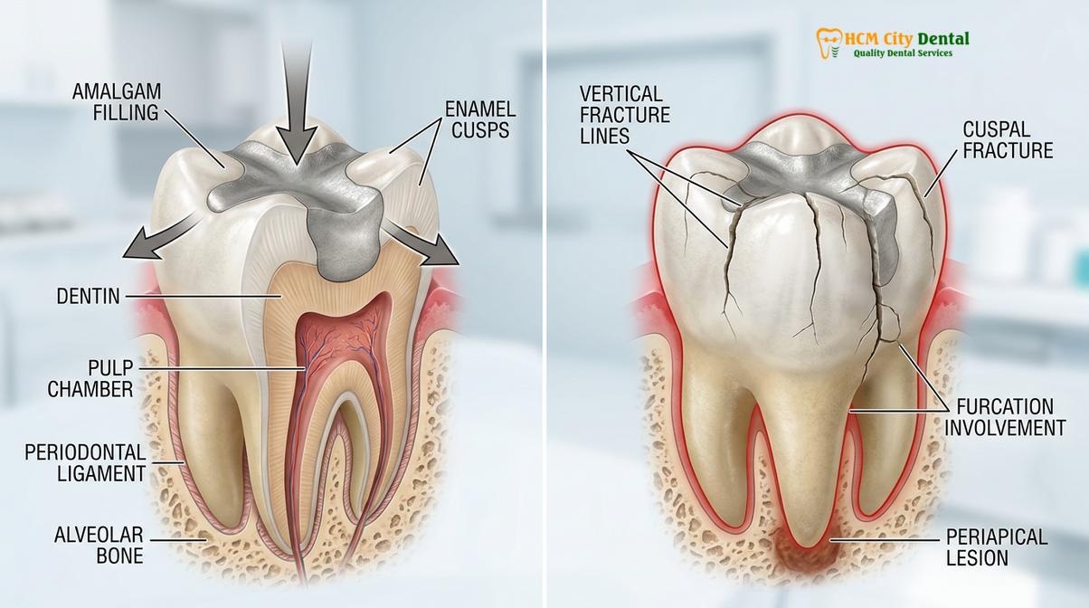
Additionally, composite resin undergoes a process called polymerization shrinkage when cured with a dental light. In massive cavities, this shrinkage creates immense stress on the remaining fragile enamel walls, pulling them inward and creating micro-cracks before the patient even leaves the dental chair[5]. Over time, this continuous wedging action and shrinkage stress cause the enamel walls to flex. Micro-cracks begin to form at the base of the cusps, leading to sharp, fleeting pain when biting down on certain foods.
“The most heartbreaking cases we see are teeth that must be extracted because a massive filling acted as a wedge and split the root down the middle. Had the tooth been protected with a crown earlier, it could have provided decades of functional service.”
If the warning signs of a cracked tooth are ignored, the micro-crack will eventually propagate into a catastrophic fracture. If the fracture extends below the bone level or splits the tooth vertically through the pulp floor, the tooth is deemed unrestorable and must be extracted. Therefore, recognizing the threshold where a filling becomes a liability rather than a cure is essential for long-term oral health.
When to Consult a Dentist: Important Clinical Signs
Recognizing the early warning signs of a failing filling can mean the difference between a simple crown placement and losing the tooth entirely.
Patients should not wait for a filling to completely fall out before seeking professional care. According to guidelines established by the Vietnam Odonto-Stomatology Association (VOSA) regarding restorative thresholds, early intervention is key to preserving natural tooth structure[6]. If you experience localized pain when chewing, sensitivity to temperature changes that lingers, or notice visible dark lines around the edges of an old filling, these are strong indicators that the restoration is failing and microleakage has begun.
Another critical sign is food impaction. If food consistently gets stuck between teeth that previously had tight contacts, it may indicate that a large filling has chipped or shifted, compromising the interproximal space. Prompt evaluation by a qualified dental professional ensures that the structural integrity of the tooth is assessed before a catastrophic fracture occurs.
How HCMC Dental Clinic Restores Failing Fillings Conservatively
Utilizing advanced diagnostics and biomimetic principles, our clinical team carefully evaluates each failing restoration to recommend the most conservative yet durable treatment.
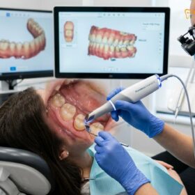
Dr. Nguyen Van Cuong and the clinical team at HCMC Dental Clinic emphasize a biomimetic approach to restorative care. By carefully evaluating the remaining healthy enamel, Dr. Cuong ensures that every treatment plan prioritizes long-term structural integrity over quick fixes. When a patient presents with a broken or leaking restoration, the first step is to conduct a meticulous assessment using high-resolution 3D CBCT imaging and intraoral cameras to map the exact extent of the decay and structural damage.
The transition from a filling to a crown is a significant step, and intermediate restorative options are always considered when clinically appropriate. If a filling has failed but the damage is confined to one or two cusps while the rest of the tooth remains robust, an onlay may be recommended. An onlay is a custom-milled ceramic restoration that covers only the damaged cusps, preserving the healthy portions of the tooth while still providing the necessary occlusal protection.
A 45-year-old patient visited HCMC Dental Clinic in Ho Chi Minh City complaining of severe pain upon biting in the lower right quadrant. Clinical examination revealed a massive, decade-old composite filling that had fractured, leaving the lingual wall of the molar severely undermined. Instead of attempting to place another large filling that would likely cause a vertical root fracture, the clinical team utilized a full-coverage ceramic crown. This provided a 360-degree ferrule, stabilizing the remaining tooth structure, eliminating the pain, and saving the tooth from extraction.

By adhering to strict biomechanical guidelines and utilizing state-of-the-art materials, the focus remains on providing restorations that not only look natural but also withstand the rigorous demands of daily function.
References
- Clinical Oral Investigations. Longevity and failure rates of large direct composite restorations in posterior teeth. (2020).
- Journal of Prosthetic Dentistry. The effect of isthmus width on the fracture resistance of maxillary premolars. (2019).
- International Endodontic Journal. Survival rates of root-filled teeth restored with crowns versus direct fillings. (2022).
- Journal of the American Dental Association. Biomechanical considerations in the restoration of endodontically treated teeth. (2021).
- Dental Materials. Polymerization shrinkage stress and cusp deflection in large composite restorations. (2018).
- Vietnam Odonto-Stomatology Association (VOSA). Guidelines on Restorative Thresholds and Structural Preservation. (2023).
If you are experiencing symptoms of a failing restoration or have concerns about the structural integrity of your teeth, contact HCMC Dental Clinic in Ho Chi Minh City today for a comprehensive evaluation. For anterior teeth requiring both structural reinforcement and cosmetic enhancement, explore our Porcelain Veneers to restore your smile’s natural strength and beauty.
For premium porcelain crowns, bridges, and advanced smile reconstructions, visit our Dental Crowns & Bridges service page at HCMC Dental Clinic in Ho Chi Minh City.