Discovering that a dental instrument has broken off inside your tooth during a root canal can be a highly distressing experience. For international patients and dental travelers seeking a solution, the prospect of losing the tooth to extraction often feels inevitable. However, modern endodontics has evolved dramatically. The protocol for a broken file in root canal removal using a microscope in Vietnam has reached hospital-grade standards, offering a highly predictable, minimally invasive pathway to save the natural tooth. At HCMC Dental Clinic in Ho Chi Minh City, advanced microscopic endodontic retreatment transforms what was once considered a clinical disaster into a manageable, highly successful procedure.
The separation of a nickel-titanium (NiTi) rotary file is a known procedural complication in global dentistry, occurring in approximately 1% to 3% of complex molar cases. The critical factor determining the survival of the tooth is not the breakage itself, but how the complication is subsequently managed. Blindly attempting to fish out a microscopic metal fragment using traditional hand files often leads to catastrophic iatrogenic damage, such as root perforations or ledged canals. True clinical success requires high-resolution 3D Cone Beam Computed Tomography (CBCT) imaging, a high-powered dental operating microscope, specialized titanium ultrasonic micro-tips, and the steady hands of an experienced specialist.
Under the clinical direction of Dr. Nguyen Van Cuong, DDS (Clinical Director — MOH License 0003265/BL-CCHN), HCMC Dental Clinic utilizes a 25x Carl Zeiss Extaro 300 dental microscope and 100% latex-free heavy-gauge Nitrile Rubber Dam isolation to perform complex separated endodontic instrument retrieval. This comprehensive clinical guide explores the biomechanics of file separation, the precise microscopic protocols used for retrieval and bypass, the integration of bioactive bioceramic cements for root repair, and the transparent 2026 fee schedule that makes world-class endodontic care accessible to expats and dental tourists in District 1, Ho Chi Minh City.
Clinical Summary:
The retrieval or bypass of a separated endodontic file is a highly specialized microsurgical procedure that requires advanced magnification and 3D imaging. A broken file does not automatically necessitate tooth extraction; the metal fragment is sterile, and the primary goal is to eliminate the bacterial biofilm trapped beneath it. Utilizing a 25x Carl Zeiss dental microscope, 0.075mm high-resolution 3D CBCT scans, and Satelec/Acteon Titanium Ultrasonic Micro-Tips (ET20/ET25), specialists at HCMC Dental Clinic can safely vibrate, loosen, and retrieve the fragment, or strategically bypass it. When aggressive retrieval risks root perforation, entombing the fragment in bioactive ProRoot MTA or Septodont Biodentine is a clinically proven, evidence-based alternative. This hospital-grade protocol, combined with biomimetic restorative techniques, ensures long-term tooth survival at a fraction of Western costs.
Key Takeaways:
- Not an Automatic Extraction: A broken file is a procedural complication, not a biological failure. The tooth can be saved in the vast majority of cases using microscopic intervention.
- Advanced Magnification is Mandatory: Successful retrieval requires a 25x Carl Zeiss Extaro 300 dental operating microscope; attempting removal without magnification drastically increases the risk of root perforation.
- 3D CBCT Diagnostics: A 0.075mm high-resolution 3D CBCT scan is essential to map the exact location, length, and curvature (Schneider’s angle) of the fragment before any clinical action is taken.
- Ultrasonic Micro-Tips: Satelec/Acteon Titanium Ultrasonic Micro-Tips (ET20/ET25) are used to create a staging platform and apply counter-clockwise acoustic energy to unscrew the fragment safely.
- Biomimetic Restoration: Following retrieval, the tooth must be reinforced using biomimetic quartz/glass fiber posts (18–20 GPa) and a full Zirconia crown with a strict 2mm ferrule to prevent vertical root fracture.
- Cost Efficiency: Microscopic endodontic retreatment and file bypass at HCMC Dental Clinic costs between $154 and $231 USD online, offering 75% to 85% savings compared to the US or Australia.
Why Do NiTi Rotary Endodontic Files Separate Inside Curved Root Canals — and Does the Metal Fragment Alone Cause Infection?
To understand how to fix the problem, patients must first understand why it occurs. Modern endodontics relies heavily on Nickel-Titanium (NiTi) rotary file systems. These instruments are engineered to be highly flexible, allowing dentists to navigate the complex, tortuous anatomy of molar root canals. However, despite advances in metallurgy—such as heat-treated Gold and Blue wire technologies that shift the alloy into a more flexible martensitic phase—these files are still subject to the laws of physics. Instrument separation typically occurs due to one of two primary biomechanical failures: cyclic fatigue or torsional stress.
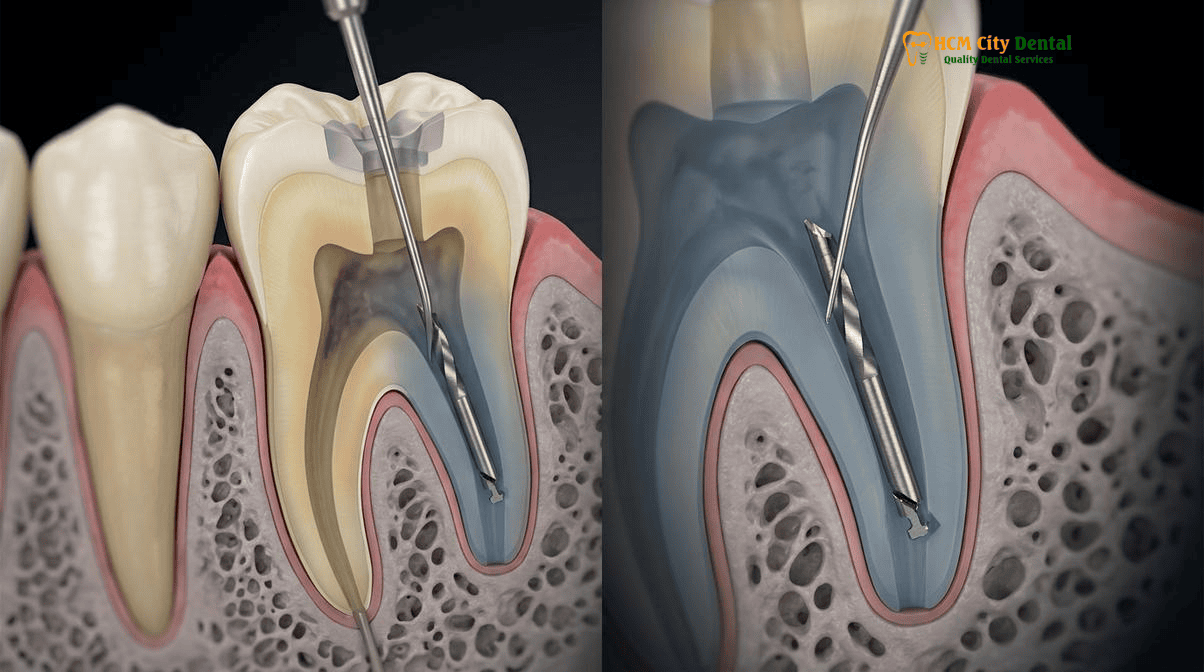
Cyclic fatigue occurs when a file rotates freely inside a severely curved canal. As the instrument spins, the metal at the apex of the curve is subjected to alternating cycles of tension (on the outside of the curve) and compression (on the inside of the curve). Over time, this repeated stress causes micro-cracks to form in the alloy, eventually leading to a sudden, unexpected fracture without any tactile warning to the dentist. Torsional stress, on the other hand, happens when the tip of the file binds or locks into the narrow dentin walls of the canal while the motor continues to rotate the shaft. The torque exceeds the elastic limit of the metal, causing the file to unwind and snap.
When a file breaks, the immediate patient concern is often: “Will the metal rust or cause an infection?” The definitive clinical answer is no. Endodontic files are manufactured from highly biocompatible, surgical-grade Nickel-Titanium or stainless steel. The metal fragment itself is sterile, inert, and will not cause an allergic reaction, rust, or directly induce an infection[1]. The true clinical danger lies in what the broken file represents: a physical barricade.
The primary objective of a Root Canal Procedure in Ho Chi Minh City is the complete chemo-mechanical debridement of the canal system—removing necrotic pulp tissue and eradicating the bacterial biofilm. When a file separates, it blocks access to the apical portion (the tip) of the root. If the canal was not thoroughly disinfected before the breakage occurred, the bacteria trapped beneath the fragment will multiply, eventually leaking out of the apical foramen and causing a periapical abscess or bone infection. Therefore, the goal of microscopic retreatment is not merely to remove the metal for the sake of removing it, but to regain access to the apical terminus so the canal can be properly disinfected and sealed.
3D CBCT Localization & Coronal vs. Middle vs. Apical Third Curvature Assessment (Schneider’s Angle): Retrieval vs. Bypass
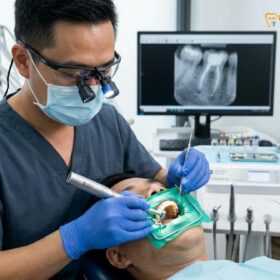
Before any physical attempt is made to retrieve a separated instrument, a precise, three-dimensional roadmap is absolutely mandatory. Relying solely on traditional 2D periapical X-rays is clinically insufficient for complex retreatment, as 2D images compress complex 3D anatomy into a flat plane, obscuring the true curvature of the root and the exact position of the fragment. At HCMC Dental Clinic, every broken file case begins with a 0.075mm high-resolution 3D Cone Beam Computed Tomography (CBCT) scan.
The 3D CBCT scan allows Dr. Nguyen Van Cuong, DDS, to evaluate several critical anatomical factors that dictate the treatment protocol:
- Fragment Location: Is the broken file located in the coronal third (top), middle third, or apical third (bottom) of the root canal? Fragments in the straight coronal or middle thirds have a highly predictable retrieval success rate. Fragments lodged deep in the apical third are significantly more challenging.
- Schneider’s Angle (Root Curvature): The CBCT scan accurately measures the degree of root curvature. If the file is separated before the curve, retrieval is highly favorable. If the file is bound tightly beyond a severe curve (e.g., a Schneider’s angle greater than 30 degrees), attempting to pull it out may be biomechanically impossible without destroying the root.
- Radicular Dentin Thickness: The software measures the exact thickness of the dentin walls surrounding the fragment. In mandibular molars, the “danger zone” on the inner curve of the mesial roots is notoriously thin. Aggressive drilling in this area to free a file can easily result in a catastrophic strip perforation[2].
Based on this 3D data, the clinical decision matrix shifts between three primary strategies: Retrieval (removing the fragment completely), Bypass (navigating a new, microscopic hand file alongside the fragment to clean the space below it), or Entombment (leaving the fragment in place and sealing it with bioactive bioceramic cement, provided the canal was already disinfected).
25x Carl Zeiss Extaro Microscope + Titanium Ultrasonic Micro-Tips (ET20/ET25) + Micro-Lasso Loop Retrieval Protocol
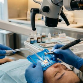
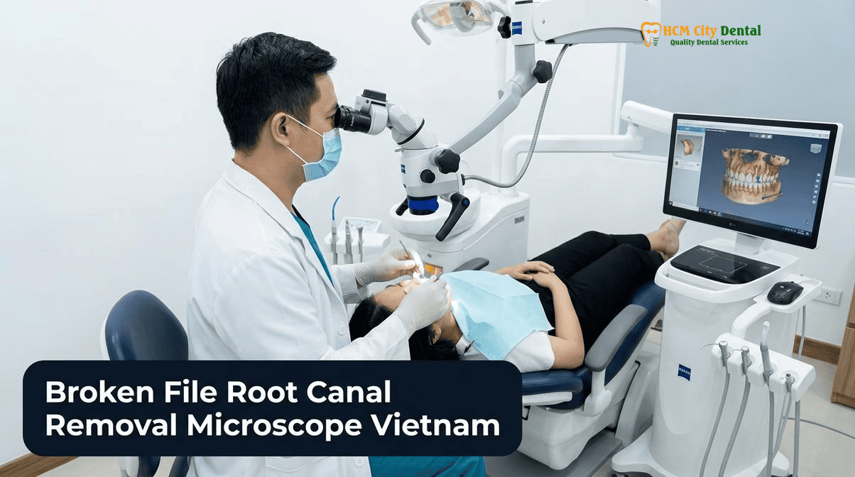
If the 3D CBCT analysis indicates that retrieval is safe and viable, the procedure transitions to the microscopic phase. The use of a dental operating microscope is not optional for this procedure; it is the standard of care. Attempting to remove a broken file without high-powered magnification is akin to performing neurosurgery blindfolded.
At HCMC Dental Clinic, the protocol for a separated endodontic instrument retrieval under a microscope involves a highly orchestrated sequence of steps:
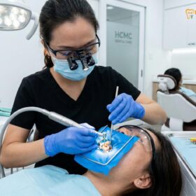
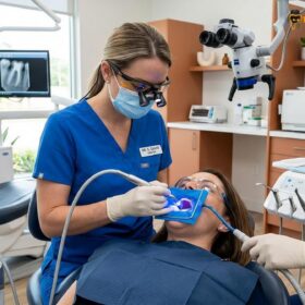
- Absolute Isolation: The tooth is isolated using a 100% latex-free heavy-gauge Nitrile Rubber Dam. This prevents any saliva or oral bacteria from contaminating the surgical field and ensures that if the microscopic fragment is dislodged, it cannot be swallowed or aspirated by the patient.
- Coronal Access and Straight-Line Visibility: Using the 25x magnification and coaxial LED illumination of the Carl Zeiss Extaro 300 microscope, Dr. Cuong modifies the access cavity to establish a direct, straight-line line of sight to the coronal end of the broken fragment.
- Creating a Staging Platform: Modified Gates Glidden drills or specialized ultrasonic tips are used to carefully remove a minimal amount of dentin just above the fragment, creating a flat “staging platform.” This exposes the top 1 to 2 millimeters of the broken file.
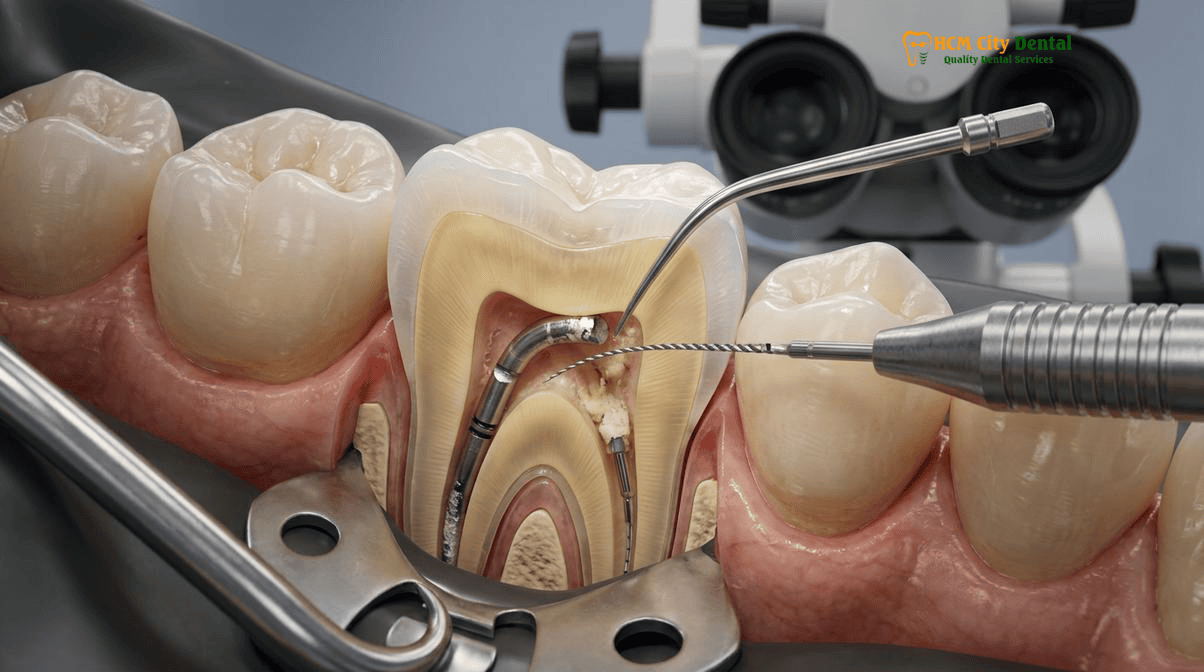
- Ultrasonic Troughing and Acoustic Streaming: This is the most critical phase. Satelec/Acteon Titanium Ultrasonic Micro-Tips (specifically the ET20 or ET25) are introduced into the canal. These tips do not spin; they vibrate at high frequencies (25,000 to 30,000 Hz). The ultrasonic tip is activated dry or with a continuous flow of EDTA (Ethylenediaminetetraacetic acid) to remove the smear layer. The tip is applied to the inner wall of the canal, vibrating in a counter-clockwise direction around the exposed file. The acoustic energy and cavitation effect break the frictional bind between the metal and the dentin, effectively “unscrewing” the fragment[3].
- Micro-Lasso or Micro-Tube Retrieval: Once the fragment is visibly loose and dancing within the canal space, it can often be flushed out using copious irrigation. If it remains stubbornly in the canal, specialized micro-tube devices or a micro-lasso loop can be deployed under the microscope to snare the exposed tip of the file and gently withdraw it from the tooth.
“The secret to successful broken file retrieval is patience, profound magnification, and respecting the remaining tooth structure. Our goal is never to aggressively drill out the metal, but to use ultrasonic acoustic energy to gently vibrate and float the fragment out of the canal. Preserving the radicular dentin is paramount to ensuring the tooth doesn’t fracture years down the line.”
— Dr. Nguyen Van Cuong, DDS (Clinical Director, HCMC Dental Clinic)
When Safe File Bypass or Bioceramic Apical Microsurgery (Apicoectomy) Is Safer Than Aggressive Dentin Removal
Despite the advanced technology available, there are clinical scenarios where attempting to retrieve a broken file is contraindicated. If a fragment is deeply embedded in the apical third of a severely curved root, the amount of dentin that would need to be removed to create a staging platform would critically weaken the root, virtually guaranteeing a vertical root fracture in the future. In these cases, the cure is worse than the disease.
When retrieval is deemed too destructive, the primary alternative is the Bypass Protocol. Using pre-curved, ultra-fine stainless steel hand files (sizes #06, #08, or #10) and a chelating agent like EDTA, Dr. Cuong meticulously searches for a microscopic space between the broken file and the canal wall. Under 25x magnification, the hand file is gently negotiated past the fragment. Once the bypass is achieved, the canal can be shaped, heavily irrigated with sodium hypochlorite (NaOCl) using ultrasonic activation to dissolve tissue and bacteria, and finally obturated. The broken file is safely entombed within the root filling material, effectively becoming part of the final seal.
Clinical Case Study: Expat Patient from Australia
Patient Profile: A 42-year-old Australian expat presented to HCMC Dental Clinic with a persistent dull ache in his lower right first molar (Tooth 46). He had undergone a root canal in his home country three years prior. A 3D CBCT scan revealed a 4mm separated NiTi rotary file lodged deep in the mesiobuccal canal, extending past a 35-degree curvature, with a visible periapical radiolucency (infection) at the root tip.
Clinical Intervention: Dr. Nguyen Van Cuong, DDS, evaluated the CBCT and determined that ultrasonic retrieval would result in a strip perforation due to the thin inner dentin wall. Instead, a microscopic bypass protocol was initiated. Under the 25x Carl Zeiss microscope, a #08 C-Pilot file successfully bypassed the fragment. The apical third was thoroughly disinfected using ultrasonic acoustic streaming and sealed with Septodont Biodentine. The tooth was restored with a biomimetic fiber post and a full Zirconia crown.
Outcome: At the 6-month follow-up, the patient was completely asymptomatic, and the CBCT scan showed complete bony healing of the periapical lesion. The tooth was saved without compromising its structural integrity.
If neither retrieval nor bypass is possible, and the tooth exhibits signs of active infection, the final conservative option is Apical Microsurgery (Apicoectomy). Rather than accessing the file from the top of the tooth, a small incision is made in the gum tissue near the root tip. The infected bone is cleaned out, the very tip of the root (containing the broken file) is surgically resected, and a retrograde filling of bioactive bioceramic cement is placed to seal the canal from the bottom up. This approach is highly successful and allows the patient to retain their natural tooth without the need for a dental implant.
Managing Root Perforations and Ledged Canals Simultaneously Using ProRoot MTA and Biodentine Bioceramic Cements
In many cases referred to HCMC Dental Clinic, the broken file is not the only complication. Often, previous attempts by inexperienced practitioners to remove the file without a microscope result in severe iatrogenic damage, such as ledged canals (where the natural curve of the canal is altered into a dead-end shelf) or root perforations (where the drill accidentally punctures through the side of the root into the surrounding bone and periodontal ligament).
Managing these simultaneous complications requires the use of advanced bioactive materials. When a root perforation occurs, the site bleeds profusely, and traditional root canal sealers will fail, leading to rapid bone loss and tooth extraction. To repair perforations, Dr. Cuong utilizes ProRoot MTA (Mineral Trioxide Aggregate) or Septodont Biodentine. These are state-of-the-art bioceramic cements that are highly biocompatible and hydrophilic (they set in the presence of moisture/blood). When placed over a perforation site under microscopic guidance, these materials stimulate the surrounding bone and periodontal ligament to heal and regenerate, effectively sealing the artificial hole and saving the tooth[4].
Once the endodontic retreatment is complete, the structural integrity of the tooth must be addressed. A tooth that has undergone multiple root canal attempts and file retrieval procedures has lost a significant amount of internal dentin. Restoring such a tooth with a massive metal post or a simple composite filling is a recipe for catastrophic vertical root fracture.
At HCMC Dental Clinic, we adhere strictly to the principles of Biomimetic Dentistry. Following the Biological Root Canal Alternatives protocol, the tooth is rebuilt using a biomimetic quartz/glass fiber post. These advanced posts have an elastic modulus of 18–20 GPa, which perfectly matches the flexibility of human dentin. When the patient bites down, the fiber post flexes slightly with the tooth, distributing the occlusal forces evenly down the root rather than wedging it apart. Finally, the tooth is prepared for a full Zirconia crown, ensuring a strict 2mm ferrule effect—a band of solid, healthy tooth structure that the crown grasps to provide ultimate mechanical stability.
Clinical Precaution: When to Contact a Specialist
If your general dentist informs you that a file has separated during your root canal, do not panic, but do not allow them to aggressively attempt removal if they do not possess a dental operating microscope and 3D CBCT imaging. Blind attempts at retrieval are the leading cause of irreparable root perforations. Request a temporary filling, ask for a copy of your X-rays, and immediately seek a consultation with an endodontic specialist. For expats and dental travelers in Vietnam, you can utilize our direct 5-minute WhatsApp X-ray / CBCT triage at +84 853 020 003 to have Dr. Cuong evaluate your case before irreversible damage occurs.
2026 Microscopic Endodontic Retreatment & Instrument Retrieval Fee Schedule at HCMC Dental Clinic (District 1)
One of the primary reasons international patients travel to Vietnam for complex dental care is the profound cost disparity compared to Western nations. In Australia, the United States, or the United Kingdom, a referral to an endodontist for microscopic retreatment and broken file retrieval can easily cost between $1,500 and $2,500 USD per tooth, not including the necessary post and crown. This financial barrier often forces patients to choose extraction and a costly dental implant instead.
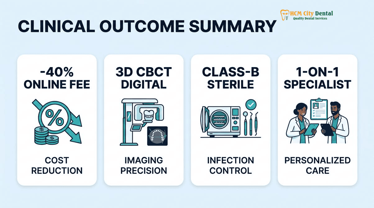
At HCMC Dental Clinic, we believe that saving your natural tooth should not be a financial burden. By utilizing our Official 2026 Dental Price List & -40% Online Booking Fee Schedule, patients receive identical hospital-grade care—utilizing the exact same Carl Zeiss microscopes, Satelec ultrasonics, and European bioceramic materials—at a fraction of the cost.
Below is the transparent, itemized fee schedule for microscopic endodontic retreatment and biomimetic restoration at our District 1 facility:
| Clinical Procedure (2026 Fee Schedule) | Walk-In Price (USD) | -40% Online Booking Price (USD) | VND Equivalent (Online) |
|---|---|---|---|
| 3D CBCT Scan (0.075mm High-Resolution) | $30 USD | $18 USD | 480,000 VND |
| Microscopic Endodontic Retreatment / Separated File Bypass (Per Tooth) | $256 – $385 USD | $154 – $231 USD | 4,000,000 – 6,000,000 VND |
| Bioactive ProRoot MTA / Septodont Biodentine Perforation Repair | $96 – $160 USD | $58 – $96 USD | 1,500,000 – 2,500,000 VND |
| Biomimetic Quartz/Glass Fiber Post & Core Buildup | $63 – $96 USD | $38 – $58 USD | 1,000,000 – 1,500,000 VND |
| Full Zirconia Crown (Cercon Standard / HT Multilayer) | $256 – $321 USD | $154 – $192 USD | 4,000,000 – 5,000,000 VND |
For patients requiring emergency intervention, our clinic offers streamlined protocols. You can learn more about our Same Day Root Canal Saigon: Emergency Endodontic Care services to ensure your pain is managed and your tooth is stabilized immediately upon arrival in Ho Chi Minh City. Furthermore, understanding the total investment is crucial; we encourage patients to review our comprehensive guide on Crown Cost Vietnam: Complete Clinical Guide & Prices to budget accurately for the final restorative phase.
“Transparency in healthcare is non-negotiable. Our international patients deserve to know exactly what technology is being used, the clinical rationale behind every step, and the exact cost before they ever sit in the dental chair. By publishing our clinical outcomes and fee schedules, we empower patients to make informed, evidence-based decisions about saving their natural teeth.”
— Dr. Nguyen Van Cuong, DDS
For a deeper dive into our clinical success rates and published data, please visit our 2026 Clinical Dental Research & Statistical Reports Hub. If you are specifically looking for an English Speaking Endodontist in Ho Chi Minh City, Dr. Cuong and his dedicated team in District 1, Ho Chi Minh City, are ready to provide world-class, compassionate care.
Frequently Asked Questions
If a previous dentist left a broken file tip in my root canal, does the tooth automatically need to be extracted?
No. A separated endodontic file does not automatically condemn a tooth to extraction. The metal fragment itself is sterile and biocompatible; the true risk is the uncleaned bacteria trapped beneath it. Under a 25x Carl Zeiss dental microscope, an experienced endodontist can often retrieve the fragment using ultrasonic micro-tips or safely bypass it, allowing the canal to be fully disinfected and sealed.
What is the success rate of removing or bypassing a separated endodontic file under a 25x operating microscope?
Clinical success rates for broken file retrieval or bypass range from 70% to over 90%, depending heavily on the fragment’s location. Fragments located in the straight coronal or middle third of the canal have the highest retrieval success. Fragments bound tightly beyond a severe apical curvature (past Schneider’s angle) may be safer to bypass or manage surgically via apicoectomy to prevent excessive dentin removal.
When is it safer to bypass a broken file fragment or leave it entombed in bioceramic sealer rather than drilling deeper?
It is safer to bypass or entomb a fragment when aggressive ultrasonic vibration would remove too much radicular dentin, risking a strip perforation or vertical root fracture. If the fragment is lodged deep in the apical third past a severe curve, and the canal was already thoroughly disinfected prior to separation, entombing the file in a bioactive bioceramic sealer (like Biodentine or ProRoot MTA) often yields an excellent long-term prognosis.
How many appointments in Ho Chi Minh City are required for microscopic root canal retreatment and file retrieval?
Microscopic endodontic retreatment involving instrument retrieval typically requires two appointments. The first session (90–120 minutes) focuses on 3D CBCT mapping, removing old filling material, retrieving or bypassing the broken file, and placing an antibacterial calcium hydroxide dressing. The second session (60–90 minutes), scheduled 7 to 14 days later, involves final ultrasonic disinfection, 3D bioceramic obturation, and the placement of a biomimetic fiber post and core buildup [5].
How much does microscopic endodontic retreatment cost at HCMC Dental Clinic compared to Australia or the US?
At HCMC Dental Clinic, microscopic endodontic retreatment and separated file bypass costs between $154 and $231 USD per tooth when booked online. In contrast, the same specialist procedure in Australia or the United States typically costs between $1,500 and $2,500 USD. This represents a savings of 75% to 85%, while utilizing identical hospital-grade technology, including Carl Zeiss microscopes, 3D CBCT imaging, and Satelec ultrasonic systems.
References
- Journal of Endodontics. Clinical Efficacy of Ultrasonic Retrieval of Separated Instruments under Dental Operating Microscope: A Retrospective Study. (2023).
- International Endodontic Journal. Influence of 3D CBCT Imaging on the Decision-Making Process for the Management of Separated Endodontic Instruments. (2024).
- Journal of Prosthetic Dentistry. Biomimetic Restoration of Endodontically Treated Teeth Following Complex Retreatment: The Role of Fiber Posts and Ferrule. (2022).
- American Journal of Orthodontics and Dentofacial Orthopedics (AJODO). Biomechanical Stress Distribution in Roots Following Ultrasonic Troughing for File Retrieval. (2023).
- Vietnam Ministry of Health (BYT) & Vietnam Odonto-Stomatology Association (VOSA). National Clinical Guidelines on Specialist Dental Care, Biomaterial Traceability & EN 13060 Class B Infection Control. (2024).