Root canal retreatment in HCMC involves removing old filling materials, disinfecting the root system, and resealing a previously treated tooth that has failed to heal. This advanced endodontic procedure saves your natural tooth, eliminates persistent infection, and prevents the need for extraction or dental implants.
Clinical Summary:
Endodontic retreatment is a highly specialized dental procedure required when an initial root canal fails due to complex anatomy, missed canals, or coronal leakage. Utilizing advanced diagnostic tools like 3D CBCT imaging and high-powered dental operating microscopes, endodontists can accurately identify the root cause of the clinical failure. The comprehensive process involves carefully disassembling existing restorations, performing meticulous gutta-percha removal, and thoroughly disinfecting the intricate root canal network to eradicate persistent bacterial biofilms. In Ho Chi Minh City, top-tier clinics employ modern bioceramic sealers and rotary instrumentation to ensure a hermetic seal, significantly improving the long-term prognosis of the compromised tooth. If non-surgical retreatment is anatomically impossible or insufficient to resolve the periapical pathology, surgical interventions such as an apicoectomy may be indicated to preserve the natural dentition.
Key Takeaways:
- Retreatment addresses persistent root infection and incomplete healing from prior endodontic procedures.
- Clinical success relies heavily on 3D CBCT imaging and high-powered dental microscopes to navigate complex anatomy.
- The procedure preserves the natural tooth structure, offering a conservative alternative to extraction and implant placement.
- Complex cases frequently require the careful disassembly of existing dental crowns and intraradicular posts.
- Apicoectomy serves as a viable surgical alternative if non-surgical retreatment methods are anatomically unviable.
Why Root Canals Can Fail: Missed Canals, Leakage, and Fractures
Initial root canal treatments can fail due to undetected accessory canals, delayed placement of a permanent restoration leading to bacterial leakage, or structural root fractures. Identifying the exact etiology is crucial for successful retreatment.
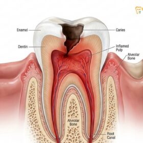
While initial endodontic therapy boasts a remarkably high success rate, a small percentage of cases may fail to heal properly or develop new infections months or even years after the original procedure. Understanding the intricate pathophysiology behind these failures is the first step in determining the appropriate clinical intervention. The root canal system is not a simple, straight tube; rather, it is a highly complex, branching network of microscopic passageways, fins, and deltas that harbor pulpal tissue and, in the case of infection, robust bacterial biofilms[1].
One of the most common reasons for endodontic failure is the presence of missed canals. Human dental anatomy is highly variable. For instance, the maxillary first molar frequently contains a second mesiobuccal canal (MB2) in over 90% of cases. Because this canal is often exceptionally narrow and calcified, it can easily be overlooked during treatment without the aid of high-level magnification and specialized illumination. When a canal is left untreated, necrotic tissue and bacteria remain sealed inside the tooth, eventually migrating to the root apex and causing a persistent root infection that manifests as periapical periodontitis.
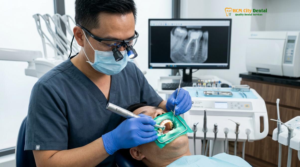
Another primary culprit is coronal leakage. After a root canal is completed, the tooth must be permanently restored—typically with a custom dental crown—to protect the fragile remaining tooth structure and seal the access cavity. If the placement of this final restoration is delayed, or if the restoration eventually margins fail due to recurrent decay, salivary bacteria can seep past the temporary filling and contaminate the previously sterilized root canal system[2]. This bacterial microleakage can compromise the gutta-percha filling material within a matter of weeks, necessitating a complete retreatment to re-establish a sterile environment.
“The ultimate goal of endodontic retreatment is not merely the removal of old filling materials, but the complete disruption of established bacterial biofilms and the re-establishment of a biologically acceptable environment for periapical healing.”
Furthermore, structural complications such as vertical root fractures or iatrogenic procedural errors (like ledging, zipping, or instrument separation) can severely compromise the outcome of the initial treatment. A vertical root fracture often presents with a deep, isolated periodontal pocket and a distinct “J-shaped” radiolucency on a radiograph. Unfortunately, teeth with true vertical root fractures generally have a poor prognosis and often require extraction. However, if the failure is strictly due to anatomical complexities or restorative leakage, non-surgical retreatment remains the gold standard for tooth preservation.
Signs Your Root Canal Has Failed: Chronic Ache and Sinus Tracts
Symptoms of a failed root canal include persistent pain when biting, prolonged sensitivity, localized gum swelling, or the presence of a pimple-like sinus tract on the gums. Some failures remain asymptomatic and are only detected via routine X-rays.
Recognizing the clinical signs of a failing root canal is essential for timely intervention. Patients often assume that once a tooth has undergone endodontic treatment and the nerve has been removed, it can no longer experience pain. However, the pain associated with a failed root canal does not originate from the inside of the tooth itself, but rather from the highly innervated periodontal ligament and the surrounding alveolar bone that encases the root tip.
The most frequent symptom reported by patients is a chronic, dull ache that exacerbates upon mastication (chewing) or the application of occlusal pressure. This tenderness indicates active inflammation in the periapical tissues, a condition known as symptomatic apical periodontitis. Unlike the sharp, lingering thermal sensitivity associated with an inflamed vital pulp, a tooth with a failed root canal will not respond to hot or cold stimuli, as the pulpal nerve fibers have already been extirpated.
Clinical Warning: Do not ignore a “pimple” on your gums, even if it is completely painless. This is a sinus tract indicating an active, draining infection that is destroying the surrounding jawbone. Prompt endodontic evaluation is required to prevent systemic spread and further structural loss.
In many cases, the infection will establish a drainage pathway through the alveolar bone and the overlying gingival tissue, presenting clinically as a parulis or “sinus tract.” This small, pimple-like bump on the gums may periodically swell, rupture, and release a foul-tasting purulent exudate (pus) into the oral cavity. While the drainage often relieves the acute pressure and pain, the underlying infection remains active and continues to erode the supporting bone structure[3].
It is also crucial to understand that a significant number of endodontic failures are entirely asymptomatic. A patient may feel perfectly fine, yet a routine panoramic or periapical radiograph may reveal a growing dark shadow (radiolucency) at the apex of the treated root. This silent progression of bone destruction highlights the critical importance of regular dental check-ups and radiographic monitoring following any major endodontic procedure. Dr. Nguyen Van Cuong frequently emphasizes to his patients that the absence of pain does not necessarily equate to the absence of disease, particularly in complex endodontic cases.
The Endodontic Retreatment Process: Step-by-Step
The retreatment workflow involves administering profound local anesthesia, isolating the tooth with a rubber dam, removing old restorative materials, thoroughly disinfecting the canal system, and placing a new biocompatible seal.
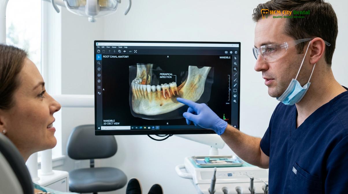
The clinical workflow for endodontic retreatment is inherently more complex and time-consuming than an initial root canal. It requires a meticulous, step-by-step approach to safely navigate the previously altered root canal anatomy while avoiding further iatrogenic damage. The procedure begins with comprehensive diagnostic imaging. At HCMC Dental Clinic, high-resolution 3D Cone Beam Computed Tomography (CBCT) is utilized to visualize the tooth in three dimensions. This advanced imaging allows the endodontist to identify missed canals, assess the extent of periapical bone loss, and evaluate the morphology of the existing root canal filling.
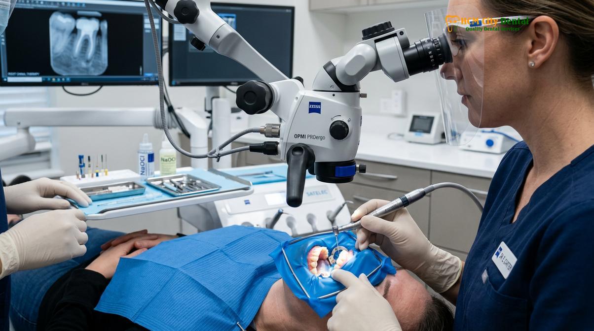
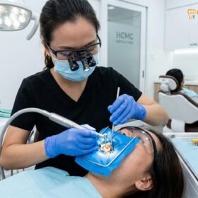
Once the treatment plan is established, profound local anesthesia is administered to ensure complete patient comfort. Even though the tooth is technically “dead,” the surrounding inflamed tissues can be highly sensitive. Following anesthesia, the tooth is strictly isolated using a dental rubber dam. This non-negotiable step prevents the ingress of salivary bacteria into the operative field and protects the patient’s airway from chemical irrigants and microscopic debris.
The endodontist then creates an access cavity through the biting surface of the tooth. If a dental crown is present, the access is typically made directly through the restorative material. The next critical phase is the complete removal of the existing obturation material, which is most commonly gutta-percha combined with an endodontic sealer. This is achieved using a combination of specialized rotary files, ultrasonic instruments, and chemical solvents like chloroform or eucalyptol, which soften the rubbery gutta-percha for easier extraction.
| Clinical Parameter | Initial Root Canal Treatment | Endodontic Retreatment |
|---|---|---|
| Primary Objective | Remove inflamed/necrotic vital pulp tissue. | Remove old filling material and eradicate persistent biofilm. |
| Procedural Complexity | Moderate; navigating virgin canal anatomy. | High; requires disassembly and bypassing blockages. |
| Diagnostic Tools | Standard 2D digital X-rays usually sufficient. | 3D CBCT imaging highly recommended. |
| Time Requirement | Typically completed in 1 to 2 appointments. | Often requires multiple extended appointments. |
| Success Rate | Very high (85% – 95%+). | High (70% – 85%), dependent on anatomical factors. |
After the canals are unblocked, the clinician must re-establish the working length of each canal using an electronic apex locator and confirm it with a radiograph. The canals are then meticulously reshaped and disinfected. This is where the true challenge of retreatment lies: eradicating the deeply entrenched bacterial biofilms that survived the initial procedure. Copious irrigation with sodium hypochlorite (bleach) and EDTA is employed, often enhanced by ultrasonic or sonic activation to drive the antimicrobial solutions deep into the dentinal tubules and lateral canals[4].
Gaining Access: Disassembling Existing Dental Crowns and Posts
To access the infected root system, the endodontist must carefully drill through or remove existing dental crowns and extract any metal or fiber posts placed during the initial restoration.
One of the most technically demanding aspects of endodontic retreatment is the disassembly of the existing coronal restoration. In many cases, the failing tooth has been heavily restored with a core buildup and a full-coverage dental crown. The clinician must decide whether to drill an access hole through the existing crown or to remove the crown entirely. Drilling through the crown is less invasive and preserves the restoration, but it limits visibility and carries a risk of fracturing porcelain or ceramic materials.
If the tooth contains an intraradicular post—a metal or fiber rod cemented deep into the root canal to provide structural support for the core buildup—the complexity of the procedure increases exponentially. Post removal is a delicate operation that requires immense patience and specialized equipment. The endodontist typically uses fine ultrasonic tips under high magnification to carefully vibrate the post, breaking the micro-mechanical retention of the dental cement without fracturing the fragile root structure.
The application of ultrasonic energy must be carefully controlled and accompanied by copious water coolant to prevent the generation of excessive heat, which could damage the surrounding periodontal ligament and alveolar bone. In some instances, specialized post-removal systems utilizing tubular drills and extraction forceps may be employed. The risk of vertical root fracture during post removal is a significant clinical concern, and patients must be fully informed of this possibility prior to the commencement of treatment.
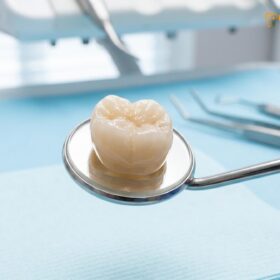
At HCMC Dental Clinic, the clinical team employs a highly conservative approach to disassembly. Dr. Nguyen Van Cuong evaluates each case individually, weighing the risks and benefits of various access strategies. If a crown must be sacrificed to gain adequate access and ensure the complete eradication of the infection, a temporary crown is fabricated to protect the tooth during the interim healing phase. Once the retreatment is deemed successful, a new, precisely fitting permanent restoration is fabricated to provide long-term structural integrity.
Locating and Disinfecting Previously Missed Canal Space
Utilizing a dental operating microscope, the specialist locates hidden canals, performs complete gutta-percha removal, and uses chemical irrigants like sodium hypochlorite to eradicate the persistent root infection.
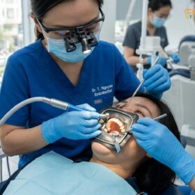
The hallmark of modern endodontic retreatment is the utilization of the dental operating microscope (DOM). The internal anatomy of a tooth is microscopic and shrouded in darkness. Operating without magnification is akin to performing surgery blindfolded. The DOM provides intense, coaxial illumination and magnifies the operative field up to 25 times, allowing the clinician to visualize intricate anatomical details that are completely invisible to the naked eye.
With the aid of the microscope, the endodontist meticulously inspects the pulpal floor for subtle color changes, developmental grooves, or tiny bleeding points that indicate the presence of previously missed canals. As mentioned earlier, the MB2 canal in maxillary molars is a frequent culprit in endodontic failures. Once located, these calcified or highly curved canals must be carefully negotiated using fine, pre-curved stainless steel hand files and specialized lubricants containing chelating agents.
Clinical Case Study: A 45-year-old patient presented to HCMC Dental Clinic in Ho Chi Minh City with chronic pain in an upper right molar, previously treated five years ago. 3D CBCT imaging revealed an untreated MB2 canal harboring a persistent root infection. Under microscopic magnification, the clinical team successfully located the calcified canal, performed thorough disinfection, and sealed the system with bioceramic materials. The patient reported complete symptom resolution within 72 hours, and follow-up radiographs at six months demonstrated excellent bone regeneration.
Following the successful negotiation of all canal spaces and the complete gutta-percha removal from the previous treatment, the focus shifts to advanced disinfection protocols. The bacterial flora in a failing root canal is often different from that of an initial infection. It is frequently dominated by resilient, Gram-positive facultative anaerobes, such as Enterococcus faecalis, which are highly resistant to standard antimicrobial agents and can survive in nutrient-deprived environments[5].
“The integration of 3D CBCT imaging and high-powered dental microscopes has fundamentally transformed endodontic retreatment from a procedure of clinical guesswork into a highly predictable, visually guided microsurgery.”
To combat these tenacious biofilms, endodontists employ a multi-faceted irrigation strategy. High-concentration sodium hypochlorite is used to dissolve organic tissue and destroy bacteria, while EDTA (Ethylenediaminetetraacetic acid) is utilized to remove the inorganic smear layer created during instrumentation, thereby opening the dentinal tubules. Advanced techniques, such as laser-assisted endodontics or multisonic ultracleaning systems, may also be integrated to agitate the irrigants, creating microscopic cavitation bubbles that physically shear the biofilm away from the canal walls. Finally, the thoroughly disinfected canal system is obturated (sealed) using modern, highly biocompatible bioceramic sealers that promote periapical tissue healing and provide an exceptional hermetic seal against future bacterial microleakage.
When Retreatment is Not Enough: Apicoectomy (Root-End Surgery) as an Alternative
If non-surgical retreatment is anatomically impossible or fails to resolve the infection, an apicoectomy is performed to surgically remove the infected root tip and seal the canal from the bottom.
While non-surgical endodontic retreatment is highly effective, there are specific clinical scenarios where it is either contraindicated or anatomically impossible. For example, if a tooth has a massive, unyielding cast metal post that cannot be safely removed without fracturing the root, or if the root canal is completely blocked by a separated instrument or severe calcification, accessing the infection from the top down is no longer a viable option. In these complex situations, a surgical approach known as an apicoectomy, or root-end surgery, becomes the treatment of choice.
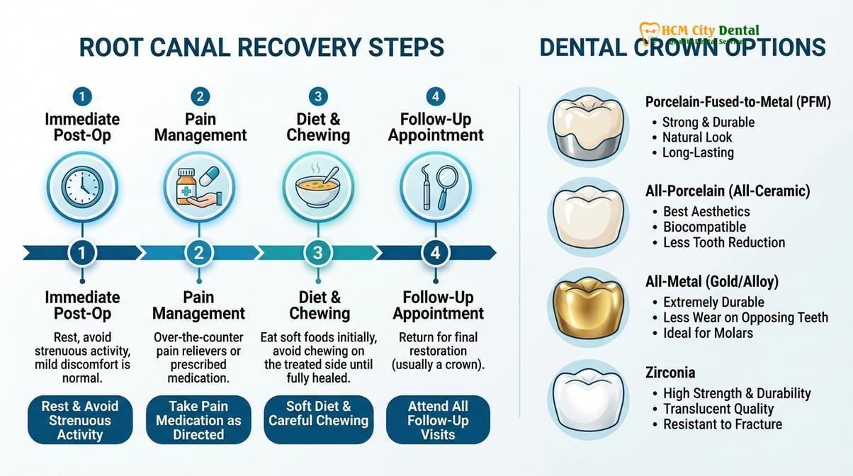
An apicoectomy bypasses the coronal aspect of the tooth entirely and addresses the infection directly at its source: the root apex. The procedure is performed under local anesthesia in a sterile surgical setting. The endodontist or oral surgeon makes a precise incision in the gum tissue adjacent to the affected tooth and gently reflects a small flap to expose the underlying infected alveolar bone. Using specialized surgical burs, a small window is created in the bone to access the root tip and the surrounding inflammatory tissue or cystic lesion.
The infected periapical tissue is meticulously curetted and removed for pathological examination. Subsequently, the terminal 3 millimeters of the root tip—which often contains a complex web of microscopic accessory canals and bacterial biofilms—is surgically resected (cut off). The resected root surface is then examined under the dental microscope to identify any structural fractures or anatomical anomalies. A microscopic ultrasonic tip is used to prepare a small cavity within the remaining root canal space, which is then sealed with a biocompatible retrograde filling material, such as Mineral Trioxide Aggregate (MTA) or a bioceramic putty[6].
This retrograde seal prevents any remaining bacteria within the canal system from escaping into the surrounding bone. The surgical site is thoroughly irrigated, and the gum tissue is carefully sutured back into place. The term “apicoectomy Saigon” is frequently searched by patients seeking this advanced surgical intervention in the region, reflecting the growing awareness of tooth-saving surgical alternatives. Over the following months, the body’s natural healing mechanisms will regenerate new bone to fill the surgical defect, restoring the tooth to full function and health.
When to See a Doctor
Determining whether a previously treated tooth requires clinical intervention is not something a patient can diagnose at home. The symptoms of endodontic failure can mimic other dental issues, such as periodontal disease, severe occlusal trauma, or temporomandibular joint (TMJ) disorders. Therefore, professional evaluation is absolutely critical.
You should schedule an immediate consultation with a dental professional if you experience any of the following symptoms associated with a tooth that has previously undergone root canal therapy:
- Persistent or worsening pain: Especially pain that is triggered by biting, chewing, or applying pressure to the tooth.
- Localized swelling: Any swelling of the gum tissue adjacent to the treated tooth, or swelling that extends to the face or neck.
- Presence of a sinus tract: A pimple-like bump on the gums that may ooze pus or cause a bad taste in your mouth.
- Mobility: If the previously treated tooth begins to feel loose or shifts in your bite.
- Systemic symptoms: If localized dental pain is accompanied by a fever, chills, or difficulty swallowing, seek emergency medical attention immediately, as this indicates a spreading infection.
Early detection and intervention significantly improve the prognosis of endodontic retreatment. Delaying care allows the infection to destroy more supporting jawbone, potentially rendering the tooth unsalvageable and necessitating extraction.
Frequently Asked Questions
Why does my root-canaled tooth hurt years later?
A previously treated tooth can hurt years later due to a recurrent infection caused by coronal leakage, undetected accessory canals, or a new structural fracture. Once bacteria re-enter the sealed root canal system, they trigger inflammation in the surrounding periapical tissues, leading to persistent pain and discomfort. This delayed failure highlights the importance of maintaining excellent oral hygiene and ensuring that the final dental crown remains intact and well-sealed.
What is the success rate of a root canal retreatment?
The success rate for endodontic retreatment is generally high, often ranging between 70% to 85%, depending on the complexity of the root anatomy and the extent of the existing infection. Utilizing advanced 3D imaging and dental microscopes significantly improves the prognosis by allowing precise disinfection and resealing. However, teeth with severe structural damage or vertical root fractures have a much lower success rate and may require alternative treatments.
How much does a root canal retreatment cost in Saigon compared to a new canal?
Root canal retreatment in Saigon typically costs 20% to 40% more than an initial root canal procedure. This price difference reflects the increased clinical time, the complexity of disassembling existing restorations, and the specialized equipment required to remove old filling materials and locate hidden anatomical structures. Despite the higher cost, retreatment remains a highly cost-effective alternative to tooth extraction and the subsequent placement of a dental implant.
Is the retreatment procedure more painful than the first root canal?
No, the retreatment procedure is not inherently more painful than the initial treatment, as it is performed under profound local anesthesia. While the tooth and surrounding gums may feel tender for a few days post-operatively due to tissue manipulation, modern anesthetic protocols ensure a comfortable experience during the procedure. Over-the-counter anti-inflammatory medications are usually sufficient to manage any mild post-operative discomfort.
How long does it take to recover from endodontic retreatment?
Initial recovery from endodontic retreatment usually takes three to five days, during which mild tenderness or sensitivity when biting is normal. However, complete radiographic healing of the periapical bone surrounding the root tip can take several months to a year, monitored through follow-up dental X-rays. Patients are advised to avoid chewing hard foods on the treated tooth until the final permanent restoration is securely in place.
References
- Journal of Endodontics. Outcomes of root canal treatment and restoration longevity. (2020).
- International Endodontic Journal. Biofilms in endodontics and disinfection strategies. (2021).
- Journal of the American Dental Association. Microscope-assisted endodontics and missed canals. (2019).
- Clinical Oral Investigations. Biocompatibility of bioceramic root canal sealers. (2022).
- British Dental Journal. Pain management and dental anxiety in endodontics. (2018).
- Oral Surgery, Oral Medicine, Oral Pathology. Indications and outcomes of apicoectomy procedures. (2021).