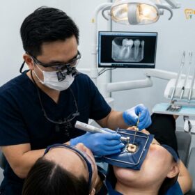
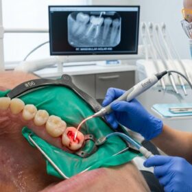
Irreversible pulpitis treatment requires the complete removal of permanently damaged dental nerve tissue through professional endodontic therapy or tooth extraction. Unlike mild, reversible inflammation, an irreversibly damaged pulp cannot heal itself, making immediate clinical intervention mandatory to eliminate severe spontaneous pain and prevent the spread of bacterial infection into the jawbone.
Clinical Summary:
Irreversible pulpitis is a severe inflammatory condition characterized by permanent damage to the dental pulp, typically resulting from deep carious lesions, repeated restorative procedures, or mechanical trauma. Because the pulp tissue is encased within rigid dentinal walls and lacks a collateral blood supply, it cannot accommodate the swelling associated with severe inflammation. This leads to increased intrapulpal pressure, venous congestion, and eventual tissue necrosis. The hallmark clinical symptom is spontaneous, lingering pain that is often exacerbated by thermal stimuli, particularly heat. Accurate diagnosis relies on a combination of patient history, radiographic evaluation, and clinical vitality testing. Treatment mandates the extirpation of the diseased tissue through root canal therapy to preserve the natural dentition, or extraction if the tooth is deemed non-restorable. Early differentiation between reversible and irreversible states is critical for determining the appropriate conservative or endodontic clinical pathway.
Key Takeaways:
- Irreversible pulpitis involves permanent nerve damage that cannot be cured with antibiotics or over-the-counter pain medications.
- Spontaneous, throbbing pain that lingers after exposure to hot or cold stimuli is the primary diagnostic indicator of irreversible damage.
- Endodontic therapy (root canal treatment) is the gold standard clinical approach to save the affected tooth and resolve the infection.
- If left untreated, the condition inevitably progresses to pulp necrosis and potentially severe periapical abscesses.
- Precise clinical pulp testing, including thermal and electric assessments, is essential to differentiate between reversible and irreversible inflammation.
Defining Pulpitis: The Tooth’s Immune Response to Cavities and Trauma
Pulpitis is the inflammation of the innermost dental tissue, triggered by bacterial invasion or trauma, representing the body’s immune response attempting to defend the tooth against external irritants.
To fully comprehend the complexities of irreversible pulpitis treatment, one must first understand the unique anatomical and physiological environment of the dental pulp. The pulp is a highly vascularized and innervated connective tissue located in the central chamber and root canals of the tooth. It is responsible for the formation of dentin, the sensory perception of thermal and mechanical stimuli, and the immunological defense of the tooth. The pulp is entirely encased within rigid, unyielding walls of dentin and enamel, with its only communication to the systemic circulation occurring through the narrow apical foramen at the tip of the root[1].
When a tooth is subjected to an irritant—most commonly bacterial ingress from dental caries, but also mechanical trauma, thermal injury from high-speed drilling without adequate water coolant, or chemical irritation from restorative materials—the pulp initiates an inflammatory response. This response is fundamentally similar to inflammation anywhere else in the body: vasodilation, increased capillary permeability, and the influx of immune cells. However, because the pulp is trapped within a rigid chamber, the resulting edema (swelling) cannot expand outward. Instead, it causes a rapid and dramatic increase in intrapulpal pressure.
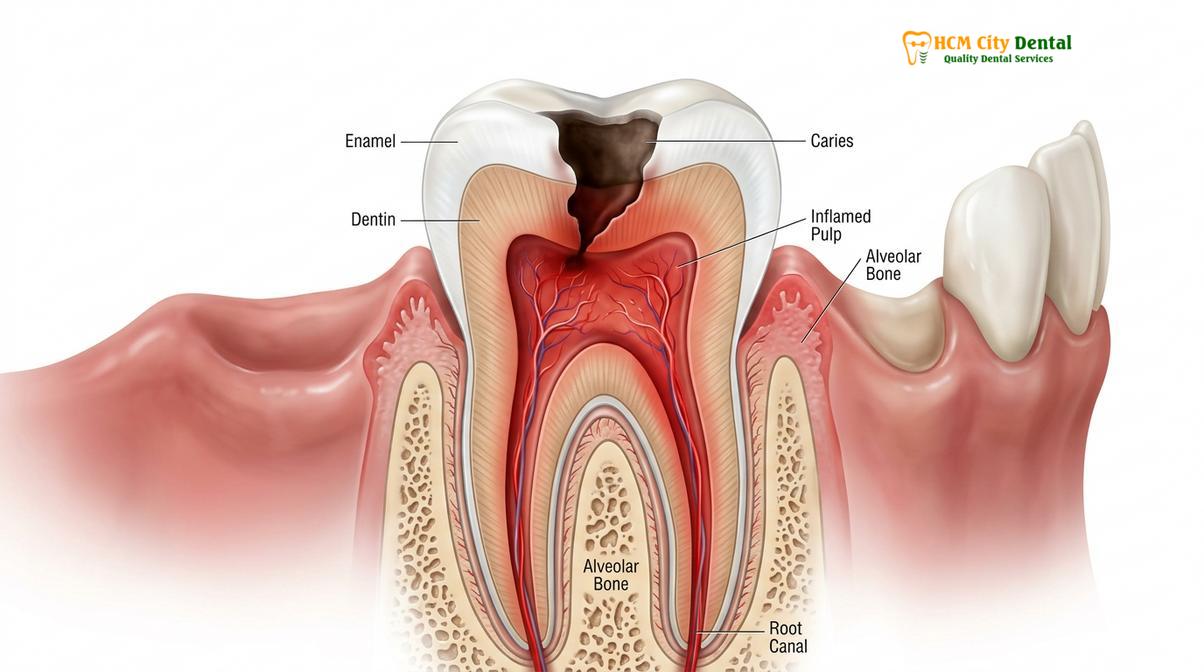
This elevated pressure compresses the delicate venules (small veins) within the pulp, leading to venous congestion and a reduction in blood flow. If the irritant is mild and transient, the pulp can clear the inflammatory exudate and return to a state of health. However, if the irritant is severe or persistent, the localized pressure exceeds the capillary perfusion pressure, leading to ischemia (lack of oxygen), micro-abscess formation, and localized tissue death. This unique “strangulation” phenomenon is the primary reason why pulpal inflammation can rapidly transition from a reversible state to an irreversible one.
Dr. Nguyen Van Cuong, a leading specialist at HCMC Dental Clinic, frequently emphasizes to patients that the dental pulp’s inability to swell makes it highly vulnerable. “The moment bacteria breach the protective dentin layer and reach the pulp, the biological clock starts ticking. Early intervention is the only way to prevent the cascade of inflammation that leads to permanent nerve death,” notes Dr. Cuong. Understanding this pathophysiology is crucial for both the clinician diagnosing the condition and the patient seeking timely care.
Reversible Pulpitis: Mild Inflammation and Fast Healing
Reversible pulpitis is an early, mild stage of nerve inflammation that can completely resolve and return to a healthy state once the underlying irritant is removed and the tooth is sealed.
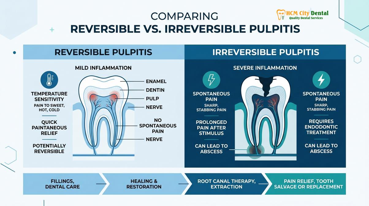
Reversible pulpitis represents the initial stage of pulpal inflammation. Histologically, it is characterized by localized hyperemia (increased blood flow) and mild edema, but without the presence of micro-abscesses or tissue necrosis. The pulp retains its structural integrity and its capacity for repair. The most critical defining feature of reversible pulpitis is that the condition is, by definition, curable without the need for endodontic therapy, provided the causative factor is promptly addressed.
The symptomatology of reversible pulpitis is highly specific. Patients typically report a sharp, transient pain that is provoked by a specific stimulus—most commonly cold temperatures, sweet foods, or acidic beverages. Crucially, this pain does not occur spontaneously; it requires a trigger. Furthermore, once the stimulus is removed, the pain subsides almost immediately, usually within a few seconds. This rapid cessation of pain indicates that the A-delta nerve fibers (responsible for sharp, localized pain) are reacting to the stimulus, but the deeper C-fibers (associated with dull, lingering, inflammatory pain) have not yet been heavily recruited[2].
“The distinction between a pain that fades instantly and one that lingers for minutes is the most critical diagnostic dividing line in endodontics. It dictates whether we can save the nerve or if we must remove it to save the tooth.”
Clinically, a tooth with reversible pulpitis will not exhibit pain upon percussion (tapping on the tooth), as the inflammation is confined entirely within the pulp chamber and has not spread to the periapical tissues (the bone and ligament surrounding the root tip). Radiographically, the periapical tissues will appear completely normal, though a deep cavity or a failing restoration may be visible.
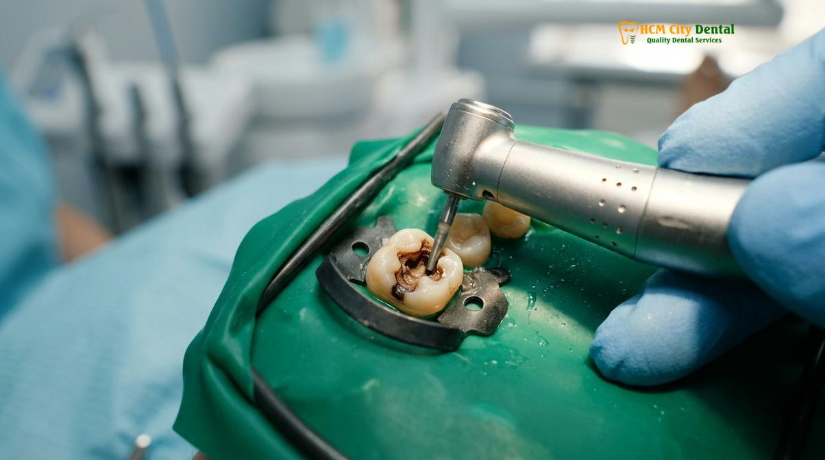
The primary goal of managing this condition is to facilitate dental pulp healing. This is achieved by removing the source of irritation—such as excavating the decayed dentin or replacing a defective filling—and placing a biocompatible restoration that seals the dentinal tubules. Once the tooth is sealed, the pulp’s internal pressure normalizes, the hyperemia subsides, and the odontoblasts (dentin-forming cells) are often stimulated to lay down a protective layer of tertiary (reparative) dentin, further insulating the nerve from future insults.
Irreversible Pulpitis: Permanent Damage and Spontaneous Pain
Irreversible pulpitis occurs when severe inflammation causes permanent tissue damage and localized necrosis, resulting in spontaneous, lingering pain that necessitates the complete removal of the dental nerve.
When the inflammatory process within the pulp crosses the threshold of no return, the diagnosis shifts to irreversible pulpitis. At this stage, the intrapulpal pressure has caused significant venous collapse, leading to localized areas of ischemia and liquefaction necrosis. The pulp tissue is actively dying, and the immune system is unable to clear the infection or repair the damage due to the compromised blood supply. The condition is progressive and will inevitably lead to total pulp necrosis if left untreated.
The clinical presentation of irreversible pulpitis is markedly different from its reversible counterpart. The most prominent symptom is spontaneous pain—pain that occurs without any external trigger. Patients often report that the pain wakes them up at night or starts unprovoked during the day. When the tooth is exposed to thermal stimuli, particularly heat, the pain is often excruciating and lingers long after the stimulus is removed, sometimes lasting for minutes or even hours. This lingering pain is a strong indicator that the unmyelinated C-fibers, which signal deep tissue damage and severe inflammation, are highly active[3].
Clinical Warning: A sudden cessation of severe, throbbing tooth pain does not mean the tooth has healed. It often indicates that the dental nerve has completely died (pulp necrosis). The bacteria will continue to multiply and spread into the jawbone, leading to a potentially dangerous abscess. Immediate dental evaluation is required even if the pain stops.
Irreversible pulpitis can be further classified into symptomatic and asymptomatic forms. Symptomatic irreversible pulpitis presents with the classic severe, lingering pain described above. Asymptomatic irreversible pulpitis, however, can be more insidious. In these cases, the inflammatory process has caused permanent damage, but the patient experiences little to no pain. This often occurs in older patients where the pulp chamber has calcified, or in cases of hyperplastic pulpitis (pulp polyp), where a large carious exposure allows the inflamed pulp tissue to expand out of the cavity, relieving the internal pressure and thereby reducing the pain.
Regardless of whether it is symptomatic or asymptomatic, the biological reality remains the same: the tissue is permanently compromised. The bacteria present in the deep carious lesion will eventually colonize the entire root canal system. Because the pulp’s immune defenses are failing, the bacteria will progress through the apical foramen and initiate an inflammatory response in the periapical bone, leading to apical periodontitis. This underscores why irreversible pulpitis treatment cannot be delayed or managed with systemic antibiotics alone.
Clinical Pulp Testing: Cold Testing, Heat Testing, and Electric Vitality
Accurate diagnosis of pulpal status requires rigorous clinical testing using thermal and electric stimuli to evaluate nerve responsiveness and differentiate between reversible and irreversible inflammation.
Because the symptoms of pulpal disease can be subjective and sometimes referred to other areas of the face or jaw, a definitive diagnosis cannot be made based on patient history alone. Clinicians must employ a battery of objective diagnostic tests to assess the vitality and inflammatory status of the pulp. These tests are designed to reproduce the patient’s symptoms in a controlled manner, allowing the dentist to pinpoint the exact offending tooth and determine the extent of the pathology.
The most reliable and frequently used diagnostic tool is the cold test. This is typically performed using a refrigerant spray, such as Endo-Ice (dichlorodifluoromethane), which reaches a temperature of approximately -26.2°C. The cold spray is applied to a cotton pellet and placed on the middle third of the facial surface of the tooth. A healthy tooth will register a mild, brief sensation of cold. A tooth with reversible pulpitis will exhibit a sharp pain that disappears immediately upon removal of the pellet. Conversely, a tooth with irreversible pulpitis will respond with a severe, intense pain that lingers for several seconds to minutes after the cold stimulus is removed.
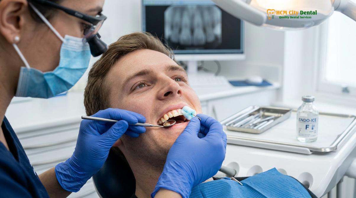
Heat testing is another critical diagnostic modality, particularly useful when a patient reports that hot foods or liquids trigger their pain. This test can be performed using heated gutta-percha, a specialized heat carrier, or by isolating the tooth and bathing it in warm water. A severe, lingering response to heat is a classic hallmark of irreversible pulpitis. Interestingly, patients with advanced irreversible pulpitis often report that cold water actually relieves their pain, as the cold causes vasoconstriction, temporarily reducing the elevated intrapulpal pressure.
Electric Pulp Testing (EPT) is utilized to determine whether the nerve fibers within the pulp are capable of conducting an electrical impulse. The EPT device delivers a gradually increasing electrical current to the tooth. If the patient feels a tingling or warming sensation, it indicates that at least some viable nerve fibers remain. If there is no response at the maximum setting, it strongly suggests that the pulp is necrotic. It is important to note that EPT assesses nerve conduction, not blood flow, and therefore cannot differentiate between reversible and irreversible pulpitis; it only confirms whether the nerve is alive or dead[4].
At advanced facilities, a comprehensive pulp vitality test HCMC protocol also includes periapical testing. Percussion testing (gently tapping the tooth with the handle of a mirror) and palpation testing (pressing on the gums near the root apex) are performed to determine if the inflammation has spread beyond the tooth and into the surrounding bone. Additionally, high-resolution digital radiographs and 3D Cone Beam Computed Tomography (CBCT) are essential to evaluate the depth of decay, the anatomy of the root canals, and the presence of any periapical bone loss.
| Diagnostic Feature | Reversible Pulpitis | Irreversible Pulpitis |
|---|---|---|
| Pain Characteristics | Sharp, transient, requires a stimulus | Spontaneous, throbbing, may wake patient at night |
| Response to Cold Test | Pain stops immediately when cold is removed | Severe pain that lingers for >10-15 seconds |
| Response to Heat Test | Usually normal or mildly sensitive | Often causes severe, lingering pain |
| Percussion Sensitivity | None (inflammation confined to pulp) | May be present if inflammation reaches the apex |
| Treatment Required | Conservative restoration (filling) | Endodontic therapy (root canal) or extraction |
Treatments for Reversible Pulpitis: Deep Cavity Removal and Protective Liners
Treatment for reversible pulpitis focuses on eliminating the bacterial irritant through careful cavity excavation and applying biocompatible materials to soothe the nerve and stimulate natural dentin repair.
When clinical testing confirms a diagnosis of reversible pulpitis, the treatment objective is highly conservative: remove the cause of the inflammation and create an environment that allows the pulp to heal itself. Because the nerve tissue is still viable and capable of repair, endodontic therapy is contraindicated. The success of this conservative approach relies heavily on meticulous clinical technique and the use of advanced biomaterials.
The procedure begins with the administration of profound local anesthesia to ensure patient comfort. The tooth is then isolated using a rubber dam. This isolation is a critical step; it prevents the bacteria present in saliva from contaminating the cavity preparation and protects the patient’s airway from debris. Once isolated, the clinician begins the process of deep cavity removal. Using high-speed and low-speed handpieces, the infected, soft, and demineralized dentin is carefully excavated.
Clinical Case Review: A 28-year-old patient presented to HCMC Dental Clinic in Ho Chi Minh City complaining of sharp pain when drinking ice water. Clinical testing revealed a lingering time of only 2 seconds, confirming reversible pulpitis. Dr. Cuong performed selective caries removal under a microscope, avoiding pulp exposure. A bioceramic liner was placed, followed by a composite restoration. At the 6-month follow-up, the tooth remained vital, asymptomatic, and showed excellent radiographic signs of secondary dentin formation.
In cases where the decay is extremely deep and approaches the pulp chamber, modern clinical protocols advocate for “selective caries removal” or “indirect pulp capping.” Instead of aggressively removing every trace of affected dentin and risking a mechanical exposure of the pulp, the dentist leaves a very thin layer of firm, affected (but not heavily infected) dentin over the nerve. This layer is then covered with a protective, bioactive liner.
The choice of liner is crucial for promoting pulpal recovery. Historically, Calcium Hydroxide was the gold standard due to its high pH, which provides an antibacterial effect and stimulates the odontoblasts to form a bridge of reparative dentin. Today, advanced bioceramic materials, such as Mineral Trioxide Aggregate (MTA) or calcium silicate-based putties (e.g., iRoot BP Plus), are frequently utilized. These materials offer superior sealing ability, excellent biocompatibility, and a strong capacity to induce tissue regeneration. Once the liner is in place, the tooth is permanently sealed with a high-quality composite resin or a ceramic restoration, preventing microleakage and allowing the pulp to return to a state of health.
Treatments for Irreversible Pulpitis: Why Endodontic Therapy is Mandatory
Because irreversibly damaged pulp cannot heal, endodontic therapy is mandatory to completely remove the infected tissue, disinfect the root canal system, and save the natural tooth structure.
When a tooth is diagnosed with irreversible pulpitis, conservative measures are no longer viable. The pulp tissue is undergoing necrosis and is heavily colonized by bacteria. If left untreated, these bacteria will inevitably migrate through the root canal system and into the periapical bone, causing an abscess, bone destruction, and severe systemic risks. Therefore, the only definitive treatments are the complete removal of the tooth (extraction) or the complete removal of the diseased pulp while retaining the tooth structure, a procedure known as endodontic therapy or root canal treatment.
Endodontic therapy is the gold standard for managing irreversible pulpitis. The procedure is highly sophisticated and relies on precise biomechanical principles. It begins with profound local anesthesia. Because irreversibly inflamed pulps can sometimes be difficult to anesthetize due to altered tissue pH and upregulated nerve receptors, clinicians may employ supplemental anesthetic techniques, such as intraligamentary or intraosseous injections, to ensure the patient is completely comfortable.
Following rubber dam isolation, an access cavity is created through the biting surface of the tooth to enter the pulp chamber. The diseased coronal pulp and the tissue within the root canals are meticulously extirpated (removed) using specialized endodontic files. Modern endodontics utilizes flexible nickel-titanium (NiTi) rotary instruments, which allow the clinician to navigate curved root canals safely and efficiently, shaping the canals to receive a filling material[5].
“The success of endodontic therapy lies not just in what we place inside the root, but in what we remove. Thorough chemical disinfection and precise shaping of the canal system are the cornerstones of predictable healing.”
Mechanical shaping alone is insufficient to clean the complex, microscopic anatomy of the root canal system. Therefore, copious chemical irrigation is employed. Sodium hypochlorite (bleach) is the primary irrigant, used for its potent antimicrobial properties and its unique ability to dissolve organic tissue remnants. This is often followed by EDTA (Ethylenediaminetetraacetic acid) to remove the smear layer—a microscopic layer of debris created during instrumentation—allowing the irrigants to penetrate deep into the dentinal tubules.
Once the canal system is thoroughly cleaned, shaped, and dried, it must be completely sealed to prevent reinfection. This process, known as obturation, typically involves filling the canals with gutta-percha (a biocompatible, rubber-like material) combined with an endodontic sealer. The sealer fills the microscopic voids between the gutta-percha and the canal walls, creating a fluid-tight seal from the apex to the coronal access.
Following the completion of the root canal, the tooth must be properly restored. Because teeth that have undergone endodontic therapy are often structurally compromised by the initial decay and the access preparation, they are more susceptible to fracture. Therefore, it is highly recommended that posterior teeth (premolars and molars) receive full-coverage restorations, such as dental crowns, to protect the remaining tooth structure and ensure long-term functional survival. Patients seeking comprehensive endodontic therapy can expect high success rates when these rigorous clinical protocols are followed.
When to See a Doctor: Important Clinical Considerations
Immediate dental evaluation is crucial if you experience spontaneous, lingering tooth pain, facial swelling, or fever, as these indicate severe pulpal infection that requires prompt professional intervention.
Recognizing the signs of pulpal distress and seeking timely medical attention is critical for preserving your natural teeth and preventing the spread of infection. You should schedule an immediate evaluation with a dental professional if you experience any of the following symptoms:
- Spontaneous Pain: A toothache that occurs without any provocation, particularly pain that wakes you up from sleep.
- Lingering Sensitivity: Severe pain following exposure to hot or cold foods that lasts for more than 10 to 15 seconds after the stimulus is removed.
- Pain on Biting: Sharp or dull pain when chewing or applying pressure to a specific tooth.
- Swelling or Abscess: Any visible swelling of the gums, face, or neck, or the presence of a pimple-like bump (sinus tract) on the gums near the affected tooth.
- Systemic Symptoms: Fever, chills, or difficulty swallowing or breathing, which indicate that a dental infection has spread beyond the oral cavity and requires emergency medical care.
It is vital to understand that dental pain is a warning sign of tissue damage. Relying on over-the-counter pain relievers or antibiotics without addressing the source of the infection will only mask the symptoms while the underlying pathology worsens. If you are experiencing these symptoms, seeking care at a specialized facility like HCMC Dental Clinic in Saigon ensures that you receive an accurate diagnosis and appropriate, evidence-based treatment. Early intervention not only relieves severe pain but also significantly increases the prognosis for saving the affected tooth.
Frequently Asked Questions
How long does it take for reversible pulpitis to heal?
Reversible pulpitis typically heals within a few days to a couple of weeks after the underlying irritant is removed and the tooth is properly restored. During this dental pulp healing phase, patients may experience mild, gradually decreasing sensitivity to cold temperatures. If symptoms persist or worsen beyond this timeframe, the condition may have progressed to irreversible pulpitis, necessitating a re-evaluation by your dentist.
Can ibuprofen heal irreversible pulpitis?
No, ibuprofen cannot heal irreversible pulpitis; it only temporarily masks the symptoms. Because the dental pulp is encased in rigid dentin and lacks a collateral blood supply, systemic medications cannot reach the site of infection in sufficient concentrations to reverse the tissue damage. Professional endodontic intervention is mandatory to physically remove the diseased nerve tissue and resolve the underlying pathology.
What happens if I ignore irreversible pulpitis?
Ignoring irreversible pulpitis will inevitably lead to pulp necrosis (death of the nerve tissue) and the spread of bacterial infection into the surrounding jawbone. This progression can cause a periapical abscess, characterized by severe facial swelling, bone destruction, and potentially life-threatening systemic infections if the bacteria enter the bloodstream or fascial spaces of the head and neck.
Is a root canal painful when treating irreversible pulpitis?
Modern root canal therapy is highly effective at eliminating pain rather than causing it. With advanced local anesthetics and precise clinical techniques, the procedure is generally comfortable and comparable to receiving a routine dental filling. The severe pain associated with irreversible pulpitis is caused by the infection itself, which the endodontic treatment is specifically designed to cure.
Can a tooth with irreversible pulpitis be saved without a root canal?
In cases of fully established irreversible pulpitis in mature permanent teeth, a root canal (or extraction) is the only definitive treatment. While vital pulp therapy (such as a pulpotomy) is sometimes utilized in very specific, early-stage cases or in young teeth with open apices, standard irreversible damage requires complete removal of the pulp to prevent recurrent infection and ensure long-term tooth survival.
References
- Journal of Endodontics. Outcomes of root canal treatment and restoration longevity. (2020).
- International Endodontic Journal. Biofilms in endodontics and disinfection strategies. (2021).
- Journal of the American Dental Association. Microscope-assisted endodontics and missed canals. (2019).
- Clinical Oral Investigations. Biocompatibility of bioceramic root canal sealers. (2022).
- British Dental Journal. Pain management and dental anxiety in endodontics. (2018).