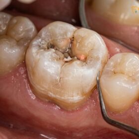
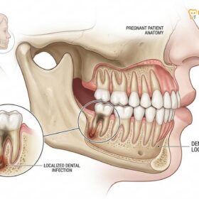
Tooth abscess and pulpitis represent two distinct, progressive stages of a severe dental infection. Pulpitis is the localized, active inflammation of the inner tooth nerve, while an abscess occurs when that infection escapes the tooth root, forming a dangerous, pus-filled pocket in the surrounding jawbone that requires immediate clinical intervention.
Clinical Summary:
Understanding the precise clinical distinction between a tooth abscess vs pulpitis is critical for effective endodontic triage and pain management. Pulpitis initiates as an acute inflammatory response within the confined pulpal chamber, most frequently triggered by deep carious lesions, microleakage from failing restorations, or mechanical trauma. If this inflammation is left untreated, the pulpal tissue inevitably undergoes liquefaction necrosis. This cellular death allows aggressive bacterial pathogens to migrate unimpeded through the root canal system and exit via the apical foramen into the periapical tissues. This migration triggers a massive immune response, resulting in a periapical abscess—a localized collection of purulent exudate (pus) trapped within the alveolar bone. Clinically, pulpitis primarily manifests with severe, localized thermal sensitivity and spontaneous, sharp pain. Conversely, an established abscess presents with constant throbbing pressure, extreme tenderness to percussion, visible facial or gingival swelling, and potential systemic involvement. Treatment protocols must shift accordingly: from vital pulp therapy or standard endodontic treatment for pulpitis, to immediate surgical drainage, complex root canal disinfection, or extraction for an abscess.
Key Takeaways:
- Progression: Pulpitis is the precursor; an abscess is the advanced complication that occurs after the tooth nerve has died.
- Pain Characteristics: Pulpitis causes sharp, lingering pain to cold or heat, whereas an abscess causes constant, throbbing pressure that worsens when lying down.
- Anatomical Location: Pulpitis is confined entirely within the hard structure of the tooth; an abscess involves the destruction of the surrounding jawbone and soft tissues.
- Visual Signs: Pulpitis rarely shows external swelling, while an abscess frequently causes visible gum boils, facial asymmetry, and swollen lymph nodes.
- Treatment Approach: Both conditions require professional intervention, but an abscess often necessitates immediate surgical drainage alongside endodontic therapy to prevent systemic spread.
The Path of Infection: From Enamel Cavity to Bone Destruction
Dental infections follow a predictable downward trajectory, starting from superficial enamel decay, penetrating the porous dentin, infecting the vital pulpal tissue, and ultimately destroying the surrounding alveolar bone.
To fully comprehend the clinical differences between a tooth abscess vs pulpitis, one must first understand the anatomical structure of a human tooth and the pathogenesis of dental caries. A healthy tooth is a highly complex organ, not merely a solid block of calcium. The outermost layer, the enamel, is the hardest substance in the human body, composed of densely packed hydroxyapatite crystals. Beneath the enamel lies the dentin, a softer, porous tissue containing thousands of microscopic channels known as dentinal tubules. At the very core of the tooth lies the pulpal chamber and root canal system, which houses the dental pulp—a delicate matrix of nerves, blood vessels, fibroblasts, and immune cells.
The journey of a dental infection almost always begins at the surface. Cariogenic bacteria, primarily Streptococcus mutans and Lactobacillus species, colonize the tooth surface within a sticky biofilm known as dental plaque. When exposed to fermentable carbohydrates from the diet, these bacteria metabolize the sugars and excrete lactic acid as a byproduct. This acid initiates the demineralization of the enamel. Because enamel contains no living cells or nerve endings, this initial stage of decay is entirely asymptomatic. Patients are usually unaware that a cavity is forming unless it is detected during a routine radiographic examination.
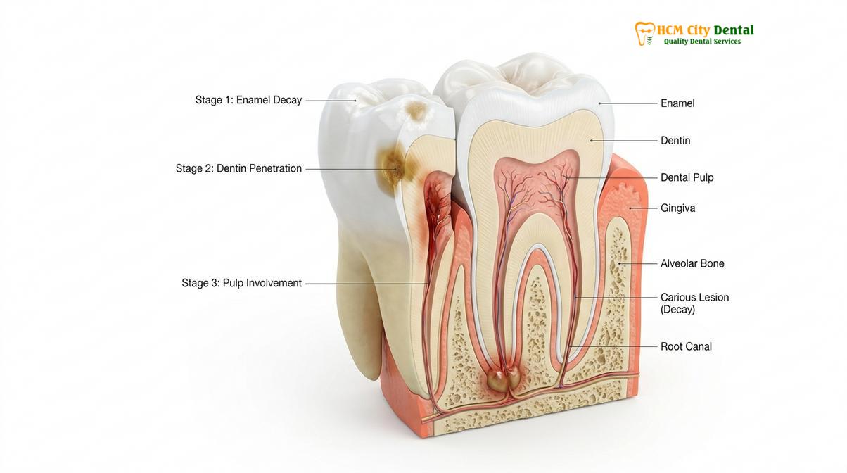
Once the bacterial acid breaches the dentinoenamel junction (DEJ) and enters the dentin, the progression of the disease accelerates significantly. The dentinal tubules provide a direct, fluid-filled highway toward the inner pulp. As bacteria and their acidic toxins travel down these tubules, they trigger a hydrodynamic response. The fluid inside the tubules shifts, stimulating the nerve endings at the periphery of the pulp. This is the stage where patients typically begin to experience mild, transient sensitivity to sweet foods or cold beverages. At this juncture, the pulp is irritated but not yet deeply infected, a condition clinically referred to as reversible pulpitis. If the decay is removed and the tooth is restored with a biocompatible filling at this stage, the pulp can heal, and the tooth will return to a normal, healthy state[1].
However, if the carious lesion is ignored, the bacterial invasion continues its relentless march inward. The physical distance between the advancing front of the bacteria and the pulpal tissue narrows. Once the bacteria physically breach the pulpal chamber, the localized irritation escalates into a full-blown, aggressive immune response. The tooth has now crossed the threshold from a simple cavity into the realm of complex endodontic disease, setting the stage for irreversible pulpitis and, eventually, a periapical abscess.
What is Pulpitis? Active Inflammation Inside the Pulpal Chamber
Pulpitis is the active immune response and severe inflammation of the tooth’s inner nerve tissue, characterized by sharp, lingering pain triggered by temperature changes or biting forces.
When bacterial pathogens finally penetrate the pulpal chamber, the body’s immune system mounts a vigorous defense. White blood cells, primarily neutrophils and macrophages, rush to the site of the infection to engulf and destroy the invading microbes. This immune response triggers the release of inflammatory mediators, such as prostaglandins, histamine, and cytokines. In any other part of the body, this inflammation would cause the tissue to swell, turn red, and become warm—a normal physiological reaction. However, the dental pulp is trapped within the rigid, unyielding walls of the dentin and enamel. It cannot expand.
Because the inflamed pulp cannot swell, the localized vasodilation leads to a rapid and dramatic increase in intrapulpal pressure. This intense pressure compresses the delicate nerve fibers within the pulp, specifically the A-delta fibers (responsible for sharp, immediate pain) and the C-fibers (responsible for dull, throbbing, unmyelinated pain). This phenomenon is the hallmark of irreversible pulpitis. The pain associated with this condition is notoriously severe, often described by patients as one of the most excruciating experiences of their lives.
“The unique anatomical confinement of the dental pulp means that even a minor inflammatory response can result in disproportionately severe pain. The inability of the tissue to expand leads to a self-strangulating cycle of pressure, ischemia, and nerve compression that defines irreversible pulpitis.”
Clinically, irreversible pulpitis presents with several distinct diagnostic markers. The most prominent symptom is spontaneous pain—pain that occurs without any provocation and frequently wakes the patient from a deep sleep. Furthermore, the tooth exhibits an exaggerated and lingering response to thermal stimuli. When a dentist applies a cold test (using a refrigerant spray like Endo-Ice) to a tooth with irreversible pulpitis, the patient will feel a sharp, intense jolt of pain that lingers for 10, 20, or even 30 seconds after the cold stimulus is removed. This lingering pain is a definitive sign that the nerve tissue is actively dying and cannot recover[2].
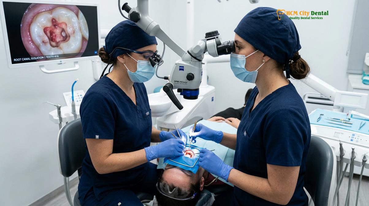
Dr. Nguyen Van Cuong, a leading specialist at HCMC Dental Clinic, frequently emphasizes the diagnostic challenges associated with this stage of infection. “Patients often present to our clinic in Ho Chi Minh City with severe, radiating pain that they cannot pinpoint to a single tooth,” notes Dr. Cuong. “Because the pulpal nerve lacks proprioceptive fibers—the fibers that tell the brain exactly where a stimulus is coming from—the pain of pulpitis can radiate to the ear, the temple, or even the opposing jaw. It requires meticulous clinical testing, including thermal diagnostics and electric pulp testing, to accurately identify the offending tooth before we can initiate advanced endodontic therapy.”
Another critical clinical consideration during the pulpitis stage is the difficulty of achieving profound local anesthesia. The intense inflammation lowers the local tissue pH, creating an acidic environment. Local anesthetics are weak bases; in an acidic environment, they become ionized and cannot effectively penetrate the nerve membrane to block the pain signals. This creates the dreaded “hot tooth” scenario, requiring advanced anesthetic techniques, such as intraligamentary or intraosseous injections, to ensure patient comfort during emergency intervention.
The Transition to Pulpal Necrosis: When the Pain Suddenly Stops
When the pulpal tissue completely dies from prolonged inflammation, the acute nerve pain temporarily ceases, creating a deceptive pain-free period before the infection spreads into the jawbone.
If irreversible pulpitis is left untreated, the intense intrapulpal pressure eventually collapses the venules and capillaries that supply blood to the tooth. This cuts off the oxygen and nutrient supply, leading to ischemia and the rapid death of the pulpal tissue. This process is known as pulpal necrosis. As the nerve fibers die, they lose their ability to transmit pain signals to the brain. Consequently, the patient may experience a sudden, miraculous cessation of the severe, spontaneous pain that had been tormenting them for days or weeks.
This pain-free period is highly deceptive and clinically dangerous. Patients often mistakenly believe that the tooth has “healed itself” or that the infection has resolved. In reality, the situation has worsened significantly. The pulpal chamber is now filled with necrotic debris—dead tissue, stagnant blood, and millions of multiplying bacteria. Because the blood supply has been severed, the body’s immune cells can no longer reach the inside of the tooth. The root canal system becomes an isolated, anaerobic incubator where highly aggressive, Gram-negative bacteria (such as Porphyromonas gingivalis and Fusobacterium nucleatum) thrive and multiply exponentially[3].
During the necrotic stage, the tooth will no longer respond to thermal testing. A cold test will yield no sensation whatsoever, confirming that the vital nerve tissue has been completely destroyed. However, the tooth may begin to feel slightly “high” or tender when biting down. This tenderness indicates that the bacterial toxins are beginning to seep out of the root tip (the apical foramen) and irritate the periodontal ligament—the shock-absorbing membrane that holds the tooth in the bone. This localized irritation is the immediate precursor to the formation of a full-blown periapical abscess.
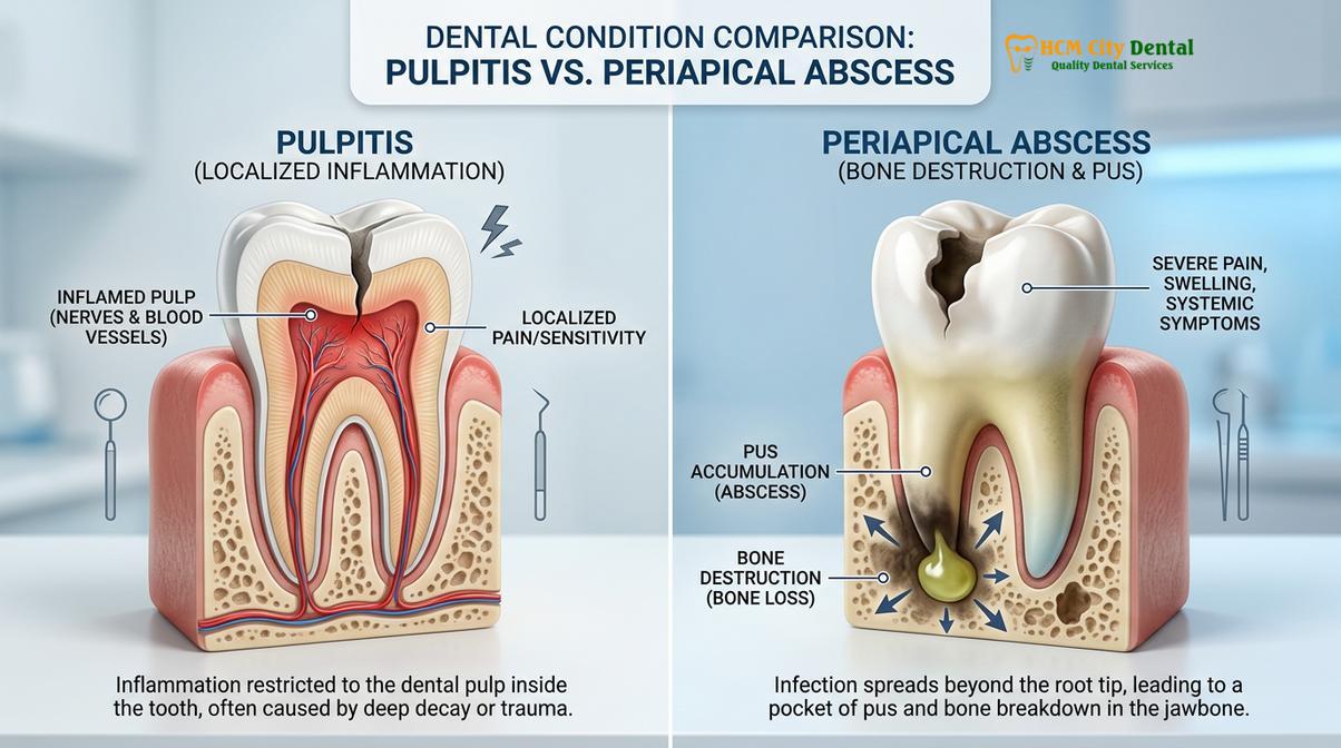
The transition from pulpitis to necrosis highlights the critical importance of timely dental intervention. Once the pulp becomes necrotic, simple vital pulp therapies are no longer viable. The entire root canal system must be meticulously cleaned, shaped, and chemically disinfected using powerful irrigants like sodium hypochlorite and EDTA to dissolve the necrotic tissue and eradicate the bacterial biofilm before the infection can establish a foothold in the surrounding jawbone.
Tooth Abscess (Periapical Infection): Pus Accumulation in the Bone
A periapical abscess develops when necrotic bacteria exit the tooth root, triggering a massive immune response that traps pus, dead tissue, and bacteria within the jawbone.
When the bacterial load within the necrotic root canal system reaches a critical mass, the pathogens and their toxic byproducts spill out through the apical foramen into the periapical tissues (the bone and ligaments surrounding the tip of the root). The body recognizes this massive breach and mounts a secondary, highly aggressive immune response to prevent the bacteria from spreading systemically into the bloodstream. Neutrophils and macrophages flood the area, engaging in a fierce battle with the invading microbes.
The casualties of this microscopic war—dead white blood cells, dead bacteria, liquefied bone tissue, and proteinaceous debris—accumulate to form purulent exudate, commonly known as pus. Because this battle is taking place deep within the dense alveolar bone of the jaw, the pus has nowhere to go. It becomes trapped, forming a pressurized, encapsulated pocket of infection known as a periapical abscess. The pressure from this expanding pocket of pus causes the severe, constant, throbbing pain that is the hallmark of an acute dental abscess.
Unlike the pain of pulpitis, which is often triggered by temperature and can radiate diffusely, the pain of an abscess is highly localized and exquisitely tender to mechanical pressure. The tooth will feel elevated in the socket, and even the slightest touch or biting force will elicit severe pain. This is because the tooth is literally being pushed upward by the pressure of the pus accumulating beneath its roots. Furthermore, the pain often worsens when the patient lies down, as the change in posture increases blood flow and hydrostatic pressure in the head and neck region.
If the acute abscess is not surgically drained, the pressurized pus will seek the path of least resistance. It will begin to eat away at the surrounding jawbone, eventually perforating the cortical plate (the hard outer layer of the bone) and spreading into the overlying soft tissues of the gums or face. This results in visible facial swelling, which can be dramatic and highly distressing for the patient. In some cases, the pus will burrow through the gums and create a small, pimple-like bump known as a sinus tract. When this tract ruptures, it allows for dental fistula drainage, releasing the trapped pus into the oral cavity. While this drainage often provides immediate relief from the severe pressure and pain, it signifies that the infection has become chronic. The source of the infection—the necrotic tooth—remains, and the abscess will continue to produce pus until the tooth is properly treated.
In our clinical experience managing complex cases of periapical abscess Saigon residents frequently encounter due to delayed care, the presence of a draining fistula is a clear indicator of long-standing necrotic infection. While the patient may not be in acute pain at the moment of presentation, the continuous destruction of the alveolar bone requires immediate and comprehensive endodontic intervention to halt the osteolytic process and promote bone regeneration.
Symptoms Comparison: Mild Ache vs. Facial Swelling and Fever
While pulpitis causes sharp, localized thermal pain, an abscess presents with constant, throbbing pressure, severe pain upon chewing, visible gum swelling, and potential systemic fever.
Accurately differentiating between a tooth abscess vs pulpitis relies heavily on a detailed analysis of the patient’s symptoms and clinical presentation. While both conditions cause significant dental pain, the nature, triggers, and associated signs of that pain are distinctly different. A thorough clinical examination, combined with high-resolution radiographic imaging, is essential for establishing the correct diagnosis and formulating an appropriate treatment plan.
The following table provides a comprehensive clinical comparison of the symptoms and diagnostic markers associated with irreversible pulpitis versus an acute periapical abscess:
| Clinical Feature | Irreversible Pulpitis | Periapical Abscess |
|---|---|---|
| Nature of Pain | Sharp, shooting, spontaneous; often wakes the patient at night. | Constant, dull, throbbing pressure; feels like a heartbeat in the tooth. |
| Thermal Sensitivity | Extreme, lingering pain to cold or hot stimuli (lasting >10 seconds). | No response to cold (nerve is dead); heat may exacerbate pressure pain. |
| Pain on Biting/Percussion | Usually mild or absent, unless inflammation has reached the root tip. | Exquisitely painful; tooth feels “high” and cannot tolerate any pressure. |
| Visible Swelling | None. The inflammation is entirely confined within the tooth structure. | Frequent. Swelling of the gums, facial asymmetry, or a draining fistula. |
| Systemic Symptoms | None. The patient feels generally well, aside from the localized tooth pain. | Possible fever, lethargy, swollen lymph nodes (lymphadenopathy). |
| Radiographic Appearance | Normal periapical bone; deep decay or large restoration visible. | Dark radiolucent halo (bone loss) visible around the apex of the root. |
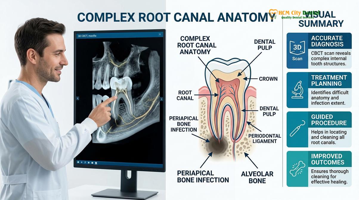
Radiographic imaging plays a pivotal role in this differential diagnosis. In cases of pure pulpitis, a standard two-dimensional periapical X-ray will typically show a deep cavity approaching the nerve, but the bone surrounding the root tip will appear completely normal and intact. This is because the infection has not yet exited the tooth. Conversely, an established periapical abscess will often present as a distinct dark circle (radiolucency) at the apex of the root, indicating that the acidic pus has dissolved the surrounding bone tissue[4]. In complex cases, particularly involving multi-rooted molars, advanced 3D Cone Beam Computed Tomography (CBCT) imaging is utilized to visualize the exact extent of the bone destruction and identify any hidden accessory canals that may be harboring necrotic debris.
Systemic Risks: Why a Dental Abscess is a Medical Emergency
Left untreated, a localized dental abscess can rapidly spread through fascial spaces, leading to life-threatening complications such as Ludwig’s angina, airway obstruction, or systemic sepsis.
While pulpitis is an agonizing condition, it is fundamentally a localized problem confined to the tooth. A dental abscess, however, represents a significant breach of the body’s defenses and carries severe systemic risks. The maxillofacial region is a complex network of interconnected fascial spaces—potential compartments between the muscles and tissues of the head and neck. When the pressurized pus from an abscess breaks through the jawbone, it can rapidly track along these fascial planes, spreading the infection far beyond the oral cavity.
If an abscess develops in a lower molar, the infection can spread downward into the submandibular, sublingual, and submental spaces. This rapid, bilateral swelling of the floor of the mouth and neck is known as Ludwig’s angina. It is a true medical emergency. The massive swelling can elevate the tongue and compress the airway, leading to asphyxiation if not treated immediately with surgical decompression and intravenous antibiotics. Similarly, an abscess in an upper tooth can spread upward toward the eyes and brain, potentially causing cavernous sinus thrombosis—a rare but highly lethal condition where a blood clot forms in the base of the brain due to the spreading bacterial infection.
Clinical Warning: A dental abscess is not merely a “bad toothache”—it is an active bacterial infection capable of systemic spread. If you experience a toothache accompanied by a high fever, difficulty swallowing (dysphagia), restricted jaw opening (trismus), or rapid swelling that extends to your eye or down your neck, you must seek immediate emergency medical or dental care. These are red-flag symptoms of a spreading fascial space infection.
Furthermore, the continuous presence of a heavy bacterial load in the highly vascularized oral tissues increases the risk of bacteremia—the presence of bacteria in the bloodstream. For healthy individuals, the immune system can usually clear transient bacteremia. However, for patients with compromised immune systems, uncontrolled diabetes, or pre-existing cardiovascular conditions, these circulating bacteria can colonize distant organs, leading to severe complications such as infective endocarditis or systemic sepsis[5].
“The transition from a localized periapical abscess to a systemic fascial space infection can occur with alarming rapidity. What begins as a throbbing toothache can evolve into a life-threatening airway compromise within a matter of hours if the purulent exudate breaches the critical anatomical boundaries of the head and neck.”
Dr. Nguyen Van Cuong strongly advises patients never to ignore facial swelling. “At HCMC Dental Clinic, we treat any visible facial swelling associated with a dental infection as a high-priority emergency,” he explains. “The primary goal is no longer just saving the tooth; it is securing the patient’s systemic health. Immediate source control through emergency dental interventions is mandatory to halt the spread of the pathogens.”
Emergency Interventions: Abscess Drainage and Emergency Root Canal
Clinical management requires immediate source control, utilizing surgical incision and drainage to relieve pressure, followed by comprehensive endodontic therapy or extraction to eradicate the bacterial reservoir.
The treatment protocols for pulpitis and a tooth abscess differ significantly in their immediate objectives, though both ultimately aim to eliminate the bacterial infection and relieve pain. For irreversible pulpitis, the treatment of choice is a pulpectomy—the complete removal of the inflamed, vital nerve tissue from the pulpal chamber and root canals. Once the inflamed tissue is removed, the severe pain resolves almost immediately. The canals are then cleaned, shaped, and sealed in a standard root canal procedure, allowing the patient to retain their natural tooth.
Treating a periapical abscess, however, is a more complex, multi-stage process. The immediate, overriding clinical objective is to achieve drainage and relieve the immense hydrostatic pressure caused by the trapped pus. If the tooth is restorable, the dentist will perform an emergency access opening, drilling through the crown of the tooth directly into the necrotic pulpal chamber. In many cases, the pressurized pus will immediately well up through the access hole—a process known as trephination. This provides profound and rapid pain relief for the patient.
Clinical Case Study: Managing an Acute Periapical Abscess
A 45-year-old patient presented to HCMC Dental Clinic in Ho Chi Minh City with severe, throbbing pain in the lower right quadrant, accompanied by visible facial swelling and a low-grade fever. Clinical examination revealed a necrotic lower first molar that was exquisitely tender to percussion. A CBCT scan confirmed a large periapical radiolucency. The clinical team immediately established profound local anesthesia using a specialized intraosseous technique. An emergency access opening was created, resulting in copious purulent drainage from the distal canal. The canals were heavily irrigated with sodium hypochlorite, and a calcium hydroxide intracanal medicament was placed to neutralize the remaining bacteria. The patient was prescribed a course of Amoxicillin to manage the systemic swelling. Upon returning one week later, the swelling had completely resolved, and the tooth was successfully obturated and restored with a custom zirconia crown, saving the natural dentition and eliminating the infection.
If the swelling is severe and localized in the soft tissues (a fluctuant swelling), the dentist may also need to perform an Incision and Drainage (I&D) procedure. This involves making a small surgical incision directly into the swollen gum tissue to allow the pus to evacuate, often placing a small rubber drain to keep the wound open and draining for several days. It is crucial to understand that while antibiotics are frequently prescribed during the management of an acute abscess to help control the systemic spread of the infection, antibiotics alone will never cure a dental abscess. The necrotic tissue inside the tooth has no blood supply, meaning the antibiotic molecules cannot reach the bacteria hiding inside the root canals. The physical source of the infection must be removed, either through comprehensive root canal therapy or, if the tooth is too severely damaged to be saved, through surgical extraction.

When to See a Doctor
Dental pain should never be ignored, as it is the body’s primary warning system that an infection is progressing. You should seek immediate clinical evaluation if you experience any of the following symptoms:
- Severe, spontaneous tooth pain that wakes you up from sleep or is not relieved by over-the-counter analgesics.
- Lingering, intense pain following exposure to hot or cold foods and beverages.
- A tooth that feels elevated, loose, or is exquisitely painful when biting down or tapped.
- Any visible swelling of the gums, face, cheek, or neck.
- The appearance of a pimple-like bump on the gums that may ooze pus or cause a bad taste in the mouth.
- Systemic symptoms accompanying a toothache, such as a fever, chills, rapid heart rate, or swollen lymph nodes.
- Difficulty swallowing, breathing, or fully opening your mouth (trismus)—these require immediate emergency medical attention.
Early intervention is the key to preserving your natural teeth and preventing life-threatening systemic complications. If you are experiencing any of these symptoms, contact a qualified dental professional immediately for a comprehensive diagnostic evaluation.
Frequently Asked Questions
Can pulpitis turn into an abscess?
Yes, untreated irreversible pulpitis will eventually progress into a tooth abscess. Once the inflamed pulpal tissue dies (necrosis), the bacteria multiply within the empty root canal and exit through the root tip, forming a pus-filled abscess in the surrounding jawbone. This transition marks the shift from a localized internal inflammation to a destructive bone infection.
Will a tooth abscess go away on its own with antibiotics?
No, a tooth abscess will never resolve completely with antibiotics alone. Antibiotics can help manage the systemic spread of the infection, but the source of the bacteria inside the necrotic tooth must be physically removed through a root canal or extraction. Because the dead tooth has no blood supply, the antibiotic medication cannot reach the bacteria hiding inside the canals.
What are the warning signs that a tooth infection has spread to the body?
Warning signs of a spreading systemic infection include a high fever, facial swelling that reaches the eyes or neck, difficulty swallowing (dysphagia), restricted jaw opening (trismus), rapid heart rate, and a general feeling of severe illness or lethargy. These symptoms indicate that the infection has breached the local tissues and entered the fascial spaces or bloodstream, requiring immediate emergency medical care.
How long does it take for an infected tooth to become an abscess?
The timeline varies significantly depending on the aggressiveness of the bacteria and the patient’s immune response. It can take anywhere from a few weeks to several months for deep decay to cause pulpitis, lead to pulpal necrosis, and finally develop into a visible periapical abscess. However, once the pulp becomes necrotic, the progression to an acute abscess can happen very rapidly.
Is it safe to extract an abscessed tooth while it is swollen?
Yes, extracting an abscessed tooth is often the fastest way to achieve profound drainage and source control. While severe swelling can make local anesthesia less effective due to tissue acidity, modern clinical protocols and advanced anesthetic techniques allow dentists to safely remove the tooth, immediately relieving the infectious pressure and initiating the healing process.
References
- Journal of Endodontics. Pathogenesis of Apical Periodontitis and Pulpal Necrosis. (2020).
- International Endodontic Journal. Diagnostic accuracy of clinical tests for pulpitis and periapical lesions. (2021).
- Journal of the American Dental Association. Management of acute dental pain and endodontic emergencies. (2019).
- Clinical Oral Investigations. Microbiology of primary and secondary endodontic infections. (2022).
- British Dental Journal. Systemic complications of odontogenic infections and fascial space spread. (2018).