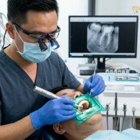
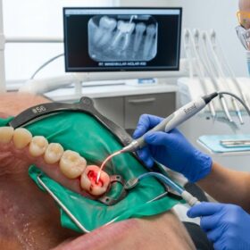
A cracked tooth root canal is a specialized endodontic procedure designed to save a damaged tooth when a fracture penetrates the inner pulp chamber. By removing infected nerve tissue and sealing the canal, dentists can alleviate severe pain and prevent the need for complete tooth extraction.
Clinical Summary:
Cracked tooth syndrome presents unique diagnostic challenges due to microscopic fractures that frequently evade standard two-dimensional X-rays. When these fissures breach the pulp chamber, endodontic therapy becomes clinically essential to halt bacterial ingress and prevent nerve necrosis. Successful retention of the natural tooth relies heavily on early intervention, precise microscopic evaluation, and immediate structural reinforcement with a custom restoration to prevent fracture propagation.
Key Takeaways:
- Not all cracks require endodontic therapy; treatment depends entirely on fracture depth and pulp involvement.
- Microscopic evaluation and specialized bite tests are critical for identifying hidden fissures that X-rays miss.
- A vertical root fracture typically renders a tooth unsavable, necessitating safe extraction and replacement.
- Endodontic treatment must be followed by immediate structural reinforcement to prevent catastrophic splitting.
- Early intervention significantly improves the long-term prognosis and biomechanical stability of a compromised tooth.
Understanding Cracked Tooth Syndrome: Symptoms and Difficulty of Diagnosis
Cracked tooth syndrome causes erratic pain during chewing and temperature sensitivity, yet diagnosis is notoriously difficult because hairline fractures rarely appear on conventional dental X-rays.
Cracked tooth syndrome is a complex clinical condition characterized by a partial fracture of a tooth that has not yet completely separated into distinct pieces. Patients typically present with a highly specific set of symptoms: sharp, sudden pain when biting down, which often intensifies at the exact moment the bite pressure is released. This phenomenon occurs because the biting force slightly opens the microscopic crack, and the sudden release causes the crack to snap shut, triggering a sharp fluid movement within the dentinal tubules that directly stimulates the underlying nerve endings[1].
In addition to mechanical pain, patients frequently experience heightened sensitivity to extreme temperatures, particularly cold. Unlike the lingering ache of an advanced infection, the pain associated with an early-stage crack is often fleeting and erratic, making it incredibly frustrating for the patient to pinpoint the exact source of their discomfort.
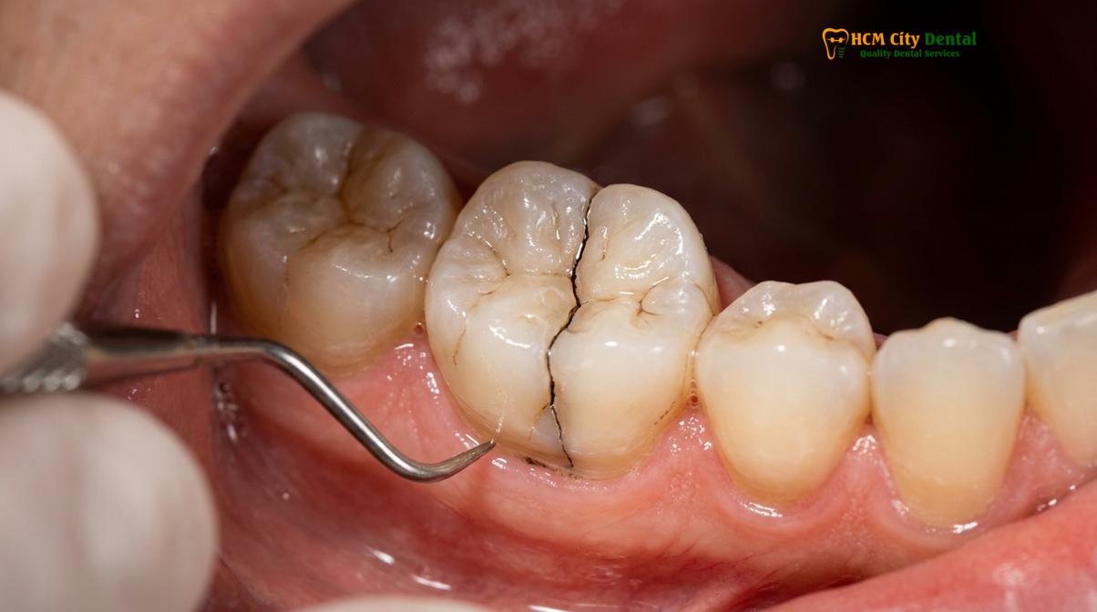
The primary difficulty in diagnosing cracked tooth syndrome lies in the limitations of standard radiographic imaging. Conventional dental X-rays provide a two-dimensional representation of a three-dimensional structure. Because cracks often propagate in a mesial-distal direction (front-to-back) parallel to the X-ray beam, they are virtually invisible on a standard radiograph unless the fracture has widened significantly or caused secondary bone loss at the root apex. Dr. Nguyen Van Cuong frequently emphasizes to patients that the absence of a visible crack on an X-ray does not rule out a structural fracture; it simply necessitates a more tactile and visually magnified diagnostic approach.
Types of Cracks: Craze Lines, Fractured Cusps, and Vertical Root Splits
Dental fractures range from superficial enamel craze lines to catastrophic vertical root splits, and identifying the specific classification dictates whether conservative restoration, endodontic therapy, or extraction is required.
Not all cracks are created equal. The human dentition, consisting of 28 to 32 adult teeth, is subjected to immense occlusal forces daily. Over time, these forces can cause various types of structural compromises. Understanding the specific type of fracture is the foundational step in formulating an effective treatment plan.
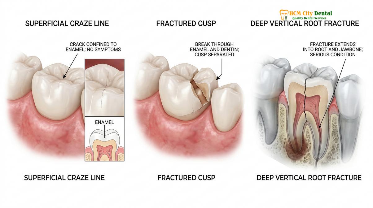
Craze Lines: These are microscopic, superficial fissures that affect only the outer enamel layer of the tooth. They are exceedingly common in adult dentition, often appearing as faint vertical lines on the front teeth. Craze lines are asymptomatic, do not require endodontic therapy, and are generally considered a cosmetic concern rather than a structural threat.
Fractured Cusps: A fractured cusp occurs when a portion of the tooth’s chewing surface weakens and breaks off. This frequently happens around large, older amalgam fillings that have expanded over time. Because a fractured cusp usually breaks off above the gum line and rarely exposes the inner pulp chamber, it typically causes minimal pain and can be effectively treated with a new filling or a protective crown without the need for a root canal.
Cracked Tooth: This classification refers to a crack that originates on the chewing surface and extends vertically toward the root. If the crack remains confined to the upper portion of the tooth, it may be stabilized. However, if the fissure deepens into the dentin and breaches the pulp, cracked tooth syndrome develops, and endodontic intervention becomes mandatory to save the tooth.
Split Tooth: A split tooth is the end-stage progression of an untreated cracked tooth. The crack has propagated entirely through the tooth, separating it into two distinct, movable segments. A split tooth cannot be saved intact; however, depending on the position of the split, a specialized endodontic procedure might salvage one healthy root of a multi-rooted molar, though extraction is often the most predictable outcome.
Vertical Root Fracture: Unlike other cracks that start at the crown, a vertical root fracture begins at the root apex and propagates upward. These fractures are particularly insidious as they show minimal early symptoms and are often only discovered when the surrounding jawbone becomes infected.
| Type of Fracture | Depth of Damage | Typical Symptoms | Standard Clinical Treatment |
|---|---|---|---|
| Craze Lines | Enamel only | None (Asymptomatic) | No treatment required; cosmetic monitoring |
| Fractured Cusp | Enamel and Dentin | Mild sensitivity, rough edge | Dental crown or composite restoration |
| Cracked Tooth | Extends toward the pulp | Sharp pain on bite release | Root canal therapy + Crown |
| Split Tooth | Complete separation | Severe pain, mobility | Extraction (or partial root amputation) |
| Vertical Root Fracture | Starts at root apex | Dull ache, localized gum swelling | Tooth extraction |
Diagnostic Methods: Transillumination, Bite Tests, and Microscope Checks
Advanced diagnostic protocols, including fiber-optic transillumination, targeted bite tests, and high-magnification microscopy, are essential for locating elusive fractures before initiating irreversible treatments.
Because standard radiographs are often inconclusive for hairline fractures, clinicians must rely on a battery of specialized diagnostic tests to confirm the presence, location, and extent of a crack. The diagnostic phase is critical; proceeding with root canal treatment without fully mapping the fracture can lead to unpredictable outcomes.
The most common functional assessment is the bite test, utilizing a specialized instrument known as a Tooth Slooth. The dentist places this small, wedge-shaped device on individual cusps of the suspected tooth and asks the patient to bite down and release. By isolating the occlusal force to a single cusp, the clinician can replicate the exact pain the patient experiences at home, thereby identifying precisely which section of the tooth is structurally compromised[2].
Visual diagnostics are equally important. Fiber-optic transillumination involves shining a high-intensity light directly through the tooth structure. In a healthy, solid tooth, the light passes through seamlessly. However, if a crack is present, the fracture line acts as a barrier, causing the light to abruptly stop or refract, clearly illuminating the fissure against the darker background of the healthy dentin.
“The integration of the dental operating microscope has revolutionized endodontic diagnostics. Magnification up to 25x allows clinicians to visually trace a fracture line across the pulpal floor, determining definitively whether a tooth can be saved or if the crack has rendered it structurally non-viable.”
Furthermore, the use of a Dental Operating Microscope (DOM) is considered the gold standard in modern endodontics. Once the tooth is accessed, the microscope allows the specialist to inspect the internal chamber under extreme magnification and bright illumination. If the crack extends deeply across the floor of the pulp chamber and down into the root orifices, the prognosis drops significantly, and the treatment plan must be adjusted accordingly.
When the Crack Reaches the Pulp: Why a Root Canal is Required
Once a fracture breaches the dentin and enters the pulp chamber, bacterial contamination leads to irreversible pulpitis, making endodontic therapy mandatory to remove diseased tissue and preserve the root.
The outer layers of a tooth—the enamel and dentin—serve as a protective fortress for the delicate inner pulp, which houses the tooth’s nerves, blood vessels, and connective tissues. When a crack propagates deep enough to breach this fortress, the sterile environment of the pulp chamber is compromised. The microscopic fissure acts as a direct highway for oral bacteria to infiltrate the inner tooth.
Once bacteria enter the pulp, they trigger a severe inflammatory response known as irreversible pulpitis. Because the pulp is encased in rigid dentin walls, the inflammation causes a rapid buildup of pressure, leading to the intense, throbbing pain that typically drives patients to seek emergency dental care. If left untreated, the bacterial infection will eventually cause the nerve tissue to undergo necrosis (die) and spread out through the root apex into the surrounding jawbone, creating a painful abscess[3].
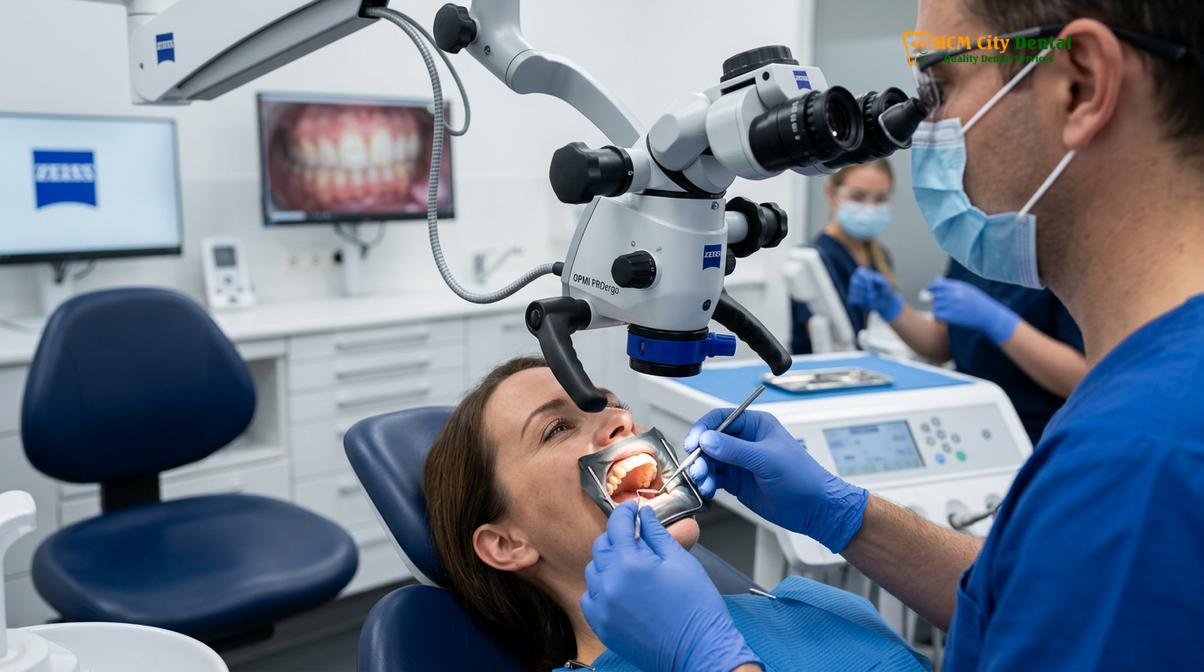
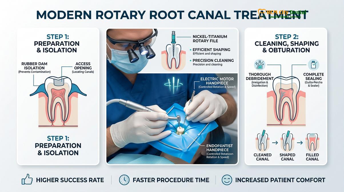
To halt this destructive process and save the natural tooth structure, a root canal procedure is clinically required. The workflow begins with profound local anesthesia to ensure patient comfort. The clinician then isolates the tooth using a rubber dam to maintain a sterile surgical field. An access opening is created, and the diseased, inflamed, or necrotic pulp tissue is meticulously removed using specialized rotary files.
Clinical Case Review: A 45-year-old patient presented to HCMC Dental Clinic in Ho Chi Minh City complaining of severe, lingering pain in a lower right molar after biting into a hard nut. Dr. Nguyen Van Cuong utilized microscopic evaluation to identify a deep central crack that had breached the pulp chamber. By performing prompt endodontic therapy to remove the inflamed nerve, followed by immediate dental crown reinforcement, the patient’s natural tooth was successfully preserved, and all pain was eliminated within 48 hours.
After the canal system is thoroughly cleaned and disinfected with antimicrobial irrigants, it is completely sealed with a biocompatible material, typically gutta-percha combined with a bioceramic sealer. This obturation process prevents any future bacterial reinfection from the oral cavity or the surrounding bone. However, the endodontic intervention only addresses the biological infection; the mechanical weakness caused by the crack must still be resolved.
Vertical Root Fractures: The Unsavable Tooth Scenario
A vertical root fracture originates at the root apex and propagates upward, fundamentally compromising the root structure and generally rendering the tooth unsavable, necessitating extraction.
While many cracked teeth can be successfully rehabilitated, a vertical root fracture represents a catastrophic structural failure. Unlike cracks that begin at the chewing surface, a vertical root fracture initiates at the very bottom of the root (the apex) and slowly splits the root upward toward the crown. These fractures are notoriously silent in their early stages, often presenting with minimal or no pain until the surrounding periodontal tissues become severely infected.
The primary reason a vertical root fracture renders a tooth unsavable is that the split creates a permanent, unsealable communication between the oral cavity and the jawbone. Bacteria continuously colonize the fracture line, leading to chronic inflammation and rapid destruction of the adjacent alveolar bone. Clinically, this often presents as a deep, isolated periodontal pocket and a distinct “J-shaped” or “halo” radiolucency on an X-ray, indicating extensive bone loss around the fractured root[4].
Clinical Warning: Attempting to retain a tooth with a confirmed vertical root fracture is highly discouraged. Prolonging extraction allows the chronic infection to destroy significant amounts of healthy jawbone, which can severely complicate future restorative options, such as the placement of a dental implant.
Because there is currently no reliable dental material or surgical technique capable of fusing a vertically split root back together while withstanding the immense forces of mastication, the standard of care is the prompt extraction of the affected tooth. Following extraction, the site is typically grafted with bone material to preserve the ridge architecture, preparing the area for a future implant-supported restoration.
Structural Reinforcement: The Necessity of an Immediate Dental Crown
Following endodontic therapy, a cracked tooth is inherently fragile; immediate structural reinforcement with a full-coverage dental crown is clinically imperative to bind the remaining tooth structure together.
Successfully completing the endodontic evaluation and nerve removal is only half the battle when treating a cracked tooth. Once the pulp is removed, the tooth loses its internal blood supply, causing the dentin to become more brittle over time. Furthermore, the access cavity required to perform the root canal, combined with the pre-existing crack, significantly compromises the tooth’s overall biomechanical integrity.
If a root-filled cracked tooth is left unprotected or simply restored with a standard composite filling, the wedge-like forces of chewing will inevitably drive the crack deeper, eventually splitting the tooth entirely and leading to extraction. Therefore, dental crown reinforcement is not an optional upgrade; it is a strict clinical necessity.
“The application of a full-coverage restoration provides a crucial ‘ferrule effect’—a 360-degree bracing mechanism that physically binds the weakened cusps together, effectively neutralizing the wedging forces that would otherwise cause a cracked tooth to fail catastrophically.”
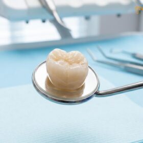
A custom-fabricated dental crown, often milled from high-strength materials like monolithic zirconia or lithium disilicate, is designed to encase the entire visible portion of the tooth down to the gum line. By distributing occlusal forces evenly across the restoration rather than allowing them to concentrate on the fracture line, the crown stabilizes the underlying tooth structure. Patients are strongly advised to proceed with crown placement immediately following the completion of their endodontic therapy to ensure the highest possible long-term success rate[5].
When to See a Doctor
Immediate clinical evaluation is necessary if you experience sharp pain upon releasing a bite, persistent thermal sensitivity, or localized gum swelling, as early intervention is key to saving a cracked tooth.
Ignoring the early warning signs of a cracked tooth can lead to rapid deterioration of the tooth’s prognosis. You should seek a professional endodontic evaluation immediately if you experience any of the following symptoms:
- Sharp, erratic pain that occurs specifically when you release your bite from food.
- A sudden, intense sensitivity to hot or cold temperatures that lingers long after the stimulus is removed.
- A localized, pimple-like swelling on the gums near the affected tooth, which may indicate an active abscess.
- A dull, constant ache that radiates to the jaw, ear, or neck.
Early diagnosis is the most critical factor in tooth preservation. If you suspect you have a cracked tooth, scheduling a comprehensive microscopic examination at a specialized facility like HCMC Dental Clinic in Saigon can provide clarity. Utilizing advanced 3D imaging and clinical diagnostics, the dental team can accurately map the fracture and implement the necessary endodontic and restorative protocols to save your natural smile before the damage becomes irreversible.
Frequently Asked Questions
Can you fix a vertical root fracture with a root canal?
No, a vertical root fracture generally cannot be fixed with a root canal. Because the fracture splits the root entirely and creates an unsealable pathway for bacteria into the jawbone, the standard clinical protocol is tooth extraction to prevent severe bone loss and systemic infection. Attempting to retain the tooth will only lead to chronic abscesses and further deterioration of the surrounding bone.
Does a crown always go on a cracked tooth after a root canal?
Yes, a dental crown is almost always required on a cracked tooth following endodontic therapy. The crown provides essential structural reinforcement, binding the fragile tooth together to prevent the existing crack from propagating further under the heavy forces of daily chewing. Without this full-coverage protection, a root-filled cracked tooth is highly susceptible to catastrophic splitting and subsequent loss.
How long can a cracked tooth stay before it becomes infected?
The timeline varies significantly, ranging from a few weeks to several months, depending on the depth of the crack and oral hygiene. Once the fissure breaches the protective enamel and dentin, bacterial infiltration into the pulp is inevitable, making prompt clinical evaluation crucial. Delaying treatment allows bacteria to multiply within the pulp chamber, eventually leading to irreversible nerve death and painful abscess formation.
Is a cracked tooth root canal painful?
Modern endodontic procedures for a cracked tooth are highly manageable and typically involve minimal discomfort. Utilizing advanced local anesthesia and precise clinical techniques, dentists ensure the area is completely numb, effectively relieving the severe pain caused by the inflamed or infected nerve. Post-operative soreness is common for a few days but is easily managed with over-the-counter anti-inflammatory medications.
How successful is endodontic treatment for a cracked tooth?
Endodontic treatment for a cracked tooth has a high success rate when the fracture is diagnosed early and has not extended below the gum line into the root structure. Success heavily relies on immediate structural reinforcement with a custom crown to stabilize the tooth and prevent future mechanical failure. Regular follow-up X-rays are recommended to monitor the healing of the surrounding bone.
References
- Journal of Endodontics. Outcomes of root canal treatment and restoration longevity. (2020).
- International Endodontic Journal. Biofilms in endodontics and disinfection strategies. (2021).
- Journal of the American Dental Association. Microscope-assisted endodontics and missed canals. (2019).
- Clinical Oral Investigations. Biocompatibility of bioceramic root canal sealers. (2022).
- British Dental Journal. Pain management and dental anxiety in endodontics. (2018).