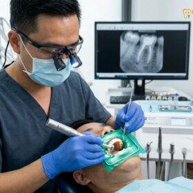
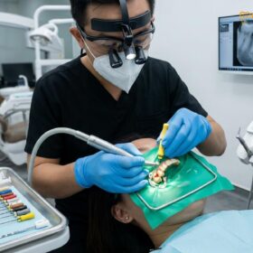
A tooth pulp vitality test is a fundamental diagnostic procedure utilized by dental professionals to evaluate the health, integrity, and functional status of the neurovascular tissue residing within the inner chamber of a tooth. By systematically applying controlled thermal, electrical, or mechanical stimuli to the tooth structure, clinicians can accurately assess whether the delicate pulpal tissue is healthy, actively inflamed, or has undergone complete necrosis. This diagnostic clarity is absolutely essential for guiding the appropriate course of clinical treatment and ensuring long-term oral health.
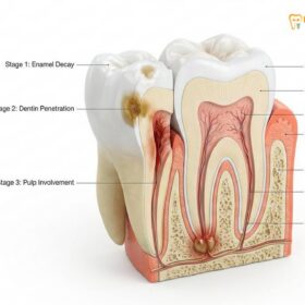
The inner core of every vital tooth contains a complex, highly organized network of blood vessels, connective tissues, specialized cells called odontoblasts, and sensitive nerve fibers. When a tooth is subjected to external trauma, deep bacterial decay, or repeated invasive restorative procedures, this enclosed tissue can become severely inflamed. Because the pulp is entirely encased within rigid walls of dentin and enamel, it cannot swell like other tissues in the body. Instead, inflammation leads to a rapid increase in internal pressure, which can eventually strangle the blood supply and cause the tissue to die. Identifying this progression early is the primary purpose of vitality testing.
Modern endodontic diagnostics rely on a combination of patient history, radiographic imaging, and direct clinical testing. While X-rays are invaluable for visualizing the hard tissues of the tooth and the surrounding jawbone, they cannot show the soft tissue of the pulp itself. Therefore, dentists must rely on the tooth pulp vitality test to indirectly evaluate the health of this hidden tissue, utilizing the sensory responses of the nerve fibers as a reliable proxy for overall pulpal health and blood flow.
Clinical Summary:
Pulp vitality testing encompasses a comprehensive suite of diagnostic tools designed to evaluate the neurovascular health of a tooth’s inner chamber. Utilizing thermal stimuli (such as extreme cold), electrical conductivity measurements, and mechanical percussion, these clinical tests help differentiate between a healthy pulp, reversible pulpitis, irreversible pulpitis, and complete pulpal necrosis. Accurate interpretation of a patient’s sensory response dictates whether conservative management is sufficient or if endodontic intervention is required, supporting evidence-based clinical outcomes and helping to prevent the spread of periapical infections into the surrounding alveolar bone.
Key Takeaways:
- Vitality tests measure the sensory nerve response to determine if the tooth pulp is alive, inflamed, or necrotic.
- Thermal testing, particularly using extreme cold refrigerants, is widely considered the most reliable primary diagnostic method in endodontics.
- Electrical testing assesses nerve fiber conductivity but does not measure actual pulpal blood flow or vascular health.
- Mechanical tests, such as percussion and palpation, evaluate the surrounding bone and periodontal ligament for secondary infection.
- Accurate diagnosis helps prevent unnecessary invasive treatments and identifies teeth that genuinely require endodontic therapy.
The Clinical Goal of Pulp Vitality Testing
The primary objective of vitality testing is to assess the neurovascular status of the dental pulp, allowing clinicians to accurately diagnose pulpal disease and formulate an appropriate, evidence-based treatment plan that preserves natural tooth structure whenever possible.
The dental pulp is the vital, living core of the tooth. It is responsible for the formation of dentin during tooth development and provides ongoing sensory and nutritional support throughout a person’s life. When a tooth is compromised by deep dental caries, structural fractures, or significant blunt force trauma, the pulp’s defensive mechanisms are triggered. The clinical challenge lies in the fact that the pulp is hidden from direct visual inspection. Therefore, dentists rely heavily on the tooth pulp vitality test to indirectly evaluate the health of this enclosed tissue[1].
In the realm of endodontics, it is critically important to distinguish between true pulpal vitality and pulpal sensitivity. True vitality refers to the presence of active, healthy blood flow within the pulp chamber, which is the ultimate and most accurate indicator of tissue health. If the blood supply is intact, the tissue is alive; if the blood supply is severed or destroyed by pressure, the tissue becomes necrotic.
However, standard clinical tests—such as thermal and electrical assessments—do not actually measure blood flow. Instead, they measure pulpal sensitivity, which is the ability of the nerve fibers within the pulp to respond to an external stimulus. While nerve response is generally a highly reliable proxy for blood flow, there are specific clinical scenarios where this correlation breaks down. For example, following a recent physical trauma to the face, the nerve fibers in a tooth may temporarily stop functioning (yielding a negative test result), even though the blood supply remains perfectly intact and the tooth is still vital.
To overcome these limitations, researchers and advanced clinical settings sometimes utilize true vitality tests, such as Laser Doppler Flowmetry (LDF) or pulse oximetry. These sophisticated devices use light wavelengths to detect the actual movement of red blood cells within the pulp chamber, providing a direct measurement of vascular health without relying on the patient’s subjective feeling of pain. While highly accurate, these technologies are currently expensive and technique-sensitive, meaning that traditional sensory testing remains the standard of care in most general dental practices.
Ultimately, the goal of performing these tests is to gather enough diagnostic data to categorize the state of the pulp accurately. By understanding whether the inflammation is reversible or irreversible, the clinician can decide whether to place a simple filling, apply a soothing medicament, or proceed with a full root canal treatment to remove the diseased tissue.
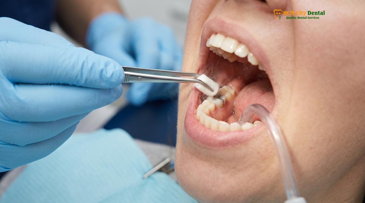
Thermal Testing: How Your Nerve Responds to Extreme Cold (Endo-Ice)
Thermal testing utilizes extreme temperature changes, most commonly a specialized cold refrigerant spray known as Endo-Ice, to trigger a sensory response from the A-delta nerve fibers within the tooth pulp.
Thermal assessment is universally recognized as the cornerstone of endodontic diagnosis. Among the various thermal methods available, the cold thermal test is the most frequently utilized and highly trusted by clinicians. This test typically involves the application of a specialized refrigerant spray, such as dichlorodifluoromethane (commonly referred to by brand names like Endo-Ice), which reaches sub-zero temperatures of approximately -26°C (-15°F). This extreme cold is applied to a small cotton pellet and placed directly onto the buccal (cheek-facing) surface of the tooth in question.
The physiological mechanism behind the cold thermal test is elegantly explained by the hydrodynamic theory of dentinal hypersensitivity, originally proposed by Dr. Martin Brännström. The dentin layer of the tooth, which lies just beneath the enamel, is composed of thousands of microscopic channels called dentinal tubules. These tubules are filled with a fluid that connects the outer environment of the tooth directly to the pulp chamber.
When the extreme cold stimulus is applied to the outer surface of the tooth, it causes a rapid, localized contraction of the fluid within these microscopic tubules. This sudden outward movement of fluid mechanically stimulates the myelinated A-delta nerve fibers located at the pulp-dentin junction. These specific nerve fibers are responsible for transmitting sharp, localized, and immediate pain signals to the brain[2].
“The application of extreme cold remains the gold standard in initial pulp testing, as it directly stimulates the myelinated nerve fibers responsible for sharp, immediate pain perception, providing a clear and immediate window into the neurovascular health of the tooth.”
During the clinical administration of the cold test, the dentist carefully monitors two critical factors: the onset of the sensation and, more importantly, the duration of the lingering effect after the cold pellet is removed. A healthy, normal tooth will register a sharp sensation of cold that dissipates almost immediately—usually within one to two seconds—once the stimulus is taken away. This indicates that the nerve fibers are functioning normally and the pulp is not in a state of chronic inflammation.
If the patient experiences a hyper-exaggerated pain response that lingers for a prolonged period (often 10 to 30 seconds or more) after the cold is removed, it strongly indicates a condition known as irreversible pulpitis. In this state, the pulp is severely inflamed and cannot heal itself. Conversely, if there is absolutely no response to the extreme cold, it suggests that the A-delta fibers have undergone complete necrosis, meaning the nerve tissue is dead.
While cold testing is the primary thermal method, heat testing is occasionally used when a patient specifically complains of severe pain triggered by hot foods or liquids. Heat testing can be performed using heated gutta-percha (a rubber-like dental material) applied to the tooth surface. A severe, lingering pain response to heat is often a late-stage sign of irreversible pulpitis or partial necrosis, as the heat causes gases from dying tissue to expand, creating immense internal pressure.
Electrical Vitality Testing (EPT): Measuring Nerve Conductivity
Electrical testing delivers a gradually increasing micro-current to the tooth structure to determine if the sensory nerve fibers within the pulp chamber can still conduct an electrical impulse.
When thermal testing yields ambiguous results, or when a secondary confirmation is required to solidify a diagnosis of pulpal necrosis, clinicians frequently turn to the electric pulp tester (EPT). This handheld, battery-operated device is specifically designed to deliver a high-frequency, pulsating electrical micro-current to the hard tissues of the tooth. The objective of the EPT is not to measure the health of the blood vessels, but strictly to determine whether the nerve fibers within the pulp are capable of generating an action potential and conducting an electrical impulse to the brain[3].
The clinical protocol for using an electric pulp tester requires meticulous attention to detail, particularly regarding moisture control. The tooth being tested must be completely isolated and dried using cotton rolls and an air syringe. If the tooth is wet, the patient’s saliva can act as a conductor, directing the electrical current away from the tooth and into the adjacent gingiva (gums) or neighboring teeth, which will result in a false-positive reading.
Once the tooth is dry, a conducting medium—typically a small dab of standard toothpaste—is placed on the metal tip of the EPT probe. The probe is then placed firmly against the natural enamel of the tooth, usually on the middle third of the facial surface. To complete the electrical circuit, the patient is often asked to hold a metal wand, or a small lip clip is attached to their mouth. The clinician then slowly increases the electrical current. The patient is instructed to raise their hand or signal the exact moment they feel a tingling, warming, or slight zapping sensation within the tooth.
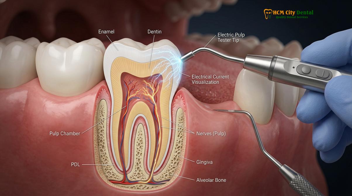
It is important to understand that the EPT primarily stimulates the A-delta fibers, similar to the cold test. It is generally ineffective at stimulating the unmyelinated C-fibers, which are located deeper in the pulp core and are responsible for dull, aching, throbbing pain. Because C-fibers can sometimes survive longer than A-delta fibers in a dying pulp, a tooth might occasionally test negative on an EPT but still contain some partially vital, inflamed tissue deep within the root canals.
While the EPT is an invaluable diagnostic tool, it is subject to both false positives and false negatives, requiring the clinician to interpret the numerical results with a high degree of clinical judgment and skepticism[4].
| Diagnostic Factor | Cold Thermal Test (Endo-Ice) | Electric Pulp Tester (EPT) |
|---|---|---|
| Primary Stimulus | Temperature drop (fluid dynamics) | Electrical micro-current |
| Nerve Fibers Targeted | A-delta fibers (sharp pain) | A-delta fibers primarily |
| Common False Positives | Stimulus touching adjacent gingiva or multi-rooted teeth with partial necrosis | Saliva conduction, metal restorations touching adjacent teeth, anxiety |
| Common False Negatives | Thick porcelain crowns insulating the tooth, calcified canals | Heavy calcification, recent trauma, immature tooth apices in children |
| Clinical Utility | Excellent for differentiating reversible vs. irreversible pulpitis | Excellent for confirming total pulpal necrosis when cold test is negative |
Historically, there were concerns about using electric pulp testers on patients with cardiac pacemakers due to the theoretical risk of electromagnetic interference. However, modern research and updated guidelines generally indicate that contemporary EPT devices are safe for use in patients with modern, shielded pacemakers, though clinicians may still consult with the patient’s cardiologist as a precautionary measure.
Mechanical Testing: Percussion and Palpation Tests for Bone Involvement
Mechanical tests evaluate the periodontal ligament and surrounding alveolar bone rather than the pulp itself, identifying if a pulpal infection has spread beyond the tooth root and into the jawbone.
While thermal and electrical tests focus entirely on the internal environment of the tooth (the pulp), mechanical testing evaluates the external supporting structures. The most common and clinically significant mechanical assessment is the tooth percussive test. This simple yet highly informative procedure involves the clinician gently tapping on the occlusal (biting) or incisal surface of the tooth, typically using the back metal or plastic handle of a standard dental mirror.
It is a common misconception among patients that the percussive test evaluates the vitality of the nerve. In reality, the tooth percussive test does not assess the pulp at all. Instead, it assesses the health and inflammatory status of the periodontal ligament (PDL)—the specialized, fibrous connective tissue that acts as a shock absorber, attaching the tooth root to the surrounding alveolar bone.
When the pulp becomes necrotic and dies, the bacteria within the root canal system multiply rapidly. The bacterial toxins, along with the body’s own inflammatory byproducts, eventually leak out through the apical foramen (the small opening at the very tip of the tooth root). This toxic leakage infiltrates the PDL and the surrounding bone, causing a condition known as symptomatic apical periodontitis[5].
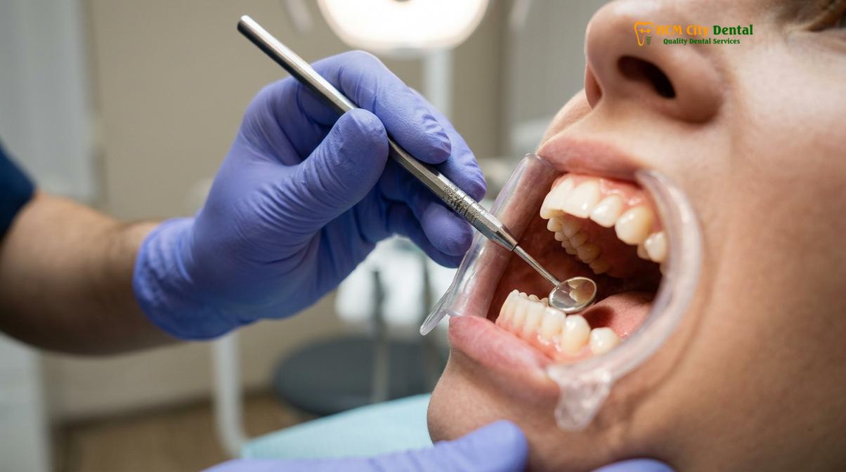
If a patient experiences sharp, localized pain during vertical percussion (tapping straight down on the tooth), it is a strong clinical indicator that the pulpal infection has breached the confines of the tooth and is actively causing inflammation in the periapical tissues. A healthy tooth will feel the pressure of the tap, but it will not register as pain.
In conjunction with percussion, clinicians routinely perform palpation tests. This involves the dentist applying firm digital pressure (using their index finger) to the mucosal tissues and gums overlying the root apex of the suspected tooth. If the patient feels tenderness or if the clinician feels a swelling or bony expansion under the gums, it further confirms that the infection has spread into the surrounding jawbone, often indicating the presence of an apical abscess.
The Test Cavity Method: The Ultimate Vitality Check (Rarely Used)
The test cavity method involves drilling a small preparation into the dentin without anesthesia to check for a pain response, a technique reserved only for complex cases where all other diagnostic tests yield inconclusive results.
In modern endodontic practice, the vast majority of diagnoses can be confidently and accurately established using a combination of patient history, high-resolution radiographic imaging, thermal testing, and electrical assessment. However, there are rare, highly complex clinical scenarios where all non-invasive tests yield ambiguous, conflicting, or entirely inconclusive results. This diagnostic dilemma most frequently occurs in teeth that are heavily restored with full-coverage dental crowns, particularly those made of thick porcelain, zirconia, or porcelain-fused-to-metal (PFM).
These heavy restorative materials act as perfect thermal and electrical insulators. They prevent the extreme cold of the Endo-Ice or the micro-current of the EPT from ever reaching the underlying natural tooth structure, resulting in a false-negative reading even if the pulp is perfectly healthy and vital. When a patient is experiencing severe, unlocalized, radiating pain, and the clinician cannot definitively identify the source tooth using standard methods, they may resort to the test cavity method.
The test cavity method is considered the ultimate, definitive test for pulpal vitality, but it is highly invasive. The procedure involves using a high-speed dental handpiece equipped with a small round bur to drill a tiny cavity directly through the enamel or the restorative material, penetrating just into the underlying dentin. Crucially, this procedure is initiated without the administration of local anesthesia.
“While non-invasive diagnostics resolve the vast majority of clinical cases, the test cavity method remains a definitive, albeit rare, diagnostic tool when heavily restored teeth completely mask underlying pulpal necrosis, allowing the clinician to confirm the diagnosis before committing to irreversible endodontic therapy.”
The objective of drilling without anesthesia is to elicit a direct response from the dentinal fluid and the underlying nerve fibers. The clinician drills very slowly and carefully. If the patient feels a sudden, sharp pain as the bur enters the dentin, the procedure is immediately halted. This positive pain response definitively confirms that the pulp is still vital and capable of transmitting sensory signals. The small test cavity is then simply restored with a standard composite filling.
Conversely, if the clinician can drill deep into the dentin—and even approach the pulp chamber itself—without the patient feeling any sensation whatsoever, it is a definitive, undeniable confirmation that the pulp is completely necrotic. At this point, the clinician can confidently administer local anesthesia and proceed directly into full root canal therapy, knowing they have accurately identified the source of the infection.
Interpreting Results: Hyper-response vs. No Response (Necrotic Pulp)
Accurate diagnosis relies on comparing the tested tooth’s sensory response against a healthy control tooth, allowing the clinician to categorize the pulp as normal, reversibly inflamed, irreversibly inflamed, or necrotic.
The true art and science of endodontic diagnosis lie not just in the mechanical execution of the tests, but in the accurate interpretation of the patient’s subjective responses. Because pain tolerance and nerve sensitivity vary wildly from person to person, a test result on a single tooth is meaningless in isolation. According to diagnostic frameworks aligned with the Vietnam Odonto-Stomatology Association (VOSA), interpreting these results requires first testing a healthy, asymptomatic control tooth (usually the same tooth on the opposite side of the mouth) to establish a baseline normal response for that specific individual[6].
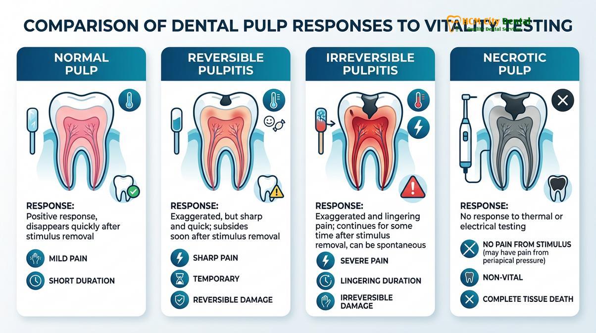
Once the baseline is established, the clinician compares the response of the suspected tooth to the control tooth. Based on these comparative results, the pulpal status is categorized into one of four primary diagnostic states, which directly dictate the required treatment:
- Normal Pulp: The tooth responds to cold or electrical stimuli with a mild, transient sensation that disappears within one to two seconds after the stimulus is removed. There is no pain on percussion or palpation. No treatment is required.
- Reversible Pulpitis: The tooth exhibits a sharp, heightened response to cold, but the pain still dissipates quickly (within a few seconds) once the stimulus is removed. This indicates mild inflammation, often caused by a recent filling or early decay. The pulp can typically heal itself if the irritant is removed and the tooth is restored conservatively.
- Irreversible Pulpitis: The tooth exhibits a severe, exaggerated, and often agonizing response to cold or heat. Crucially, the pain lingers for a prolonged period (often 30 seconds to several minutes) after the stimulus is removed. The pulp is damaged beyond repair, is actively dying, and requires immediate endodontic intervention to relieve the pain.
- Necrotic Pulp: The tooth yields absolutely no response to extreme cold or maximum electrical current. The nerve tissue is dead. This is often accompanied by a positive (painful) response to percussion, indicating that the infection has spread to the bone. A root canal or extraction is mandatory to remove the necrotic debris.
When to See a Doctor for Pulp Vitality Assessment
Recognizing the early warning signs of pulpal distress can help you seek timely professional care, potentially saving your natural tooth from extraction and preventing systemic infection.
Understanding when to seek professional dental evaluation is crucial for preserving your oral health and avoiding unnecessary pain. You should schedule an appointment for a comprehensive vitality assessment if you experience prolonged sensitivity to hot or cold foods, especially if the pain lingers long after the temperature trigger has been swallowed or removed from your mouth. Spontaneous throbbing pain that occurs without any apparent cause, or pain that wakes you up in the middle of the night, are strong, classic indicators of irreversible nerve damage that requires immediate attention.
Additionally, if you notice localized swelling in your gums (often appearing like a small pimple on the gum line), a persistent bad taste or odor in your mouth, or sharp pain when biting down or tapping on a specific tooth, these are definitive signs that an infection may have already spread beyond the pulp chamber and into the surrounding bone. Early diagnostic testing allows your dentist to identify the exact source of the problem and intervene before the infection causes extensive, irreversible damage to the supporting jawbone.

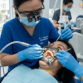
Dr. Nguyen Van Cuong, a leading specialist in restorative and endodontic care, emphasizes that accurate vitality testing is the absolute cornerstone of successful Root Canal therapy. By meticulously evaluating nerve health and utilizing a combination of thermal, electrical, and mechanical diagnostics, Dr. Cuong ensures that patients only receive endodontic intervention when it is clinically necessary, prioritizing the preservation of natural tooth structure and ensuring long-term functional success.
References
- Journal of Endodontics. Diagnostic accuracy of cold and electrical tests in determining pulp vitality. (2020).
- International Endodontic Journal. The hydrodynamic theory of dentinal hypersensitivity and pulp testing. (2019).
- Journal of the American Dental Association. Clinical evaluation of pulp and periapical disease. (2021).
- Clinical Oral Investigations. False positives and false negatives in electric pulp testing. (2018).
- British Dental Journal. Endodontic diagnosis: the role of thermal and mechanical testing. (2022).
- Vietnam Odonto-Stomatology Association (VOSA). National guidelines on endodontic diagnostics and pulp vitality assessment. (2023).
If you are experiencing severe tooth pain, prolonged sensitivity to temperature, or discomfort when chewing, accurate diagnosis is the critical first step toward lasting relief. Contact HCMC Dental Clinic in Ho Chi Minh City today to schedule a comprehensive vitality evaluation and discuss whether a Root Canal or other targeted restorative treatment is the right solution to restore your oral health.