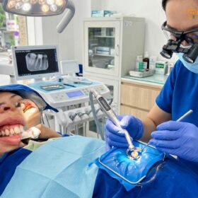
A microscope root canal in Vietnam utilizes advanced dental magnification to treat complex endodontic infections with extreme precision. By illuminating hidden canals and micro-fractures, this specialized procedure significantly increases tooth preservation rates compared to traditional methods, offering international patients world-class clinical outcomes at highly accessible costs.
Clinical Summary:
Microscope-assisted endodontics represents the gold standard in modern root canal therapy. Utilizing high-powered magnification (up to 25x) and intense coaxial illumination, endodontists can visualize the intricate internal anatomy of the tooth root system. This technology is critical for identifying accessory canals, navigating severe curvatures, and managing complex anatomical variations that are invisible to the naked eye. At HCMC Dental Clinic, the integration of microscopic technology with 3D CBCT imaging ensures thorough debridement and precise obturation, minimizing the risk of reinfection and maximizing the long-term survival of the natural tooth structure. This visual approach shifts endodontics from a tactile, blind procedure to a highly predictable microsurgical discipline.
Key Takeaways:
- Magnification up to 25x reveals hidden anatomy, including the frequently missed MB2 canal in maxillary molars.
- Intense coaxial illumination allows for the safe retrieval of fractured instruments and precise repair of root perforations.
- Enhanced visualization significantly reduces the removal of healthy dentin, preserving the tooth’s structural integrity.
- Clinical documentation through integrated cameras provides transparent treatment verification for patients.
- Microscope-assisted procedures offer higher long-term success rates compared to traditional tactile-based endodontics.
Why Magnification is Essential in Modern Endodontics
Dental operating microscopes provide up to 25x magnification and intense coaxial lighting, allowing endodontists to transition from tactile guesswork to precise visual control during complex root canal therapy.
The human tooth is a marvel of biological engineering, but its internal anatomy presents significant challenges for dental professionals. The pulp chamber and the root canal system are microscopic spaces encased in hard dentin. Historically, advanced root canal therapy was performed largely by tactile sensation—dentists relied on the “feel” of the instruments as they navigated the dark, narrow canals. While this traditional approach has saved millions of teeth, it inherently carries a higher risk of missing complex anatomical structures, leading to persistent infections and treatment failures.
The introduction of the dental operating microscope revolutionized the field of endodontics. Unlike standard dental loupes, which typically offer 2.5x to 4.5x magnification, a clinical microscope provides adjustable magnification ranging from 3x for general orientation up to 25x for detailed inspection of the apical third of the root[1]. This extreme magnification is coupled with coaxial illumination—a specialized lighting system where the light source runs parallel to the clinician’s line of sight. This eliminates shadows and projects intense, focused light deep into the narrowest root canals, illuminating areas that were previously shrouded in darkness.
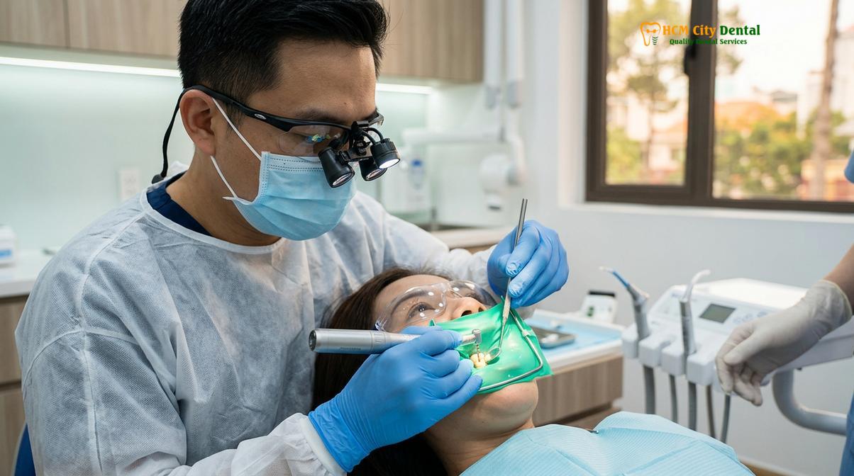
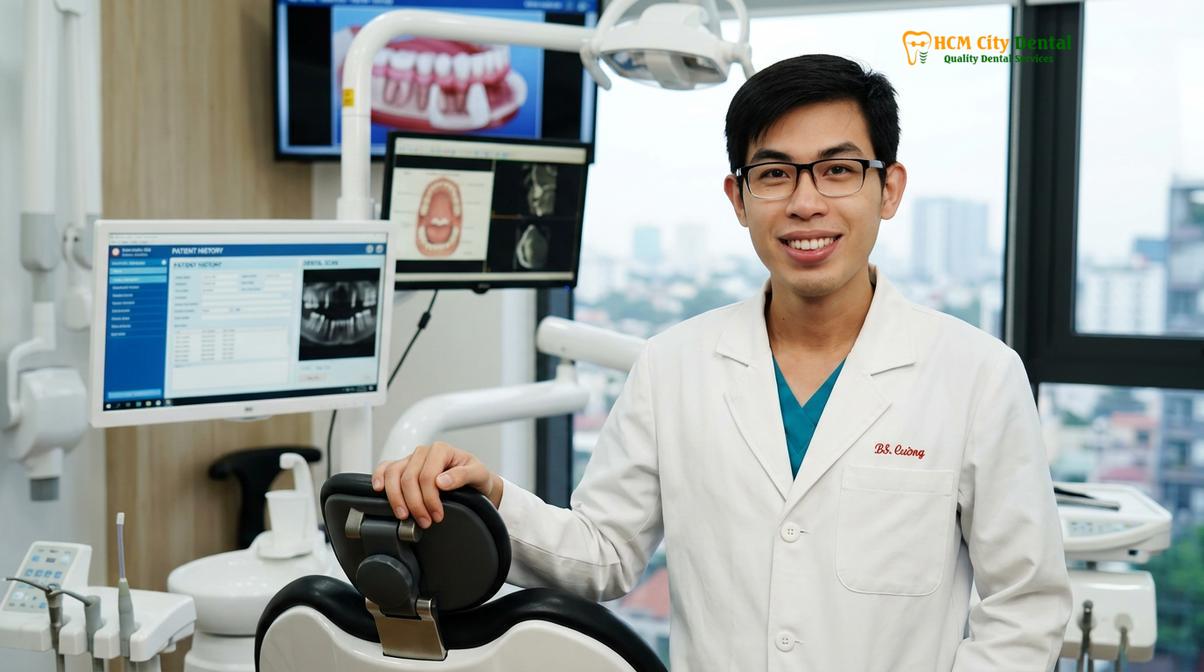
According to Dr. Nguyen Van Cuong, a leading specialist at HCMC Dental Clinic, the transition from tactile-based to visually-guided endodontics is the most significant advancement in saving severely compromised teeth. When a clinician can clearly see the pulpal floor, they can practice minimally invasive endodontics. Instead of removing large amounts of healthy tooth structure to gain access and visibility, the dentist can create a conservative access cavity. This preservation of pericervical dentin—the crucial tooth structure near the gumline—is vital for the long-term structural integrity of the tooth, reducing the risk of future fractures.
“The integration of high-powered magnification in endodontics has fundamentally shifted our clinical paradigm. We are no longer operating in the dark; we can visually confirm the complete removal of necrotic tissue and the precise sealing of the root canal system, dramatically improving the predictability of our outcomes.”
Furthermore, the microscope aids in the identification of micro-fractures and cracks within the tooth structure. A tooth may present with symptoms of irreversible pulpitis, but if a vertical root fracture extends deep into the canal, the tooth may be structurally unsavable. Identifying these fractures early under high magnification prevents the patient from undergoing a futile root canal procedure, allowing the clinician to pivot to alternative treatment plans, such as extraction and dental implant placement, in a timely manner.
Under the Microscope: Locating Hidden and Accessory Canals
High-powered magnification is crucial for identifying microscopic accessory canals, isthmuses, and apical ramifications that harbor infection and cause treatment failure if left unsealed.
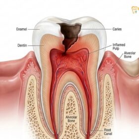
A common misconception among patients is that a tooth root contains a single, straight tube resembling a plumbing pipe. In reality, the root canal system is a highly complex, branching network more akin to the root system of a tree. Alongside the main canals, there are often lateral canals, accessory canals, fins, webs, and isthmuses (narrow connections between two main canals). When the dental pulp becomes infected, bacteria and their associated biofilms infiltrate this entire microscopic labyrinth[2].
If a dentist only cleans and seals the main canals, leaving the accessory anatomy untouched, the remaining bacteria will eventually multiply, leading to a recurrent periapical infection (a dental abscess). This is where the utilization of a high-end Zeiss dental microscope becomes clinically indispensable. Under intense magnification, the clinician can meticulously inspect the pulpal floor for the tiny orifices of these accessory canals. They often appear as subtle color changes in the dentin or tiny bleeding points that are entirely invisible to the naked eye.
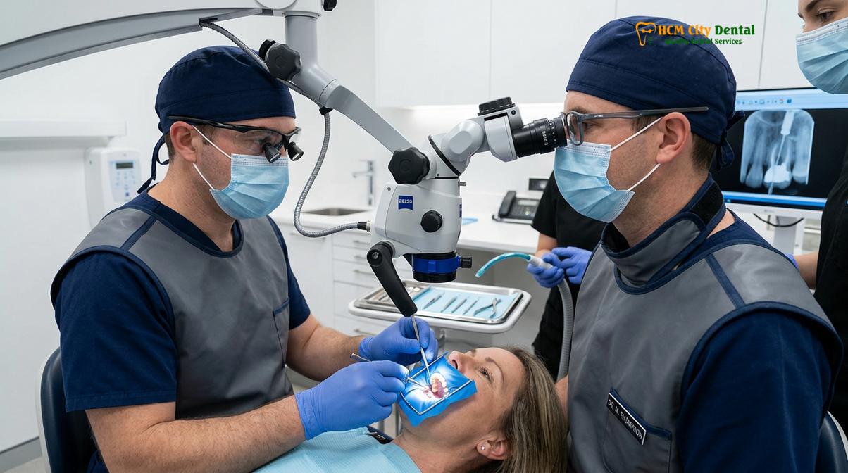
Once identified, these hidden canals must be carefully negotiated and cleaned. Because they are often extremely narrow and tortuous, standard endodontic files may not easily penetrate them. Clinicians utilize specialized ultrasonic instruments under microscopic guidance to gently trough the dentin, removing microscopic calcifications and opening the pathway to the hidden anatomy. This process requires a delicate touch and continuous visual feedback to avoid perforating the side of the root.
The microscope also plays a critical role during the irrigation phase of the root canal. Chemical disinfection using solutions like sodium hypochlorite is essential for dissolving organic tissue and killing bacteria in areas that mechanical files cannot reach. Under the microscope, the clinician can observe the acoustic streaming and cavitation effects of ultrasonic irrigation, visually confirming that the disinfecting solutions are actively penetrating the complex web of the root canal system, ensuring a thoroughly debrided environment before the final filling material is placed.
The Elusive MB2 Canal: The Primary Cause of Molar Treatment Failure
The second mesiobuccal (MB2) canal in upper molars is present in a vast majority of cases but is frequently missed without a microscope, leading to persistent infection.
When discussing endodontic complexity, maxillary (upper) first and second molars present the greatest anatomical challenges. Historically, these teeth were taught to have three roots and three canals. However, extensive clinical research and the widespread adoption of microscopic endodontics have revealed that a fourth canal—the second mesiobuccal or MB2 canal—is present in up to 90% of maxillary first molars and a significant percentage of second molars[3].
The missed MB2 canal is widely recognized in endodontic literature as the leading cause of treatment failure and the primary reason patients require root canal retreatment in upper molars. The difficulty lies in its location and size. The orifice of the MB2 canal is typically located on the pulpal floor, slightly palatal to the main mesiobuccal (MB1) canal. However, it is frequently hidden beneath a shelf of dentin or heavily calcified, making it virtually undetectable without specialized equipment.
Clinical Case Review: Managing a Missed MB2 Canal
A patient presented to HCMC Dental Clinic in Ho Chi Minh City complaining of persistent, dull aching in an upper right molar that had undergone root canal therapy two years prior at a different facility. A 3D CBCT scan revealed a periapical radiolucency (bone infection) associated with the mesiobuccal root. Dr. Nguyen Van Cuong initiated retreatment under the operating microscope. By carefully removing a small dentinal triangle using ultrasonic tips at 16x magnification, a previously untreated, highly infected MB2 canal was located, cleaned, and sealed. The patient’s symptoms resolved completely within days, and follow-up imaging confirmed bone healing, successfully saving the natural tooth.
The clinical workflow for locating the MB2 canal requires patience and precision. After locating the primary canals, the clinician uses the microscope to trace the developmental groove connecting the MB1 and palatal canals. Using specialized ultrasonic tips, the clinician carefully shaves away the overlying dentin layer by layer. This must be done under high magnification to differentiate between the lighter-colored secondary dentin and the darker pulpal floor, ensuring that the search for the canal does not result in an accidental perforation of the tooth root.
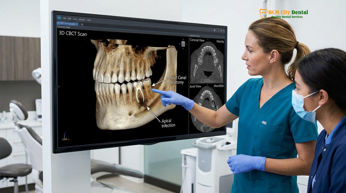
Once the MB2 orifice is exposed, negotiating it presents another hurdle. These canals are often extremely narrow, curved, and may merge with the MB1 canal or exit through a separate apical foramen. The use of highly flexible, heat-treated nickel-titanium (NiTi) rotary files, combined with continuous microscopic visualization, allows the endodontist to safely navigate and shape this elusive canal, ensuring comprehensive disinfection of the molar’s root system.
Micro-Surgical Applications: Removing Broken Instruments and Repairing Perforations
Microscopic visualization combined with specialized ultrasonic tips enables the safe retrieval of separated files and the precise sealing of iatrogenic root perforations using biocompatible materials.
Endodontic therapy involves working with extremely fine, delicate instruments inside narrow, curved spaces. Despite the best techniques and modern metallurgy, the separation (breakage) of an endodontic file inside a root canal is a known clinical complication. When a file fractures, it blocks the canal, preventing thorough disinfection and sealing of the space beyond the fragment. In the past, a separated instrument often meant the tooth had to be extracted or subjected to invasive apical surgery.
Today, fractured instrument retrieval is a highly specialized micro-surgical procedure made possible by the dental operating microscope. The clinician first establishes straight-line access to the fragment. Under high magnification (often 15x to 20x), the clinician can actually see the top of the broken file deep within the canal. Using ultra-fine ultrasonic tips, the dentist carefully removes a microscopic amount of dentin around the fragment to create a staging platform. The ultrasonic vibration is then applied directly to the file, loosening it from the canal walls until it can be safely flushed out or grasped with micro-forceps.
Another complex scenario managed under the microscope is the repair of root perforations. A perforation occurs when an instrument accidentally breaches the side of the tooth root, creating an artificial communication between the root canal system and the surrounding periodontal tissues. If left untreated, a perforation will rapidly lead to severe inflammation, bone loss, and eventual tooth loss.
Repairing a perforation requires absolute visual control. The clinician must clean the perforation site without pushing debris into the surrounding bone. Under the microscope, biocompatible materials—such as Mineral Trioxide Aggregate (MTA) or advanced bioceramic putties—are meticulously packed into the defect[4]. These materials are highly tissue-tolerant and promote the regeneration of bone and periodontal ligaments. The microscope ensures that the bioceramic material is placed exactly at the site of the defect, achieving a fluid-tight seal without blocking the main root canal pathway.
Enhanced Ergonomics and Documentation for Patient Verification
Dental microscopes improve the clinician’s posture during lengthy procedures and allow for high-resolution video documentation, enhancing patient communication and clinical transparency.
While the clinical benefits of magnification for the patient are profound, the ergonomic benefits for the dental practitioner are equally significant. Dentistry is a physically demanding profession, and traditional endodontics often requires the clinician to bend over the patient, contorting their neck and back to peer into the oral cavity. Over time, this poor posture leads to severe musculoskeletal disorders, fatigue, and a potential decrease in clinical precision during long, complex procedures.
The operating microscope fundamentally changes the clinician’s posture. The dentist sits completely upright, looking straight ahead into the binoculars, while the microscope’s optics and angled mirrors bring the surgical field directly to their eyes. This ergonomic advantage means the clinician remains comfortable, focused, and steady, even during a two-hour molar retreatment. This sustained focus directly translates to safer, more precise care for the patient.
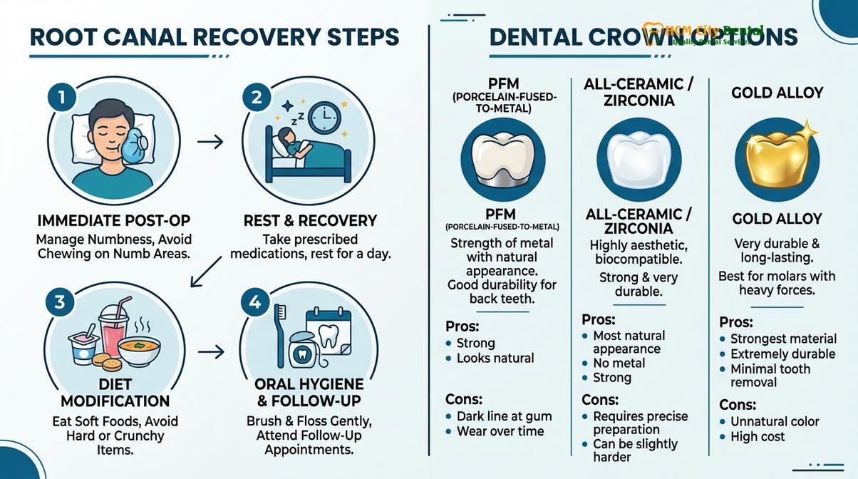
Furthermore, modern dental microscopes are equipped with integrated high-definition cameras and video recording systems. This technology fosters an unprecedented level of transparency and patient education. International patients traveling to Saigon for dental care highly value this documentation. The clinician can capture images of the hidden canals, the retrieved instrument, or the repaired crack, and display them on a monitor.
| Feature | Traditional Endodontics | Microscope-Assisted Endodontics |
|---|---|---|
| Visual Record | Limited to 2D X-rays before and after treatment. | High-resolution intra-canal photos and live video feeds. |
| Patient Communication | Verbal explanations of complex internal anatomy. | Visual proof of cracks, extra canals, and clean preparations. |
| Diagnostic Verification | Relies on tactile feedback and radiographic interpretation. | Direct visual confirmation of tissue removal and sealing. |
| Referral Collaboration | Basic written reports and standard radiographs. | Detailed visual reports for comprehensive multidisciplinary care. |
Showing a patient the internal reality of their tooth demystifies the procedure. When a patient can see the hairline crack that is causing their pain, or the extra MB2 canal that was previously missed, they gain a deeper understanding of their diagnosis and the value of the specialized treatment they are receiving. This visual evidence builds profound trust between the patient and the dental team.
Microscope-Assisted Endodontics vs. Traditional Blind Procedures
Traditional endodontics relies heavily on tactile sensation and 2D imaging, whereas microscope-assisted therapy provides direct visual confirmation, significantly improving the thoroughness of disinfection and long-term tooth survival.
To fully appreciate the value of microscope root canal therapy, it is helpful to contrast it directly with traditional methods. In a traditional “blind” procedure, the dentist relies on their knowledge of average tooth anatomy and the tactile feedback from their instruments. They use 2D X-rays to estimate the length and curvature of the roots. While this method is often successful for straightforward, single-rooted front teeth, it becomes highly unpredictable when dealing with the complex, multi-rooted anatomy of molars.
Traditional access cavities are often cut larger to allow more light into the tooth, which unnecessarily weakens the crown. Furthermore, without magnification, it is nearly impossible to visually confirm that all necrotic pulp tissue, bacteria, and previous filling materials (in the case of retreatments) have been completely removed from the canal walls. The clinician must assume the canal is clean based on the protocol followed, rather than direct visual evidence.
“The difference between traditional and microscopic endodontics is the difference between navigating a complex maze in the dark by feeling the walls, versus walking through the same maze with a high-powered flashlight and a detailed map. The visual confirmation ensures no anatomical variation is left untreated.”
Microscope-assisted endodontics, especially when combined with 3D Cone Beam Computed Tomography (CBCT), eliminates this guesswork. The CBCT scan provides the “map” of the root canal system, revealing the exact number of canals, their curvatures, and any surrounding bone pathology before the procedure even begins. The microscope then provides the “flashlight,” allowing the clinician to execute the treatment plan with pinpoint accuracy.
This combination of 3D imaging and microscopic visualization allows for directed dentin conservation. The clinician only removes the exact amount of tooth structure necessary to access the infected areas. The thoroughness of the disinfection is visually verified, and the final obturation (filling) of the canals is performed under direct sight, ensuring a dense, void-free seal that prevents bacterial reinfection. Consequently, the long-term survival rate of teeth treated with this advanced microsurgical approach is exceptionally high, providing patients with a reliable foundation for their final dental crown restorations.
When to See a Doctor for Endodontic Evaluation
Patients experiencing severe spontaneous pain, prolonged sensitivity to temperature, or swelling around a previously treated tooth should seek immediate evaluation by an endodontic specialist.
Recognizing the signs of pulpal infection or the failure of a previous root canal is crucial for timely intervention. Dental pulp does not heal itself once it becomes irreversibly inflamed or necrotic. Delaying treatment allows the bacterial infection to spread from the confines of the tooth root into the surrounding jawbone, creating a periapical abscess.
Important Clinical Warning: An untreated dental abscess is not merely a localized tooth problem; it is an active bacterial infection in the bone. If left untreated, the infection can spread to adjacent anatomical spaces, potentially causing severe facial swelling, systemic illness, and in rare cases, life-threatening complications. Prompt clinical evaluation is mandatory.
You should schedule a consultation with a dental professional if you experience any of the following symptoms:
- Spontaneous Pain: A severe, throbbing toothache that occurs without any trigger, often waking you up at night.
- Prolonged Sensitivity: Pain from hot or cold foods that lingers long after the stimulus is removed.
- Pain on Biting: Sharp pain when applying chewing pressure to a specific tooth, which may indicate an infection at the root tip or a cracked tooth.
- Gum Swelling: A raised, pimple-like bump on the gums (a sinus tract) that may occasionally drain pus and leave a bad taste in the mouth.
- Discoloration: A tooth that gradually darkens or turns gray, indicating that the internal nerve tissue has died following trauma.
If you have a tooth that was previously treated but is now symptomatic again, a specialized evaluation is highly recommended. At HCMC Dental Clinic, our specialists utilize 3D imaging and microscopic examination to determine the exact cause of the failure and assess whether the tooth is a viable candidate for microsurgical retreatment. A personalized clinical examination is the only way to establish an accurate diagnosis and formulate a safe, effective treatment plan.
Frequently Asked Questions
Find clear, expert answers to the most common questions patients have regarding microscope-assisted root canal therapy and endodontic care in Vietnam.
Does every dentist use a microscope for root canals in Vietnam?
No, not every dentist in Vietnam uses a microscope for root canals, as it requires specialized training and significant capital investment. This advanced equipment is typically reserved for endodontic specialists and high-tier dental institutions focused on complex tooth preservation and retreatment cases. General dental clinics often rely on standard loupes or the naked eye for routine procedures.
How much extra does microscope endodontics cost in HCMC?
Microscope-assisted root canals in HCMC typically cost moderately more than traditional treatments, reflecting the advanced technology and specialist expertise required. However, this initial investment is highly cost-effective when it prevents the need for future retreatments, extractions, or expensive dental implant replacements. The precise cost varies based on the tooth’s anatomical complexity and the number of canals involved.
Can a microscope save a tooth that was declared unsavable?
Yes, a microscope can often save teeth deemed unsavable by identifying hidden canals or allowing for the repair of complex root perforations. By providing extreme magnification, specialists can address micro-anatomical issues that traditional methods cannot. However, teeth with severe vertical root fractures or catastrophic loss of structural dentin generally remain unsavable regardless of the magnification used.
Is a microscope root canal procedure painful?
No, a microscope root canal is not painful, as it is performed under profound local anesthesia using modern delivery techniques. The extreme precision afforded by the microscope actually minimizes trauma to surrounding healthy tissues, often resulting in a more comfortable postoperative recovery. Patients typically experience immense relief from their pre-existing toothache once the infected tissue is removed[5].
How long does a microscope-assisted root canal take?
A microscope-assisted root canal typically takes between 60 to 90 minutes per session, depending on the anatomical complexity and the number of canals present. Complex molar retreatments or cases involving fractured instrument retrieval may require multiple extended appointments to ensure complete disinfection and healing before the final restoration is placed. Your specialist will outline the expected timeline during your consultation.
References
- Journal of Endodontics. Outcomes of root canal treatment and restoration longevity. (2020).
- International Endodontic Journal. Biofilms in endodontics and disinfection strategies. (2021).
- Journal of the American Dental Association. Microscope-assisted endodontics and missed canals. (2019).
- Clinical Oral Investigations. Biocompatibility of bioceramic root canal sealers. (2022).
- British Dental Journal. Pain management and dental anxiety in endodontics. (2018).