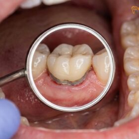
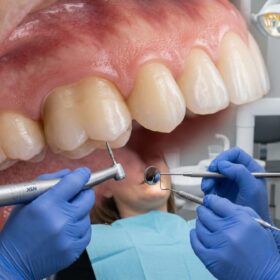
A dental crown is typically required after a root canal to restore structural integrity, prevent bacterial microleakage, and protect the weakened tooth from fracturing under chewing forces. While anterior teeth may sometimes only need a filling, molars and premolars almost universally require full-coverage crowns for long-term survival.
Clinical Summary:
Following endodontic therapy, a tooth undergoes significant biomechanical and histological changes. The removal of the internal neurovascular bundle (the pulp) deprives the dentin of its natural moisture supply, altering the collagen cross-linking and reducing the tooth’s modulus of elasticity. Furthermore, the physical access cavity required to perform the root canal inherently compromises the structural roof of the pulp chamber, drastically reducing the tooth’s resistance to occlusal forces. To mitigate the high risk of catastrophic vertical root fractures and to establish a hermetic seal against oral bacteria, prosthodontic intervention is necessary. A full-coverage dental crown acts as a protective ferrule, binding the remaining tooth structure together, distributing masticatory loads evenly, and ensuring the long-term clinical success of the underlying endodontic treatment.
Key Takeaways:
- Root-canaled teeth lose their internal blood supply, making the dentin more susceptible to structural fatigue over time.
- Posterior teeth (molars and premolars) bear the brunt of chewing forces and almost always require a crown after endodontic therapy.
- A crown provides a crucial “ferrule effect,” acting like a physical hoop that prevents the tooth from splitting under pressure.
- Coronal microleakage—bacteria seeping through a temporary filling—is a leading cause of root canal failure, which a permanent crown prevents.
- Delaying the placement of a permanent crown significantly increases the risk of irreversible tooth fracture and the need for extraction.
Loss of Vitality: Why a Root-Canaled Tooth Becomes Dehydrated and Brittle
Removing the dental pulp eliminates the tooth’s blood supply, leading to a gradual loss of internal moisture and a reduction in the dentin’s natural flexibility.
To fully comprehend why a root canal treatment is usually followed by the placement of a dental crown, one must first understand the profound histological and biomechanical changes that occur within the tooth once the pulp is removed. The dental pulp is the living heart of the tooth, housing a complex network of nerves, blood vessels, and connective tissues. Its primary function during the developmental stages of life is to form dentin, the hard tissue that makes up the bulk of the tooth structure. In a mature tooth, the pulp continues to serve a vital role by providing hydration and sensory feedback.
When a tooth becomes infected or deeply decayed, endodontic therapy is performed to meticulously clean out this necrotic or inflamed tissue. While this procedure is highly effective at eliminating pain and eradicating infection, it inherently leaves the tooth pulpless, or “non-vital.” The immediate consequence of this loss of vitality is the cessation of fluid flow through the dentinal tubules. In a healthy, vital tooth, odontoblastic processes extend into these microscopic tubules, maintaining a constant outward flow of dentinal fluid that keeps the collagen matrix of the dentin hydrated and resilient.
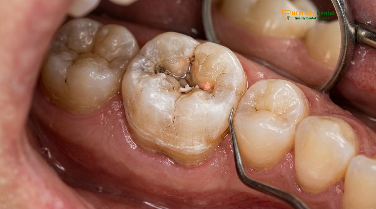
Without this continuous hydration, the dentin undergoes a slow but progressive desiccation process. The collagen fibers within the dentin matrix, which normally provide the tooth with a degree of tensile strength and flexibility, begin to degrade and lose their cross-linking integrity. As the moisture content drops, the modulus of elasticity of the tooth changes. It transforms from a dynamic, shock-absorbing structure into a more rigid and unyielding one. This histological shift significantly increases the risk of a brittle tooth fracture, especially when the tooth is subjected to the repetitive, high-impact forces of daily mastication.
Furthermore, the architectural compromise extends beyond mere dehydration. The very act of accessing the root canal system requires the dentist to drill through the occlusal (biting) surface of the tooth, removing the roof of the pulp chamber. The pulp chamber roof acts much like the keystone of an arch; once it is removed, the structural integrity of the entire coronal portion of the tooth is severely diminished. The remaining walls of the tooth are left unsupported, making them highly susceptible to flexing and ultimately fracturing under load. Dr. Nguyen Van Cuong frequently emphasizes to patients that a root canal saves the foundation of the tooth, but it is the crown that rebuilds and protects the house.
The Mechanics of Chewing: Protecting the Cusp from High Vertical Forces
Molars endure immense occlusal pressure during mastication, and a crown distributes these forces evenly to prevent the unsupported cusps from snapping under the strain.
The human masticatory system is an incredibly powerful biomechanical engine. The muscles of mastication, particularly the masseter and temporalis muscles, are capable of generating extraordinary forces during the chewing cycle. In the posterior region of the mouth—where the molars and premolars reside—these forces are magnified. A healthy adult can exert anywhere from 200 to 600 Newtons of force on their back teeth during normal chewing, and this number can spike significantly higher during episodes of bruxism (teeth grinding) or when accidentally biting down on a hard object like a seed or a piece of ice.
When a tooth is intact, its natural anatomy is perfectly designed to handle these loads. The cusps (the raised points on the biting surface) and the ridges work in harmony to crush and grind food, while the intact enamel and dentin distribute the stress down through the roots and into the surrounding alveolar bone. However, an endodontically treated tooth has lost a significant portion of its central core. The structural void left by the access cavity and the removal of decayed tissue means that the remaining cusps are no longer tied together by a solid central mass.
“The biomechanical behavior of an endodontically treated tooth is fundamentally altered. Without the central structural integrity provided by the pulp chamber roof and the internal dentin, the remaining cusps act as independent, unsupported cantilevers that are highly vulnerable to shear forces and catastrophic failure.”
When vertical forces are applied to these unsupported cusps, they tend to deflect outward. This phenomenon, known as cuspal flexure, creates immense stress concentrations at the base of the cusps, near the gumline. Over time, this repetitive micro-bending leads to material fatigue within the dentin. Micro-cracks begin to propagate, often unnoticed by the patient because the tooth lacks a nerve to signal pain. Eventually, a critical threshold is reached, and a piece of the tooth fractures off. If the fracture is superficial, the tooth might still be salvageable. However, if the fracture propagates subgingivally (below the gumline) or splits the root, the tooth is deemed non-restorable and must be extracted.
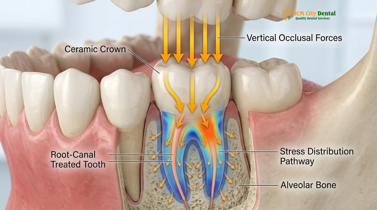
To counteract this dangerous biomechanical reality, a full-coverage dental crown is employed. A crown is designed to encapsulate the entire visible portion of the tooth, effectively tying the remaining cusps together. By covering the occlusal surface entirely, the crown alters the way forces are transmitted. Instead of the cusps being wedged apart by the opposing teeth, the crown absorbs the impact and distributes the vertical loading strength evenly across the entire remaining tooth structure and down the long axis of the root. This protective mechanism is absolutely critical for the survival of posterior teeth that have undergone root canal therapy.
Structural Integrity: How a Crown Binds the Tooth Together to Prevent Splitting
A full-coverage crown acts like a protective helmet, creating a ferrule effect that binds the remaining tooth structure together and prevents catastrophic vertical splitting.
The concept of structural integrity in post-endodontic restorations revolves heavily around a prosthodontic principle known as the “ferrule effect.” The term ferrule comes from the Latin words *ferrum* (iron) and *viria* (bracelet), and in engineering, it refers to a metal ring or cap placed around a shaft to reinforce it and prevent it from splitting. In dentistry, a ferrule is a band of restorative material—specifically, the margins of the dental crown—that completely encircles the external surface of the remaining healthy tooth structure.
For a crown to be highly effective, it must grasp a minimum of 1.5 to 2.0 millimeters of solid, healthy dentin above the gumline, extending 360 degrees around the tooth. This collar of tooth structure provides the necessary resistance form to counteract lateral (side-to-side) forces. When a patient chews, the jaw does not merely move up and down; it moves in a complex, multi-directional pattern that introduces significant shear and tipping forces to the teeth. Without an adequate ferrule, these lateral forces would be transmitted directly to the core buildup or the root itself, drastically increasing the likelihood of a root fracture or the dislodgement of the restoration.
Clinical Case Review: HCMC Dental Clinic
A 45-year-old patient presented to HCMC Dental Clinic in Ho Chi Minh City with a history of a root canal performed on a lower first molar three years prior. The patient had opted for a large composite filling instead of a crown due to time constraints. Upon clinical examination and CBCT imaging, Dr. Cuong identified a deep vertical fracture extending from the central fossa down into the mesial root, caused by years of unprotected occlusal loading. Unfortunately, the tooth was non-restorable and required extraction followed by implant placement. This case strongly highlights the critical necessity of full-coverage protection for endodontically treated posterior teeth.
To prepare a tooth for a crown, the dentist must first rebuild the missing internal structure. This is achieved through a procedure known as a core buildup. Using advanced composite resins, the dentist fills the void left by the root canal access cavity, creating a solid foundation. In cases where the tooth structure is severely compromised, a post may be cemented into one of the cleaned root canals to help anchor the core material. However, it is crucial to understand that a post does not strengthen the root; its sole purpose is to retain the core buildup. The true strength and protection come from the overlying crown.
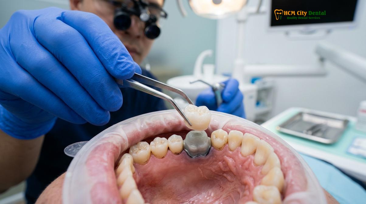
Once the core is established, the dentist meticulously shapes the outer surface of the tooth, creating a precise margin at or slightly below the gumline. The crown is then custom-fabricated to fit intimately over this preparation. When the crown is permanently cemented into place, it acts as an external splint. It creates a state of “hoop stress,” tightly binding the remaining walls of the tooth and the core buildup into a single, cohesive unit. This unified structure is vastly superior at resisting the complex biomechanical forces of the oral environment compared to a tooth restored with merely an intracoronal (inside the tooth) filling.
Microleakage Prevention: The Crown as a Final Seal Against Saliva Bacteria
Beyond physical protection, a crown provides an essential hermetic seal that stops oral bacteria from reinfecting the vulnerable root canal system.
While the biomechanical protection offered by a crown is often the most discussed benefit, its role in maintaining the biological success of the root canal is equally, if not more, important. The oral cavity is a hostile environment, teeming with billions of bacteria representing hundreds of different species. When a root canal is performed, the primary goal is to eliminate these bacteria from the internal spaces of the tooth and seal the canals with a biocompatible material, typically gutta-percha and an endodontic sealer.
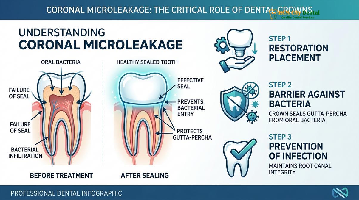
However, the gutta-percha filling alone is not designed to withstand exposure to the oral environment. If the temporary filling placed after the root canal degrades, washes out, or fractures, the underlying gutta-percha is exposed to saliva. Saliva acts as a transport medium for bacteria, allowing them to rapidly colonize the access cavity. This phenomenon is known in endodontics as coronal microleakage. Clinical studies have demonstrated that if gutta-percha is exposed to natural saliva, bacteria can penetrate the entire length of the root canal system and reach the bone at the tip of the root in as little as 30 days.[1]
When coronal microleakage occurs, the root canal system becomes reinfected. The bacteria establish complex biofilms within the dentinal tubules and the irregularities of the canal walls, leading to a recurrence of apical periodontitis (infection of the bone surrounding the root). This often necessitates a complex and costly root canal retreatment, or in severe cases, extraction of the tooth. Therefore, the long-term success of endodontic therapy is inextricably linked to the quality of the coronal restoration.[2]
| Restoration Type | Seal Integrity | Microleakage Risk | Clinical Recommendation |
|---|---|---|---|
| Temporary Filling (Cavit/IRM) | Poor (degrades rapidly) | Very High after 2-4 weeks | Strictly for short-term use (1-3 weeks max) |
| Direct Composite Filling | Moderate to Good | Moderate (margins may degrade over time) | Acceptable for anterior teeth with minimal structure loss |
| Full-Coverage Crown (Zirconia/Porcelain) | Excellent (Hermetic Seal) | Very Low | Gold standard for all posterior teeth and compromised anteriors |
A precisely fabricated dental crown provides the ultimate defense against coronal microleakage. The margins of the crown are designed to fit seamlessly against the prepared tooth structure, and the use of modern, insoluble dental cements creates a virtually impenetrable barrier. By encapsulating the entire coronal portion of the tooth, the crown ensures that the sterile environment achieved during the root canal procedure is maintained indefinitely. Dr. Cuong advises patients that investing in a high-quality root canal is only half the battle; securing that investment with a well-fitting, permanent crown is what guarantees the long-term health of the tooth.
Molar vs. Anterior Teeth: When Can You Choose a Filling Over a Crown?
While posterior teeth almost always require crowns due to heavy chewing forces, intact front teeth treated with root canals can sometimes be restored with just a composite filling.
A common question among patients is whether every single tooth that undergoes a root canal absolutely requires a crown. The clinical answer depends heavily on the anatomical location of the tooth within the dental arch and the amount of remaining healthy tooth structure. The biomechanical demands placed on anterior teeth (incisors and canines) are fundamentally different from those placed on posterior teeth (premolars and molars).
Anterior teeth are primarily designed for incising (tearing and cutting) food, rather than the heavy crushing and grinding performed by the molars. The forces applied to front teeth are generally lighter and are directed at different angles. Furthermore, the access cavity for a root canal on an anterior tooth is typically made through the lingual surface (the back of the tooth), leaving the entire facial (front) enamel and the incisal edge largely intact. If an anterior tooth has not been significantly weakened by large cavities or previous trauma, the structural integrity may remain sufficient even after endodontic therapy.
In such highly specific cases, a conservative approach may be indicated. The endodontic access hole can be meticulously sealed with a bonded composite resin filling. This restores the internal anatomy and provides an adequate seal without the need to aggressively reduce the outer enamel for a crown. However, if the anterior tooth is heavily discolored (a common side effect of pulpal necrosis), has large existing fillings, or exhibits significant structural compromise, a full-coverage restoration is still required. In some aesthetic cases, a patient might explore options like Porcelain Veneers to restore the facial appearance, though a full crown is often more appropriate for a heavily compromised root-canaled tooth.
Important Clinical Warning
Patients should never assume that a temporary filling is sufficient for long-term function. Delaying the placement of a permanent restoration (whether a definitive composite or a crown) beyond the recommended 30-day window drastically increases the risk of bacterial reinfection and catastrophic tooth fracture. Always follow your dentist’s specific restorative timeline.
Conversely, the rules for posterior teeth are much more rigid. As discussed earlier, the compressive and shear forces in the back of the mouth are immense. The anatomy of molars, with their multiple cusps and deep fissures, makes them highly susceptible to splitting once the central core is removed. Therefore, the standard of care dictates that virtually all endodontically treated premolars and molars must receive full-coverage protection. When discussing materials, patients often weigh the benefits of a porcelain crown vs zirconia HCMC clinics offer. Zirconia is frequently the material of choice for molars due to its unparalleled flexural strength and fracture toughness, ensuring the tooth can withstand decades of heavy chewing.
Clinical Success Rates: Longevity of Crowned vs. Uncrowned Treated Teeth
Clinical studies consistently show that root-canaled posterior teeth restored with crowns have a significantly higher long-term survival rate compared to those left uncrowned.
The necessity of a crown following a root canal is not merely a matter of clinical opinion; it is a protocol deeply rooted in evidence-based dentistry. Decades of retrospective studies and clinical trials have evaluated the long-term survival rates of endodontically treated teeth, comparing those that received full-coverage restorations against those that were restored with only direct fillings. The data is overwhelmingly clear and consistent across the global dental literature.[3]
One of the most frequently cited studies in endodontic literature, conducted by Aquilino and Caplan, investigated the survival rates of endodontically treated teeth over a multi-year period. The findings were stark: teeth that did not receive a crown after root canal therapy were lost at a rate six times higher than those that were properly crowned. The primary causes of failure in the uncrowned group were catastrophic vertical root fractures and severe coronal microleakage leading to non-restorable decay or recurrent apical periodontitis.[4]
“The placement of a full-coverage coronal restoration is the single most significant factor in the long-term survival of endodontically treated posterior teeth. The endodontic treatment heals the disease, but the prosthodontic restoration ensures the tooth remains functional within the oral cavity.”
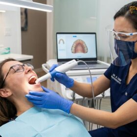
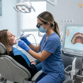
When a root canal is performed to a high standard and subsequently protected by a well-fitting, precision-milled crown, the long-term prognosis is excellent. Success rates for this combined therapy routinely exceed 90% over a ten-year period. The integration of advanced technologies, such as 3D CBCT imaging for precise endodontic diagnosis and CAD/CAM digital scanning for flawless crown fabrication, has further elevated these success rates. At facilities like HCMC Dental Clinic, the workflow is designed to minimize the time between the completion of the root canal and the delivery of the final crown, thereby mitigating the risks of interim fracture or leakage.[5]
Ultimately, the decision to invest in a crown is an investment in the longevity of the natural dentition. While modern dental implants offer an excellent solution for missing teeth, the preservation of a natural tooth through endodontic and prosthodontic therapy remains the gold standard of care whenever clinically viable. The proprioception (sensory feedback during chewing) and the natural periodontal ligament attachment of a saved tooth cannot be perfectly replicated by any artificial substitute.
When to See a Doctor for Post-Endodontic Restoration
After your root canal procedure is completed, you will typically be sent home with a temporary filling sealing the access cavity. It is imperative to schedule your follow-up appointment for the permanent crown preparation as directed by your dental provider, usually within 1 to 3 weeks. You should contact your dentist immediately if you experience any of the following symptoms before your crown appointment:
- The temporary filling falls out completely or feels significantly worn down.
- You experience a sharp, cracking sensation when biting down on the treated tooth.
- There is visible swelling in the gums surrounding the treated tooth, or a pimple-like bump appears on the gum tissue.
- You feel severe, throbbing pain that is not alleviated by over-the-counter analgesics (mild tenderness for a few days post-op is normal, but severe pain is not).
- Your bite feels uneven, or the temporary filling feels “high” when you close your teeth together.
Prompt clinical evaluation based on these signs can prevent the loss of the tooth and ensure the endodontic treatment remains uncompromised.
Frequently Asked Questions
Can I get away with just a composite filling instead of a crown?
For posterior teeth (molars and premolars), a composite filling is rarely sufficient due to heavy chewing forces. However, intact anterior teeth (incisors and canines) with minimal structural loss may sometimes be successfully restored with a high-quality composite filling instead of a full crown. Your dentist must evaluate the specific biomechanical demands and the amount of remaining healthy dentin before recommending a filling over a crown. In most cases involving back teeth, a filling will eventually lead to a catastrophic fracture.
How long can I wait after a root canal to get my permanent crown?
It is clinically recommended to place the permanent crown within 30 days of completing the root canal. Delaying the restoration increases the risk of bacterial microleakage contaminating the root canal system and significantly elevates the danger of a catastrophic tooth fracture. Temporary fillings are highly porous and begin to degrade rapidly in the oral environment. Waiting several months leaves the brittle, hollowed-out tooth completely unprotected against the immense forces of daily chewing.
Does getting a crown on a root-canaled tooth hurt?
No, preparing and placing a crown on a root-canaled tooth is generally painless. Because the nerve tissue has already been removed during the endodontic procedure, the tooth itself cannot feel thermal sensitivity or drilling pain, though the surrounding gums may feel mild, temporary pressure. Local anesthesia is often still used to ensure the gum tissue remains comfortable while the dentist shapes the tooth margin and takes the necessary digital or physical impressions.
What is the best material for a crown after a root canal?
The ideal material depends on the tooth’s location and functional requirements. Zirconia is highly recommended for molars due to its exceptional fracture resistance, while lithium disilicate (porcelain) is often preferred for front teeth because of its superior optical properties and natural translucency. Porcelain-fused-to-metal (PFM) crowns are also a durable option, though modern monolithic ceramics like zirconia have largely become the standard of care due to their combination of immense strength and excellent biocompatibility.
How do I care for my new crown after the procedure?
Care for your crowned tooth exactly like a natural tooth by brushing twice daily and flossing meticulously around the margins. Additionally, wearing a custom night guard is highly recommended if you suffer from bruxism (teeth grinding) to protect the ceramic material from excessive wear. Regular professional cleanings and examinations are also crucial to ensure the gum tissue around the crown remains healthy and to verify that the margins remain perfectly sealed against bacterial ingress.
References
- Journal of Endodontics. Biomechanical principles of post-endodontic restorations. (2021).
- International Endodontic Journal. Coronal microleakage and its impact on endodontic success. (2020).
- Journal of Prosthetic Dentistry. Survival rates of crowned versus uncrowned endodontically treated teeth. (2019).
- Clinical Oral Investigations. Fracture resistance of teeth restored with various crown materials. (2022).
- American Association of Endodontists. Guidelines for the restoration of endodontically treated teeth. (2018).
For premium porcelain crowns, bridges, and advanced smile reconstructions, visit our Dental Crowns & Bridges service page at HCMC Dental Clinic in Ho Chi Minh City.