Full mouth reconstruction for TMJ in Vietnam is a comprehensive clinical protocol designed to rebuild collapsed bites, restore the vertical dimension of occlusion, and eliminate chronic jaw pain. By utilizing advanced prosthodontics and neuromuscular dentistry, specialists permanently realign the jaw to its optimal physiological resting position.
Clinical Summary:
A full mouth reconstruction for temporomandibular joint (TMJ) disorders involves the strategic rebuilding of all teeth to correct severe bite collapse and restore the vertical dimension of occlusion. This complex bite rehabilitation utilizes neuromuscular mapping, custom orthotics, and fixed prosthetics like zirconia crowns or dental implants to stabilize the jaw. By addressing the root mechanical failure of the bite, this comprehensive treatment permanently relieves chronic facial pain, muscle spasms, and joint degeneration, offering a highly predictable functional and aesthetic restoration for patients suffering from advanced occlusal disease.
Key Takeaways:
- Bite collapse directly forces the temporomandibular joint out of its natural socket, triggering chronic pain and disc displacement.
- Restoring the vertical dimension of occlusion (VDO) is the foundational step in successful TMJ rehabilitation.
- Neuromuscular mapping ensures the new bite is designed around relaxed jaw muscles, not the habitual broken bite.
- Zirconia crowns and implant-supported bridges provide the rigid posterior support necessary to protect the TMJ from crushing forces.
- Comprehensive treatment in Vietnam offers international-standard digital dentistry and prosthodontics at a highly accessible price point.
Bite Collapse: The Direct Path to Chronic TMJ Degeneration
Bite collapse occurs when missing or severely worn teeth fail to support the jaw, forcing the temporomandibular joint into an unnatural, compressed position that causes chronic pain and structural joint damage.
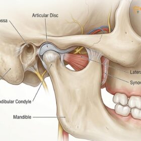
The temporomandibular joint (TMJ) is one of the most complex joints in the human body, functioning as a sliding hinge that connects the mandible (lower jaw) to the temporal bone of the skull. In a healthy masticatory system, the teeth, the jaw muscles, and the TMJs work in perfect harmony. The posterior teeth (molars and premolars) act as the foundational pillars of the mouth. They are designed to absorb the heavy vertical forces of chewing, thereby protecting the delicate structures of the TMJ from excessive compression. However, when these posterior teeth are lost due to decay, periodontal disease, or severe wear from chronic grinding, the structural integrity of the bite is compromised.
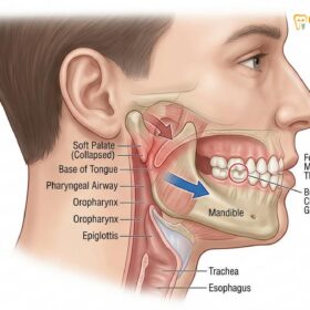
This loss of posterior support leads to a condition known clinically as “bite collapse.” Without the molars to hold the jaws apart at their proper distance, the mandible over-closes. As the lower jaw rotates upward and backward further than nature intended, the condyle (the rounded end of the jawbone) is driven deep into the glenoid fossa (the socket in the skull). This unnatural posterior-superior displacement compresses the highly innervated retrodiscal tissues, leading to immediate and severe inflammation. Over time, this constant mechanical trauma forces the protective articular disc out of place, resulting in the clicking, popping, and locking sensations characteristic of advanced temporomandibular joint disorders (TMD)[1].
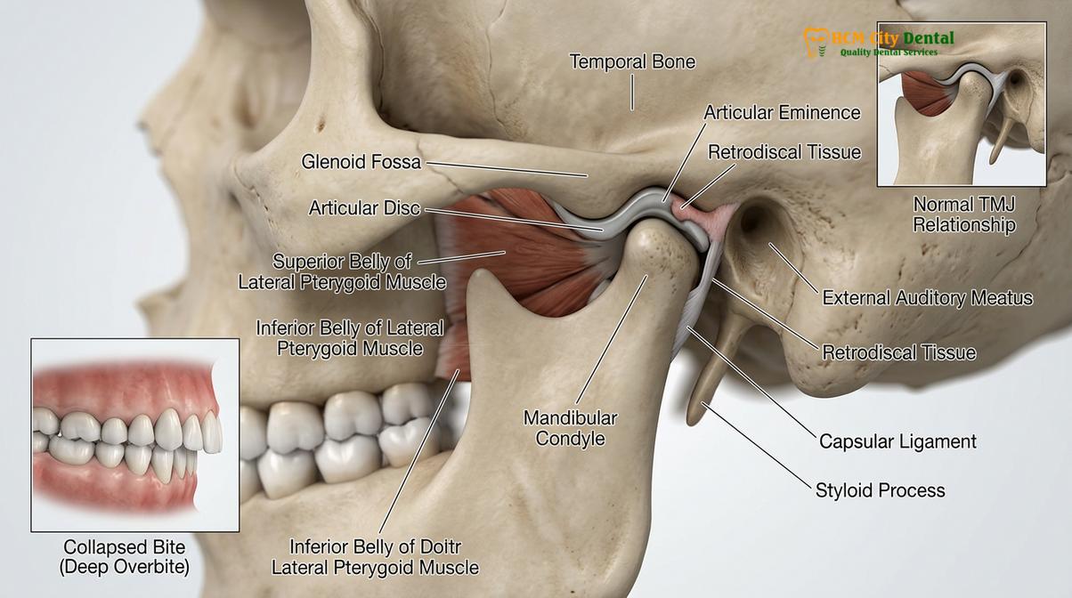
Furthermore, bite collapse initiates a destructive domino effect across the entire dental arch. As the back teeth fail, the heavy forces of mastication are transferred to the anterior teeth (incisors and canines). These front teeth are anatomically designed for shearing food, not for bearing heavy vertical loads. Consequently, they begin to flare outward, chip, and wear down rapidly. The muscles of mastication, particularly the masseter and temporalis muscles, are forced into a state of chronic hyperactivity as they struggle to find a stable resting position for the jaw. This muscular overcompensation is the primary source of the severe tension headaches, neck pain, and facial spasms reported by patients suffering from occlusal disease.
Clinical Warning: Ignoring the early signs of bite collapse, such as accelerated tooth wear or mild joint clicking, can lead to irreversible damage to the TMJ articular disc. Once the disc is severely perforated or the condylar bone begins to degenerate (osteoarthritis), conservative dental treatments may no longer be sufficient, and invasive joint surgery may become necessary.
Early intervention is critical. In many cases, the root cause of this destruction begins early in life. Conditions such as pediatric bruxism can set the stage for a lifetime of occlusal instability if left unaddressed. When adult patients present with advanced bite collapse, a simple filling or a single crown is entirely inadequate. The entire system has failed, and the only viable medical solution is a full mouth reconstruction to rebuild the lost architecture of the bite.
Defining the Vertical Dimension of Occlusion (VDO)
The vertical dimension of occlusion is the precise anatomical distance between the upper and lower jaws at rest, which dictates facial height, muscle tension, and proper TMJ alignment.
At the core of any successful full mouth reconstruction is the restoration of the Vertical Dimension of Occlusion (VDO). VDO is defined as the distance between two arbitrary anatomical points—typically one on the tip of the nose and one on the chin—when the upper and lower teeth are in maximum intercuspation (fully biting together). In a healthy patient, this dimension is maintained by the eruption and presence of the natural teeth. It provides the optimal length-tension relationship for the muscles of the face and neck, allowing them to function efficiently and rest comfortably.
When a patient suffers from severe tooth wear or tooth loss, the VDO decreases. This loss of vertical height has profound physiological and aesthetic consequences. Aesthetically, a collapsed VDO results in a prematurely aged appearance. The lower third of the face appears shortened, the chin protrudes forward (creating a pseudo-Class III malocclusion), the lips thin out, and deep wrinkles form at the corners of the mouth (angular cheilitis). Physiologically, the reduced VDO forces the jaw muscles to contract beyond their optimal resting length, leading to chronic fatigue, lactic acid buildup, and severe myofascial pain[2].
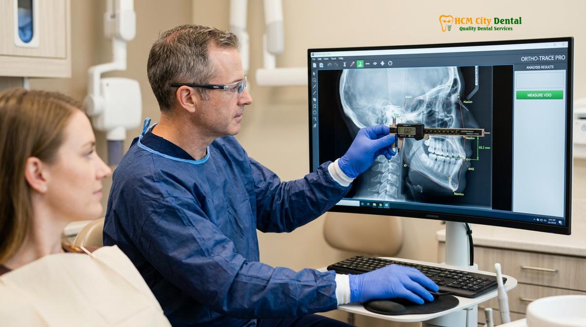
Restoring the VDO is not a matter of guesswork; it requires precise clinical diagnostics. Prosthodontists utilize a variety of techniques to determine the patient’s optimal physiological VDO before any teeth are prepared. One common method is the evaluation of the “freeway space” or interocclusal rest space. When the jaw muscles are completely relaxed, the teeth should not be touching; there should be a gap of approximately 2 to 4 millimeters between the upper and lower arches. By measuring the resting facial height and subtracting this ideal freeway space, the dentist can calculate the target VDO for the reconstruction.
“Restoring the vertical dimension of occlusion is the architectural foundation of TMJ therapy. If we build beautiful ceramic crowns but fail to establish the correct vertical height, the jaw muscles will remain in spasm, and the temporomandibular joint will continue to degenerate. Precision in measuring the VDO dictates the long-term survival of both the joint and the restorations.”
— Dr. Nguyen Van Cuong, Lead Prosthodontist
Advanced digital dentistry has revolutionized the accuracy of VDO restoration. At leading facilities like HCMC Dental Clinic in Ho Chi Minh City, specialists use Cone Beam Computed Tomography (CBCT) and digital cephalometric tracing to analyze the skeletal relationship of the jaws. This data is combined with intraoral optical scans to create a highly accurate 3D model of the patient’s skull and dentition. Through meticulous bite adjustment and digital planning, the dental team can virtually open the bite on the computer screen, ensuring that the new VDO will decompress the TMJ and harmonize with the patient’s unique facial anatomy.
Neuromuscular Phase: Mapping the New Bite Before Ceramic Placement
Before any permanent teeth are fabricated, neuromuscular mapping utilizes digital sensors and temporary orthotics to locate and test the jaw’s optimal, pain-free resting position.
One of the most critical errors in traditional dentistry is attempting to rebuild a patient’s teeth based on their existing, dysfunctional bite. If a patient has been suffering from bite collapse and TMJ pain for years, their jaw muscles have adapted to a pathological position. This is known as the habitual bite. If a dentist simply places new crowns to fit this habitual bite, they are merely cementing the dysfunction into place. To achieve true complex bite rehabilitation, the dentist must bypass the habitual bite and find the jaw’s true physiological resting position—a process known as neuromuscular dentistry.
The neuromuscular phase begins with muscle deprogramming. Because the jaw muscles are chronically tense and accustomed to forcing the jaw into the collapsed position, they must be relaxed before an accurate bite registration can be taken. This is often achieved using Transcutaneous Electrical Nerve Stimulation (TENS). A specialized ultra-low frequency TENS unit delivers mild electrical impulses to the facial muscles, causing them to gently twitch and relax. After 45 to 60 minutes of TENS therapy, the muscles are flushed of lactic acid and return to their natural, unstrained length.
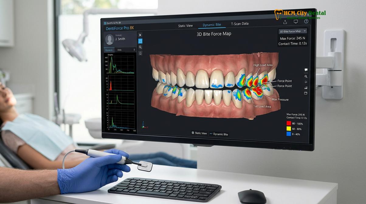
Once the muscles are relaxed, the dentist captures the new bite position. This is where dental crowns bridge mapping becomes essential. Using computerized occlusal analysis systems, the dentist can measure the exact force and timing of the teeth coming together. The software displays a dynamic 3D map of the bite, highlighting areas of premature contact or excessive force. This data allows the dentist to design a bite where all posterior teeth hit simultaneously and with equal intensity, providing perfect stability for the TMJ[3].
Clinical Case Study: Neuromuscular Rehabilitation
A 52-year-old patient presented to HCMC Dental Clinic in Ho Chi Minh City with severe bruxism, chronic migraines, and a heavily worn dentition resulting in a 4mm loss of VDO. Instead of immediately preparing the teeth for crowns, the clinical team initiated a neuromuscular phase. Following TENS deprogramming, a custom fixed orthotic was bonded to the lower teeth, opening the bite to the newly calculated VDO. Within three weeks of wearing the orthotic, the patient reported a 90% reduction in TMJ pain and headaches. Only after this new jaw position was proven successful over a two-month testing period did the team proceed to finalize the full mouth reconstruction with permanent zirconia restorations.
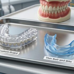
During this testing phase, the patient is usually fitted with a custom orthotic device or provisional (temporary) crowns. This allows the patient to “test drive” the new VDO and jaw position in their daily life. They can evaluate the aesthetics, speech, and chewing function, while the dentist monitors the TMJ for any signs of residual inflammation. Understanding the difference between a therapeutic orthotic used during this phase and a standard protective guard is crucial; patients can learn more by reviewing the clinical differences between an occlusal splint vs night guard.
Restoring Anterior Guidance and Posterior Support with Zirconia Crowns
Once the optimal bite is verified, specialists use ultra-durable zirconia crowns and implant bridges to permanently lock in the new vertical dimension and provide stable posterior support.
After the neuromuscular phase has successfully established a pain-free, stable jaw position, the final phase of the full mouth reconstruction begins. This phase involves translating the temporary bite into permanent, highly durable ceramic restorations. The biomechanical goals of this phase are twofold: establishing proper anterior guidance and securing rigid posterior support.
Anterior guidance refers to the dynamic relationship of the front teeth during jaw movement. When the lower jaw slides forward or side-to-side, the lower incisors and canines should glide smoothly against the back surfaces of the upper front teeth. This gliding action must immediately separate (disclude) the posterior teeth. If the back teeth rub together during these lateral movements—a condition known as posterior interference—it triggers hyperactive muscle contractions and severe stress on the TMJ. By precisely engineering the shape and angle of the new anterior crowns, the prosthodontist ensures that the front teeth protect the back teeth during movement.
Conversely, posterior support is the mechanism by which the back teeth protect the front teeth and the TMJ during heavy vertical chewing. The molars and premolars must interlock perfectly to absorb the crushing forces of mastication. To achieve this, the choice of dental materials is paramount. In modern full mouth reconstructions, monolithic zirconia has become the gold standard. Zirconia is a biocompatible ceramic that possesses exceptional flexural strength, making it highly resistant to fracture even in patients with a history of severe bruxism[4].
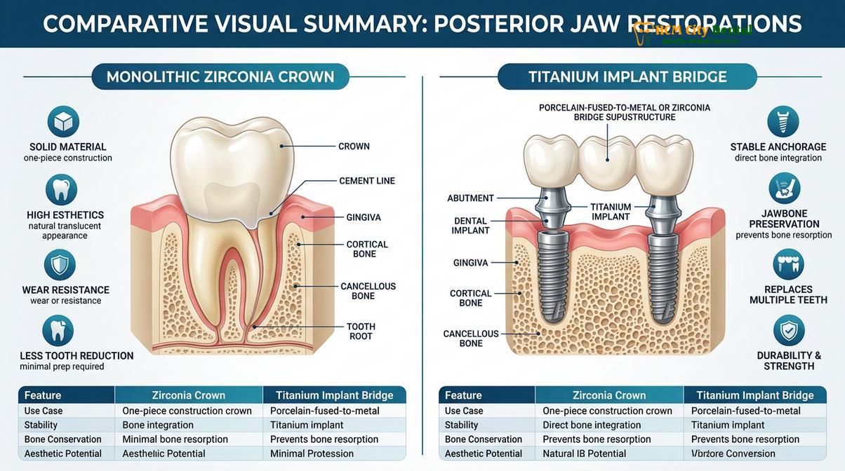
For patients who are missing multiple teeth or have teeth that are too structurally compromised to support crowns, dental implants are integrated into the reconstruction. Techniques such as the All-on-4 or All-on-6 protocols utilize titanium implants anchored directly into the jawbone to support a full arch of zirconia teeth. This approach not only restores the VDO but also prevents the progressive bone loss that occurs after tooth extraction.
| Material / Approach | Primary Indication | Biomechanical Advantage for TMJ | Durability |
|---|---|---|---|
| Monolithic Zirconia Crowns | Restoring severely worn but structurally sound natural teeth. | Exceptional compressive strength; maintains precise VDO without wearing down over time. | Very High (15-20+ years) |
| Titanium Implant Bridges (All-on-6) | Complete arch replacement for missing or failing teeth. | Provides rigid, unyielding posterior support; halts alveolar bone resorption. | Permanent (Implants), 15+ years (Prosthetic) |
| Lithium Disilicate (E-max) | Anterior teeth requiring high aesthetic translucency. | Excellent for establishing smooth anterior guidance; highly aesthetic. | High (10-15 years) |
The integration of these materials requires absolute precision. The digital bite map created during the neuromuscular phase is sent to a CAD/CAM (Computer-Aided Design and Computer-Aided Manufacturing) milling machine. The zirconia crowns are milled to microscopic tolerances, ensuring that when they are bonded to the teeth, the bite forces are distributed perfectly evenly. Following the final placement, patients are strongly advised to protect their investment. While the zirconia is incredibly strong, the underlying bone and the TMJ still require protection from nocturnal grinding. The use of a specialized hard acrylic night guard is typically prescribed to ensure long-term success.
Important Clinical Considerations
While full mouth reconstruction offers a profound, life-changing solution for patients with severe occlusal disease, it is a highly complex medical procedure that requires careful patient selection and rigorous diagnostics. Not all jaw pain is caused by bite collapse. Systemic conditions such as rheumatoid arthritis, autoimmune disorders, or primary tumors of the jaw can present with symptoms similar to mechanical TMJ dysfunction. Therefore, a comprehensive differential diagnosis is mandatory before initiating irreversible prosthodontic treatment.
Patients should seek immediate specialist evaluation if they experience “red flag” symptoms. These include a sudden inability to open or close the mouth (closed lock or open lock), severe, sharp pain radiating through the face that is unresponsive to basic analgesics, or loud, grinding crepitus (a sound like crushing gravel) in the joint, which indicates active bone-on-bone degeneration.
“The decision to proceed with a full mouth reconstruction must be evidence-based. We do not cut teeth simply to make them look white; we reconstruct the dental arch to save the joint. If a patient’s TMJ pain is primarily muscular and their bite is structurally sound, conservative therapies must be exhausted first. Reconstruction is reserved for cases where the mechanical foundation has unequivocally failed.”
— Clinical Consensus on Advanced Prosthodontics
For patients whose TMJ pain is primarily driven by muscular hyperactivity rather than severe structural bite collapse, less invasive interventions may be appropriate as a first-line therapy. Treatments such as masseter Botox injections can temporarily paralyze the overactive muscles, providing significant pain relief and breaking the cycle of bruxism. Additionally, specialized anterior deprogrammers, such as the NTI device, can be utilized to prevent the posterior teeth from engaging during sleep, thereby reducing the load on the TMJ[5]. However, if the VDO is fundamentally lost, these conservative measures will only provide temporary symptomatic relief, and a full structural reconstruction will ultimately be required.
Patients considering this level of comprehensive care are encouraged to undergo a thorough clinical examination. At facilities like HCMC Dental Clinic, the focus remains on evidence-based, predictable outcomes that prioritize the long-term health of the temporomandibular joint alongside the aesthetic restoration of the smile.
Frequently Asked Questions
What is a full mouth reconstruction for TMJ?
A full mouth reconstruction for TMJ is a comprehensive dental protocol that rebuilds all teeth to correct bite collapse and restore optimal neuromuscular jaw alignment. By utilizing crowns, bridges, or implants, this treatment permanently stabilizes the temporomandibular joint, eliminating chronic pain and restoring proper masticatory function. The process involves meticulous digital planning to ensure the new dental arches provide the exact vertical height required to decompress the jaw joint.
How long does full mouth rehabilitation take in Vietnam?
Full mouth rehabilitation in Vietnam typically requires two to three clinical phases spanning several weeks to a few months. The initial neuromuscular mapping and provisional phase takes one to two weeks, followed by a testing period, and concludes with the final placement of permanent ceramic restorations. The exact timeline depends heavily on whether dental implants or bone grafting are required, which can extend the healing phase.
Can crowns and veneers fix TMJ permanently?
Crowns and veneers can permanently resolve TMJ issues if the underlying cause is a collapsed bite or improper occlusion. By precisely engineering these restorations to support the correct vertical dimension of occlusion, they provide the rigid structural support necessary to keep the jaw joint in its healthy physiological position. However, they must be designed using neuromuscular principles to ensure they harmonize with the jaw muscles.
Is neuromuscular mapping mandatory before placing permanent crowns?
Yes, neuromuscular mapping is a mandatory clinical step in complex TMJ cases. It ensures that the new bite is designed around the relaxed, optimal position of the jaw muscles rather than the patient’s habitual, dysfunctional bite, preventing the recurrence of joint pain and restoration failure. Skipping this step often leads to the new ceramic crowns fracturing or the patient experiencing worsened muscular spasms.
How do I protect my new bite after a full mouth reconstruction?
Protecting your new bite requires the nightly use of a custom-calibrated orthotic device to prevent bruxism from damaging the new ceramics. Regular clinical follow-ups and occlusal adjustments are also necessary to ensure the bite forces remain evenly distributed across the restored dental arches. Patients can learn more about long-term maintenance by exploring comprehensive Nightguards and TMJ management protocols.
References
- Journal of the American Dental Association. Diagnostic protocols for temporomandibular joint degeneration and bite collapse. (2018).
- Journal of Prosthetic Dentistry. The role of vertical dimension of occlusion in temporomandibular disorders. (2021).
- International Journal of Prosthodontics. Neuromuscular dentistry and computerized occlusal analysis in full mouth rehabilitation. (2020).
- Clinical Oral Implants Research. Zirconia vs Titanium in full arch implant-supported prostheses: A biomechanical review. (2022).
- Journal of Oral Rehabilitation. Efficacy of anterior guidance and posterior support in TMJ stabilization. (2019).