Gingival hyperplasia treatment focuses on reducing excessive gum tissue through improved oral hygiene, medication adjustments, or surgical interventions like gingivectomy. By addressing the root cause—whether drug-induced, inflammatory, or genetic—dentists can restore healthy gingival contours and prevent secondary periodontal complications.
Clinical Summary:
Gingival hyperplasia, clinically referred to as gum overgrowth or gingival enlargement, is a pathological condition characterized by the excessive proliferation of gingival tissues. It is frequently triggered by specific systemic medications (such as anticonvulsants, immunosuppressants, and calcium channel blockers), hormonal fluctuations, or chronic plaque-induced inflammation. First-line treatment involves non-surgical periodontal therapy, rigorous plaque control, and pharmacological substitution in consultation with a physician. Refractory or severe fibrotic cases require surgical excision via traditional scalpel gingivectomy or advanced laser contouring to restore physiological architecture, eliminate pseudopockets, and facilitate proper oral hygiene maintenance.
Key Takeaways:
- Gingival hyperplasia is an abnormal overgrowth of gum tissue that can obscure the teeth and impair oral function.
- Common triggers include severe plaque accumulation, hormonal shifts, and specific prescription medications.
- First-line therapy emphasizes rigorous mechanical plaque control and potential medication substitution by a physician.
- Surgical removal (gingivectomy) is highly effective for severe, fibrotic tissue enlargement that resists conservative care.
- Untreated overgrowth traps pathogenic bacteria, significantly increasing the risk of advanced periodontal disease and bone loss.
What is Gingival Hyperplasia (Gum Overgrowth)?
Gingival hyperplasia is the abnormal, excessive growth of gum tissue that alters the natural architecture of the periodontium. It requires clinical intervention to prevent functional and aesthetic complications.
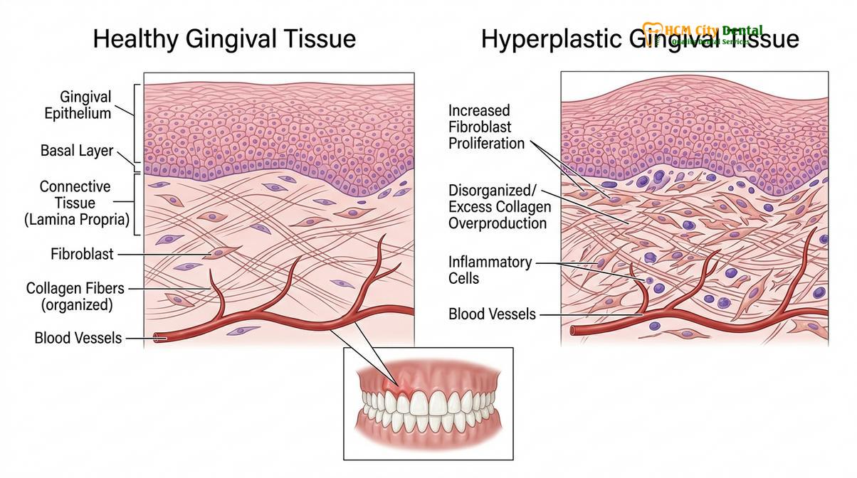
Gingival hyperplasia, also known in the medical community as gingival overgrowth or gingival enlargement, is a condition where the gingival tissues (gums) become swollen, enlarged, and grow excessively over the clinical crowns of the teeth. Unlike standard gingivitis, which primarily involves inflammation and vascular engorgement, hyperplasia often involves an actual increase in the number of cells (fibroblasts) and an overproduction of the extracellular matrix, particularly collagen [1]. This results in a dense, fibrotic tissue mass that alters the normal scalloped architecture of the gum line.
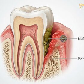
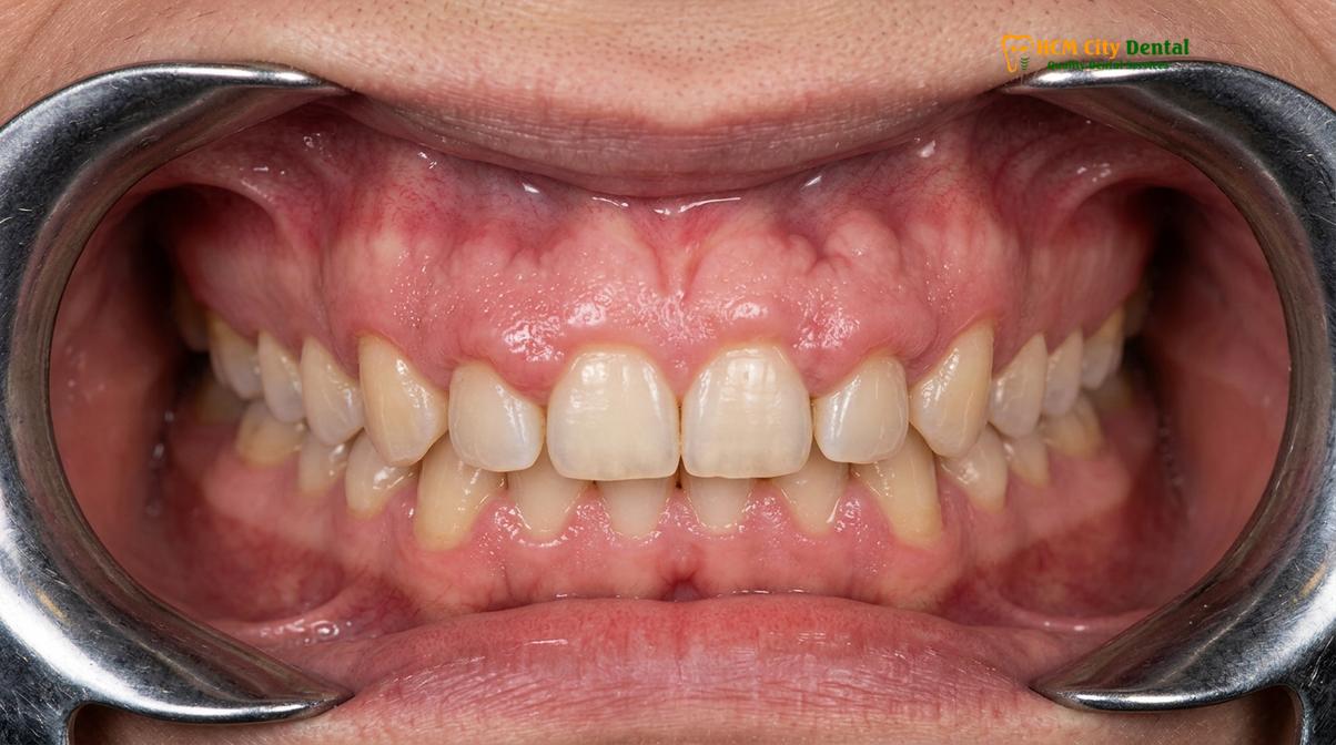
The condition can range from mild, where the interdental papillae (the gum tissue between the teeth) become slightly bulbous, to severe, where the hyperplastic tissue completely covers the crowns of the teeth, interfering with mastication (chewing) and speech. When seeking General Dentistry care, early identification of this condition is paramount. If left unchecked, the overgrown tissue creates deep “pseudopockets”—spaces between the gum and tooth that are not caused by bone loss but by the upward growth of the gum. These pockets become highly retentive for dental plaque and calculus, creating a vicious cycle of inflammation and further tissue enlargement.
According to Dr. Nguyen Van Cuong, a leading specialist at HCMC Dental Clinic, “Understanding the precise etiology of gingival enlargement is the cornerstone of effective treatment. We must differentiate between purely inflammatory enlargement, which responds well to deep cleaning, and fibrotic overgrowth, which often requires surgical intervention.” This diagnostic differentiation is critical because the approach to enlarged gums treatment varies drastically based on the underlying cellular pathology.
Histological and Pathological Mechanisms
To fully comprehend gingival hyperplasia treatment, one must look at the condition at a microscopic level. The gingival connective tissue is primarily composed of collagen fibers produced by fibroblasts. In a healthy state, there is a delicate balance between collagen synthesis and collagen degradation (mediated by enzymes called matrix metalloproteinases, or MMPs). In cases of hyperplasia, this homeostasis is disrupted. Certain triggers cause fibroblasts to become hyperactive, synthesizing excessive amounts of collagen while simultaneously downregulating the production of degradative enzymes. The result is a net accumulation of dense, fibrous connective tissue.
Furthermore, the epithelial layer covering the gums may undergo acanthosis (thickening) and elongate its rete pegs deep into the underlying connective tissue. This structural alteration makes the gums highly resilient but also highly prone to trapping subgingival bacteria. The presence of these bacteria induces a secondary inflammatory response, meaning that most cases of gingival hyperplasia present with a combined etiology: a primary fibrotic overgrowth complicated by secondary plaque-induced inflammation.
Medications That Cause Drug-Induced Gingival Enlargement
Drug-induced gingival enlargement is a well-documented side effect of certain anticonvulsants, immunosuppressants, and calcium channel blockers. Management often requires collaboration between your dentist and prescribing physician.
One of the most prevalent and clinically significant causes of gum overgrowth is the systemic administration of specific pharmacological agents. This condition, specifically termed drug induced gingival enlargement (DIGO), is a well-recognized adverse effect associated with three main classes of medications. The pathogenesis of DIGO is complex and multifactorial, involving an interaction between the drug, the host’s cellular response, and the presence of dental plaque [2].
Clinical Warning: Never discontinue or alter the dosage of your prescribed systemic medications without explicit authorization from your primary care physician or cardiologist. Dentists and physicians must collaborate to find a safe pharmacological alternative that manages your systemic health while mitigating gingival side effects.
1. Anticonvulsants (Phenytoin)
Phenytoin, commonly marketed under the brand name Dilantin, is a widely used medication for the management of epilepsy and other seizure disorders. It was the first drug historically linked to gingival overgrowth. Clinical studies indicate that approximately 50% of patients on long-term phenytoin therapy will develop some degree of gingival hyperplasia. The drug is believed to directly stimulate gingival fibroblasts, altering their metabolism and leading to an accumulation of inactive collagenase, which prevents the normal breakdown of connective tissue. The overgrowth typically begins in the interdental papillae of the anterior teeth and can progress to form massive tissue folds that obscure the teeth entirely.
2. Immunosuppressants (Cyclosporine)
Cyclosporine is a potent immunosuppressive agent used primarily to prevent organ rejection in transplant recipients and to manage severe autoimmune diseases like rheumatoid arthritis and psoriasis. Gingival overgrowth occurs in about 25% to 30% of patients taking this medication. Cyclosporine affects the gingival tissues by altering the function of fibroblasts and increasing the expression of transforming growth factor-beta (TGF-β), a cytokine that strongly promotes collagen synthesis. Unlike phenytoin-induced overgrowth, cyclosporine-induced hyperplasia is often highly vascularized, meaning the gums may appear more red, swollen, and prone to bleeding upon probing.
3. Calcium Channel Blockers (Nifedipine, Amlodipine, Verapamil)
Calcium channel blockers (CCBs) are a broad class of medications used to treat hypertension (high blood pressure), angina, and cardiac arrhythmias. Drugs such as nifedipine, amlodipine, and diltiazem are frequently implicated in gingival enlargement. These medications work by inhibiting the influx of calcium ions into cardiac and smooth muscle cells. However, in the gingival tissues, this alteration in intracellular calcium levels interferes with the secretion of collagenase by fibroblasts, leading to collagen accumulation. The prevalence of CCB-induced overgrowth varies but is particularly high with nifedipine. Because hypertension is so common globally, CCBs represent one of the most frequently encountered causes of drug-induced hyperplasia in modern dental practice.
Other Causes: Inflammation, Pregnancy, and Genetic Disorders
Beyond medications, gum overgrowth can be triggered by chronic plaque accumulation, hormonal shifts during pregnancy, or rare hereditary fibromatosis conditions. Identifying the exact etiology dictates the treatment protocol.
While medications are a primary culprit, understanding all gum overgrowth causes is essential for comprehensive clinical management. The periodontium is highly sensitive to local environmental factors, systemic hormonal fluctuations, and genetic predispositions. When a patient presents with enlarged gums, a thorough medical and dental history is required to rule out these alternative etiologies.
Plaque-Induced Inflammatory Hyperplasia
The most common, albeit often less fibrotic, cause of gingival enlargement is chronic plaque-induced gingivitis. When dental plaque—a sticky biofilm composed of bacteria—is allowed to accumulate along the gingival margin due to inadequate oral hygiene, the host immune system mounts an inflammatory response. Inflammatory mediators such as interleukins and tumor necrosis factor-alpha (TNF-α) cause vasodilation and increased capillary permeability [3]. This leads to edema (swelling) and an increase in the size of the gingival tissues. Unlike drug-induced overgrowth, which is firm and pale pink, inflammatory hyperplasia is typically soft, erythematous (red), and bleeds easily. This condition is highly responsive to professional Deep Cleaning Teeth procedures and improved home care.
“In many cases of mild to moderate gingival enlargement, the primary driver is simply the chronic presence of subgingival calculus. Removing this local irritant is always our first therapeutic step before considering any surgical intervention.”
— Dr. Nguyen Van Cuong, Clinical Director
Hormonal Influences: Pregnancy and Puberty
Systemic hormonal changes can profoundly exaggerate the gingival tissue’s response to local plaque. During puberty and pregnancy, there is a significant surge in circulating levels of estrogen and progesterone. These hormones can alter the microbial composition of dental plaque (favoring the growth of certain pathogens like *Prevotella intermedia*) and increase the vascular permeability of the gingival tissues. “Pregnancy gingivitis” often presents as generalized gingival enlargement, and in some cases, a localized, highly vascular mass known as a pyogenic granuloma (or pregnancy tumor) may develop. These hormonal overgrowths typically regress postpartum once hormone levels stabilize, provided that meticulous oral hygiene is maintained.
Hereditary Gingival Fibromatosis (HGF)
Hereditary gingival fibromatosis is a rare, genetically determined condition characterized by a slow, progressive, and benign enlargement of the gingival tissues. It can occur as an isolated condition or as part of a broader genetic syndrome. The overgrowth usually begins during the eruption of the permanent dentition and can be so severe that it completely covers the teeth, preventing normal eruption and causing significant functional and aesthetic impairment. The tissue in HGF is extremely dense, firm, and normal in color. Because the etiology is genetic, non-surgical therapies are largely ineffective, and repeated surgical excisions are often required throughout the patient’s life to maintain oral function.
Symptoms and How Overgrown Gums Impact Oral Health
Overgrown gums create deep pockets that trap bacteria, leading to bleeding, halitosis, and structural damage to the underlying alveolar bone if left untreated.
The clinical manifestations of gingival hyperplasia extend far beyond cosmetic concerns. While a “gummy smile” or the appearance of abnormally small teeth is often the primary reason patients seek consultation, the functional and pathological consequences of the condition are severe. The altered topography of the gums creates an environment highly conducive to disease progression.
One of the most immediate impacts is the formation of pseudopockets. In a healthy mouth, the gingival sulcus (the space between the tooth and gum) is typically 1 to 3 millimeters deep. In hyperplastic conditions, the upward growth of the tissue can create pocket depths of 5, 7, or even 10 millimeters, without any initial loss of the underlying bone. These deep crevices are impossible for a patient to clean with a standard toothbrush or dental floss. Consequently, anaerobic bacteria proliferate in these oxygen-deprived environments, leading to chronic Bad Breath Gum Disease and persistent inflammation.
| Clinical Parameter | Healthy Gingiva | Hyperplastic Gingiva (Overgrowth) |
|---|---|---|
| Tissue Contour | Scalloped, knife-edged margins hugging the teeth. | Bulbous, rolled margins; blunted or enlarged papillae. |
| Tissue Consistency | Firm, resilient, tightly bound to underlying bone. | Can be dense and fibrotic (drug-induced) or soft and edematous (inflammatory). |
| Pocket Depth | 1 – 3 mm (Physiological sulcus). | 4 – 10+ mm (Pseudopockets trapping bacteria). |
| Bleeding on Probing | Absent. | Frequent, especially if secondary inflammation is present. |
| Crown Visibility | Full clinical crown is visible. | Partial or near-complete coverage of the clinical crown. |
As the condition progresses, patients frequently experience Swollen Gums Around Tooth margins that bleed spontaneously or during mastication. The physical bulk of the tissue can interfere with proper chewing, leading to nutritional challenges, and can alter phonetics, causing speech impediments. Furthermore, the chronic retention of plaque within the pseudopockets eventually leads to true periodontal disease, where the inflammatory process begins to destroy the periodontal ligament and alveolar bone, ultimately resulting in tooth mobility and loss.
Medical Management: Changing Prescriptions and Hygiene
Initial treatment focuses on non-surgical methods, including rigorous professional dental cleanings, antimicrobial rinses, and consulting your physician to substitute offending medications.
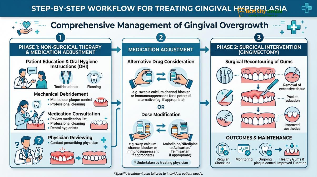
The management of gingival hyperplasia is highly systematic, beginning with the most conservative, non-invasive therapies. Phase I therapy, or cause-related therapy, aims to eliminate all local inflammatory factors and address any systemic contributors. This phase is critical because even if surgical intervention is eventually required, operating on highly inflamed, plaque-laden tissue increases the risk of intraoperative bleeding, delayed healing, and post-operative infection [4].
Professional Scaling and Root Planing (SRP)
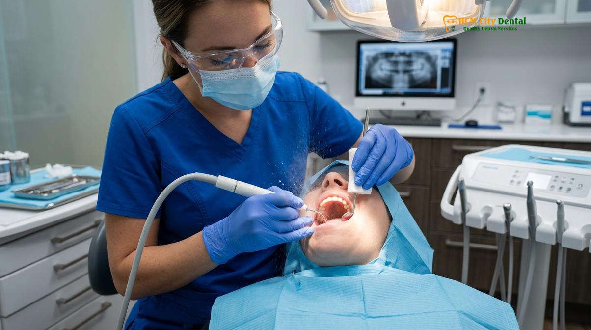
The cornerstone of non-surgical management is meticulous mechanical debridement. Scaling and root planing involves the use of specialized ultrasonic instruments and hand curettes to remove plaque, calculus (tartar), and bacterial endotoxins from the tooth surfaces, both above and below the gumline. By eliminating these local irritants, the secondary inflammatory component of the hyperplasia is resolved. In cases where the overgrowth is purely inflammatory, SRP combined with excellent home care may be the only treatment required to restore normal gingival contours.
Pharmacological Substitution and Adjuncts
For patients with drug-induced gingival enlargement, the most effective medical intervention is the cessation or substitution of the offending medication. This must be coordinated directly with the patient’s prescribing physician. For example, a patient developing severe hyperplasia from nifedipine might be safely transitioned to a different class of antihypertensive, such as an ACE inhibitor or an angiotensin II receptor blocker (ARB), which do not carry the same risk of gingival overgrowth. If substitution is successful, the gingival tissue often begins to regress spontaneously within a few weeks to months.
In situations where medication substitution is not medically feasible (e.g., a transplant patient strictly stabilized on cyclosporine), pharmacological adjuncts may be utilized. Clinical evidence suggests that short courses of specific antibiotics, such as azithromycin, can help reduce cyclosporine-induced overgrowth. Azithromycin is believed to inhibit the proliferation of fibroblasts and reduce collagen synthesis. Additionally, the daily use of prescription antimicrobial mouthwashes containing 0.12% chlorhexidine gluconate is highly recommended to control the bacterial bioburden within the deep pseudopockets.
Surgical Treatment: Gingivectomy and Laser Contouring
When non-surgical methods fail or tissue becomes highly fibrotic, surgical removal via traditional scalpel gingivectomy or precision laser contouring is necessary to restore gum health.
When gingival hyperplasia is severe, highly fibrotic, or unresponsive to non-surgical therapy and medication adjustments, surgical intervention becomes the definitive treatment. The goal of surgery is to excise the excess tissue, eliminate pseudopockets, restore a physiological gingival contour, and provide the patient with an oral architecture that can be effectively maintained through daily hygiene. The primary surgical procedure for this condition is a gingivectomy.
Clinical Case Review: A 45-year-old patient presented to HCMC Dental Clinic in Ho Chi Minh City with severe, fibrotic gingival overgrowth induced by long-term amlodipine use. The tissue covered 50% of the anterior teeth, causing aesthetic distress and chronic bleeding. After consulting with the patient’s cardiologist to adjust the medication, the dental team performed a comprehensive scaling followed by a precision laser gingivectomy. Within four weeks, the patient exhibited fully healed, tightly adapted gingival margins with zero bleeding on probing, successfully restoring both function and confidence.
Traditional Scalpel Gingivectomy
The traditional approach to removing hyperplastic tissue is the scalpel gingivectomy. This procedure is performed under profound local anesthesia. The periodontist or oral surgeon uses specialized periodontal knives (such as Kirkland and Orban knives) to make a beveled incision, carefully excising the excess fibrotic tissue. The remaining tissue is then contoured (gingivoplasty) to recreate the natural scalloped appearance of the gum line. Following the excision, a periodontal dressing (a putty-like bandage) is often placed over the surgical site to protect the raw tissue, control post-operative bleeding, and minimize discomfort during the initial healing phase. While highly effective, traditional scalpel surgery can be associated with moderate post-operative discomfort and bleeding [5]. For more complex cases involving underlying bone recontouring, a full Gum Flap Surgery may be indicated.
Advanced Laser-Assisted Contouring
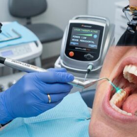
Modern periodontal practice increasingly relies on laser technology for the management of gingival overgrowth. Devices such as diode lasers, Nd:YAG, and CO2 lasers offer significant advantages over traditional scalpels. When performing Laser Gum Treatment for hyperplasia, the laser energy precisely ablates (vaporizes) the excess tissue while simultaneously coagulating blood vessels and sealing nerve endings. This dual action results in a virtually bloodless surgical field, significantly reduced post-operative pain, and minimal swelling. Furthermore, the laser sterilizes the surgical pocket, reducing the risk of post-operative infection. Patients seeking gingivectomy surgery saigon often prefer laser contouring due to its accelerated recovery timeline and the frequent elimination of the need for sutures or periodontal dressings.
When to See a Doctor
Gingival hyperplasia is not a condition that resolves with over-the-counter remedies or aggressive brushing; in fact, brushing hyperplastic tissue too vigorously can exacerbate inflammation and cause severe bleeding. It is imperative to seek professional dental evaluation if you notice any abnormal changes in the size, shape, or texture of your gums. Early intervention is the key to preventing irreversible damage to the underlying alveolar bone.
“Patients should never ignore gums that are progressively enlarging or covering their teeth. What begins as a minor aesthetic annoyance can rapidly evolve into severe periodontitis if the bacterial reservoirs within the pseudopockets are not professionally managed.”
— Clinical Consensus, Periodontal Health Guidelines
You should schedule an immediate consultation at a qualified facility, such as HCMC Dental Clinic, if you experience any of the following clinical warning signs:
- Gum tissue that has grown over the crowns of your teeth, altering your smile or bite.
- Persistent, spontaneous bleeding from the gums, or heavy bleeding during normal brushing and flossing.
- Gums that appear bright red, swollen, or feel tender and painful to the touch.
- Chronic bad breath (halitosis) or a persistent metallic taste in the mouth that does not improve with hygiene.
- Difficulty chewing food due to the bulk of the gum tissue interfering with your bite.
- You have recently started a new medication (especially for blood pressure, seizures, or immunosuppression) and notice subsequent changes in your gum architecture.
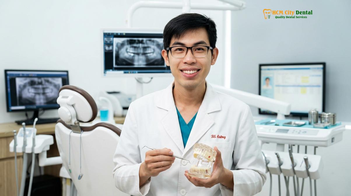
During your consultation, the clinical team will perform a comprehensive periodontal charting, measuring the depth of the pockets around each tooth, and take necessary radiographs to assess the health of the underlying bone. Based on these diagnostics, a personalized treatment plan will be formulated, ranging from conservative deep cleaning to advanced laser contouring, ensuring the optimal restoration of your oral health.
Frequently Asked Questions
Can swollen gums grow over teeth?
Yes, in severe cases of gingival hyperplasia, the overgrown gum tissue can proliferate extensively enough to partially or completely cover the clinical crowns of the teeth. This advanced overgrowth creates deep pseudopockets that trap bacteria, making oral hygiene nearly impossible and necessitating professional dental intervention. Over time, this physical barrier not only affects aesthetics but severely impairs chewing function and speech.
What medications cause gum overgrowth?
The three primary classes of medications known to cause drug-induced gingival enlargement are anticonvulsants (like phenytoin), immunosuppressants (such as cyclosporine), and calcium channel blockers (including nifedipine and amlodipine). These drugs alter cellular metabolism, leading to an overproduction of collagen in the gingival tissues. If you suspect your medication is causing this issue, your dentist will collaborate with your physician to explore safe pharmacological alternatives.
How do dentists treat gingival hyperplasia?
Dentists treat gingival hyperplasia through a phased approach starting with non-surgical deep cleaning and improved oral hygiene. If the condition is drug-induced, a physician may adjust medications; if the tissue is highly fibrotic and unresponsive, surgical removal via gingivectomy or laser contouring is performed. The exact protocol is tailored to the underlying etiology, ensuring both the symptoms and the root cause are addressed.
Is gum overgrowth surgery painful?
Gum overgrowth surgery is performed under local anesthesia, ensuring the procedure itself is virtually painless. Post-operative discomfort is typically mild to moderate and can be effectively managed with over-the-counter analgesics, warm saltwater rinses, and adherence to your dentist’s specific aftercare instructions. The use of modern dental lasers further minimizes post-operative pain, swelling, and bleeding compared to traditional scalpel methods.
Can gingival hyperplasia return after surgery?
Yes, gingival hyperplasia can recur after surgical removal if the underlying cause is not resolved. Recurrence is common if the patient continues taking the inducing medication without meticulous plaque control, or if underlying genetic and inflammatory factors are not continuously managed through regular dental maintenance. Strict adherence to a 3-to-4-month periodontal recall schedule is critical to preventing relapse.
References
- Journal of Periodontology. Pathogenesis and clinical management of drug-induced gingival overgrowth. (2021).
- International Journal of Dentistry. The role of calcium channel blockers in gingival hyperplasia: A clinical review. (2020).
- Clinical Oral Investigations. Inflammatory mechanisms in plaque-induced gingival enlargement. (2019).
- Journal of the American Dental Association. Non-surgical periodontal therapy and its impact on gingival health. (2022).
- British Dental Journal. Comparative efficacy of laser versus scalpel gingivectomy in hyperplastic tissue management. (2018).
For pricing, booking, and a free clinical assessment, visit our Gum Disease Treatment service page at HCMC Dental Clinic in Ho Chi Minh City.