Aggressive periodontitis is a severe, rapidly progressing form of gum disease that primarily affects healthy young individuals. It is characterized by rapid attachment loss and bone destruction around the teeth, often driven by specific bacterial pathogens and genetic immune system vulnerabilities rather than heavy plaque accumulation.
Clinical Summary:
Aggressive periodontitis represents a highly destructive category of periodontal disease characterized by rapid clinical attachment loss and alveolar bone destruction. Unlike chronic forms of gum disease, it frequently presents in adolescents and young adults who are otherwise medically healthy and often exhibit minimal dental plaque or calculus. The pathogenesis is heavily linked to highly virulent microorganisms, predominantly Aggregatibacter actinomycetemcomitans and Porphyromonas gingivalis, coupled with localized host immune defects such as impaired neutrophil chemotaxis. Due to the tissue-invasive nature of these pathogens, mechanical debridement alone is often insufficient. Successful clinical management requires early diagnosis, aggressive mechanical therapy, adjunctive systemic antibiotics, and frequently, advanced periodontal surgery to halt disease progression and regenerate lost supportive tissues.
Key Takeaways:
- Characterized by rapid, severe destruction of the periodontal ligament and alveolar bone in young, healthy patients.
- Strongly associated with specific virulent bacteria, notably Aggregatibacter actinomycetemcomitans.
- Familial aggregation is common, indicating a significant genetic predisposition and immune system vulnerability.
- Clinical presentation often shows tissue destruction that is disproportionate to the amount of visible plaque.
- Treatment protocols mandate a combination of mechanical scaling, systemic antibiotics, and rigorous maintenance.
What is Aggressive Periodontitis?
Aggressive periodontitis is a distinct periodontal condition marked by rapid destruction of the periodontal ligament and alveolar bone, typically affecting younger, otherwise healthy patients.
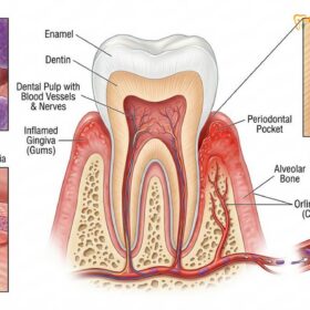
Aggressive periodontitis is a complex, multifactorial inflammatory disease of the supporting structures of the teeth. Historically classified under various terms such as early-onset periodontitis or juvenile periodontitis, the clinical entity is defined by its exceptionally rapid rate of disease progression. Patients afflicted with this condition experience a swift degradation of the periodontal ligament and the surrounding alveolar bone, which serve as the critical foundation for dental stability. What makes this condition particularly alarming from a clinical perspective is that it predominantly strikes individuals who are systemically healthy, often during puberty or early adulthood.
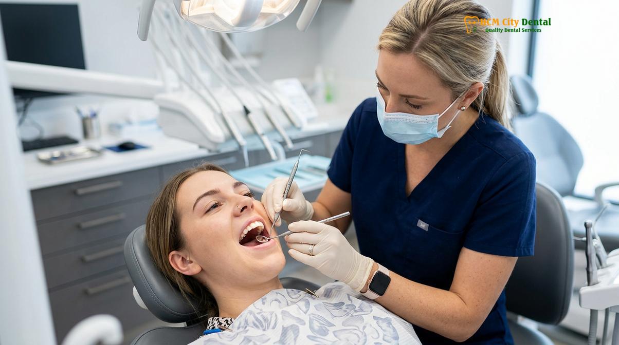
The etiology of aggressive periodontitis is deeply rooted in a specific microbiological profile. While chronic gum disease is generally associated with a broad spectrum of plaque bacteria, aggressive cases are frequently dominated by highly virulent, tissue-invasive pathogens. The most notorious of these is Aggregatibacter actinomycetemcomitans (Aa). This Gram-negative, facultative anaerobe possesses an array of virulence factors that allow it to colonize the subgingival pocket, evade host defenses, and initiate profound tissue destruction [1]. One of its primary weapons is a potent leukotoxin, which actively targets and destroys human neutrophils and macrophages—the very white blood cells dispatched by the immune system to fight the infection.
In addition to A. actinomycetemcomitans, other pathogens such as Porphyromonas gingivalis and Capnocytophaga species are frequently isolated from active, destructive lesions. These bacteria produce collagenases and other proteolytic enzymes that directly break down the connective tissue of the gums. Furthermore, they stimulate the host’s immune cells to release pro-inflammatory cytokines, such as Interleukin-1 beta (IL-1β) and Tumor Necrosis Factor-alpha (TNF-α), which inadvertently trigger osteoclasts (bone-resorbing cells) to break down the alveolar bone. This hyper-inflammatory response is a hallmark of the disease’s pathogenesis.
Understanding the nuances of comprehensive gum treatment is essential when addressing such virulent infections. Because these bacteria can physically invade the gingival connective tissues—moving beyond the surface of the tooth root—superficial cleaning is entirely ineffective. The bacteria residing within the tissue act as a reservoir, ready to recolonize the periodontal pocket immediately after standard cleaning procedures. This tissue-invasive characteristic is the primary reason why aggressive periodontitis requires a fundamentally different therapeutic approach compared to standard gingivitis or chronic periodontitis.
How Aggressive Periodontitis Differs from Chronic Type
Unlike chronic periodontitis which progresses slowly with heavy plaque, the aggressive form causes rapid tissue destruction that is often disproportionate to the minimal amount of dental plaque present.
Distinguishing aggressive periodontitis from its chronic counterpart is a critical step in periodontal diagnostics. While both conditions ultimately lead to the loss of tooth-supporting structures, their clinical presentation, rate of progression, and underlying host-microbe interactions are vastly different. Chronic periodontitis is generally considered a disease of aging, developing slowly over decades as a direct result of long-term plaque and calculus (tartar) accumulation. In contrast, aggressive periodontitis can progress three to four times faster, causing devastating bone loss in a matter of months or a few short years.
One of the most striking clinical differences is the plaque-to-destruction ratio. In chronic cases, the amount of bone loss is typically commensurate with the volume of local irritants (plaque and calculus) and the patient’s oral hygiene habits. However, in patients suffering from aggressive periodontitis, clinicians frequently observe severe, deep periodontal pockets and massive bone loss despite the patient having relatively clean teeth and minimal visible plaque. This disproportionate destruction highlights that the disease is driven more by the high virulence of specific bacteria and the patient’s hyper-responsive or defective immune system, rather than mere plaque volume [2].
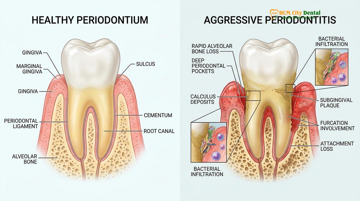
To fully understand the clinical divergence, we must examine the host immune response. Patients with chronic periodontitis generally have a normal, albeit overwhelmed, immune response to a massive bacterial load. Conversely, patients with aggressive periodontitis often exhibit specific, localized immune defects. For instance, a significant percentage of these patients demonstrate abnormalities in neutrophil chemotaxis—the process by which white blood cells navigate toward the site of infection. Because the neutrophils are sluggish or misdirected, the bacteria are allowed to multiply and invade tissues unchecked.
| Clinical Feature | Aggressive Periodontitis | Chronic Periodontitis |
|---|---|---|
| Age of Onset | Typically under 30 years (often puberty) | Usually over 35 years |
| Rate of Progression | Rapid (3 to 4 times faster) | Slow to moderate |
| Plaque/Calculus Levels | Often minimal, disproportionate to destruction | Abundant, correlates with destruction |
| Primary Pathogens | A. actinomycetemcomitans, P. gingivalis | Mixed complex flora (Red complex) |
| Familial Aggregation | Highly common (strong genetic link) | Less pronounced |
Because of these fundamental differences, the standard approach of deep cleaning teeth via scaling and root planing must be heavily modified. While mechanical debridement remains the cornerstone of therapy, it is rarely sufficient on its own for aggressive cases. The rapid progression demands immediate, intensive intervention to stabilize the dentition before the teeth become unsalvageable.
Localized vs Generalized Aggressive Periodontitis in Youth
The localized form primarily targets first molars and incisors with minimal inflammation, while the generalized form affects at least three other permanent teeth with more pronounced gingival inflammation.
Within the spectrum of aggressive periodontal diseases, clinicians recognize two distinct phenotypic presentations: Localized Aggressive Periodontitis (LAP) and Generalized Aggressive Periodontitis (GAP). While both share the hallmark of rapid tissue destruction in young individuals, their clinical patterns, age of onset, and immunological profiles differ significantly. Understanding these differences is vital for formulating an effective juvenile periodontitis treatment plan.
Localized Aggressive Periodontitis (LAP) typically has its onset around the time of puberty. It presents with a highly specific and somewhat perplexing pattern of bone loss: it almost exclusively targets the first permanent molars and the incisors. A patient with LAP may have severe, deep vertical bone defects around these specific teeth, while the premolars and second molars remain completely healthy and unaffected. Clinically, the gingival tissues in LAP patients often appear surprisingly healthy, with minimal signs of clinical inflammation, redness, or swelling, which can easily lead to missed diagnoses if periodontal probing and radiographic examinations are not routinely performed [3].
“The localized form of aggressive periodontitis presents a unique diagnostic challenge; the gingival tissues may appear deceptively pristine while profound, silent bone destruction occurs rapidly around the first molars and incisors.”
Immunologically, patients with LAP often mount a very robust, specific antibody response to the infecting pathogens. They typically produce high titers of IgG2 antibodies directed against A. actinomycetemcomitans. It is hypothesized that this strong antibody response is what localizes the infection, preventing the bacteria from spreading to the rest of the dentition. The disease remains confined to the teeth that erupted first (incisors and first molars), which were colonized by the bacteria before the immune system could mount its localized defense.
Conversely, Generalized Aggressive Periodontitis (GAP) usually affects individuals in their late teens or twenties. As the name implies, the destruction is widespread. By definition, GAP involves generalized interproximal attachment loss affecting at least three permanent teeth other than the first molars and incisors. Unlike LAP, the gingival tissues in GAP patients frequently exhibit acute signs of inflammation. The gums may appear fiery red, swollen, and bleed profusely upon probing. During active phases of the disease, patients may even experience systemic symptoms such as malaise or weight loss, alongside severe oral manifestations.
Patients with GAP generally lack the protective, high-titer antibody response seen in LAP. Their inability to mount an effective IgG2 defense allows the periodontal pathogens to proliferate unchecked and spread throughout the entire mouth. The generalized nature of the destruction makes GAP particularly devastating, often requiring extensive general dentistry and specialized periodontal interventions to prevent full-mouth edentulism at a young age.
The Strong Genetic and Familial Link
A hallmark of aggressive periodontitis is its strong familial aggregation, heavily influenced by genetic anomalies that impair neutrophil chemotaxis and phagocytosis in the host immune response.
One of the most defining characteristics of aggressive periodontitis is its profound tendency to run in families. When a young patient is diagnosed with this condition, it is a standard clinical protocol to strongly recommend periodontal screenings for all siblings and close relatives. This familial aggregation is not merely a result of shared environmental factors or transmission of bacteria within a household; it is deeply rooted in genetic susceptibility.
Extensive genetic research has demonstrated that aggressive periodontitis, particularly the localized form, often follows an autosomal dominant mode of inheritance, though autosomal recessive patterns have also been observed depending on the population studied. The genetic anomalies primarily affect the host’s immune system, specifically the function of polymorphonuclear leukocytes (neutrophils). Neutrophils are the first responders of the immune system, tasked with migrating to the site of infection (chemotaxis) and engulfing and destroying bacteria (phagocytosis).
Clinical Case Review: Familial Aggregation
A 16-year-old patient presented to HCMC Dental Clinic in Ho Chi Minh City with severe mobility in the lower first molars. Radiographs revealed advanced vertical bone loss characteristic of localized aggressive periodontitis. Recognizing the genetic implications, Dr. Nguyen Van Cuong immediately requested examinations for the patient’s two younger siblings. Despite having no outward symptoms, the 14-year-old sibling was found to have early-stage pocketing around the incisors. Early intervention for the sibling prevented the severe bone destruction experienced by the older teenager, highlighting the critical importance of family screening in genetic gum disease family history.
In many patients with aggressive periodontitis, there is a genetically determined defect in neutrophil chemotaxis. The white blood cells are essentially “blinded” or sluggish, unable to efficiently navigate the chemical gradients to reach the periodontal pocket where the bacteria are multiplying. Furthermore, genetic variations (polymorphisms) in the genes encoding for various cytokines, such as Interleukin-1 (IL-1) and Fc gamma receptors (FcγR), can lead to a hyper-inflammatory response. When the immune system finally does respond, it overreacts, releasing excessive amounts of enzymes and inflammatory mediators that destroy the patient’s own bone and connective tissue in an attempt to clear the infection.
This genetic predisposition means that even with immaculate oral hygiene, susceptible individuals may still develop the disease if the specific pathogens are introduced into their oral microbiome. It underscores the necessity of personalized medicine in periodontics, where understanding a patient’s genetic background is just as important as evaluating their clinical symptoms.
Rapid Bone Destruction: The Warning Signs
Early warning signs include increasing tooth mobility, drifting of incisors, and deep periodontal pockets, often occurring before significant pain or visible tartar buildup alerts the patient.
Because aggressive periodontitis progresses so swiftly, recognizing the early warning signs is paramount for tooth preservation. Unfortunately, the disease is often “silent” in its early stages, causing profound structural damage before the patient experiences any significant pain. The rapid bone loss in young adults is frequently discovered incidentally during routine radiographic examinations, emphasizing the need for regular dental check-ups.
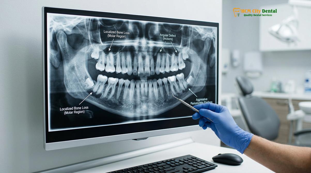
One of the earliest and most common clinical signs reported by patients is tooth mobility. A young adult may notice that their teeth feel slightly loose when chewing or that they can shift the teeth with their tongue. This mobility is a direct result of the rapid destruction of the periodontal ligament fibers that anchor the tooth to the bone. Alongside mobility, pathologic migration of the teeth is highly indicative of aggressive disease. Patients may observe that their front teeth (incisors) are beginning to flare outward or that new gaps (diastemas) are forming between teeth that were previously straight [4].
Clinical Warning:
Tooth mobility, shifting, or the sudden appearance of gaps between teeth in adolescents or young adults should never be ignored or attributed merely to orthodontic shifting. These are classic signs of rapid attachment loss and require immediate periodontal probing and radiographic evaluation to rule out aggressive periodontitis.
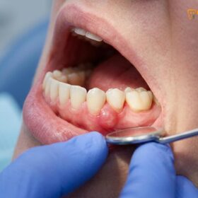
During a clinical examination, a dentist will use a periodontal probe to measure the depth of the sulcus (the space between the gum and the tooth). In a healthy mouth, these depths are typically 1 to 3 millimeters. In aggressive periodontitis, probing depths can rapidly increase to 6, 8, or even 10 millimeters or more. These deep pockets create an anaerobic (oxygen-free) environment that is highly conducive to the proliferation of the virulent bacteria driving the disease. Furthermore, patients may experience swollen gums around the tooth, particularly in the generalized form of the disease, accompanied by spontaneous bleeding or purulent exudate (pus) expressing from the gumline.
Radiographically, the bone loss patterns in aggressive periodontitis are highly distinctive. In the localized form, X-rays typically reveal classic “arc-shaped” or vertical bone defects extending down the roots of the first molars and incisors. The crestal lamina dura—the dense layer of bone lining the tooth socket—is rapidly obliterated. Identifying these specific radiographic markers early is the key to implementing therapies that can halt the destruction and potentially regenerate the lost bone.
Surgical and Antibiotic Protocols for Aggressive Cases
Successful management requires a combined approach of mechanical debridement, systemic antibiotics to eradicate tissue-invasive bacteria, and often periodontal flap surgery to regenerate lost bone.
The treatment of aggressive periodontitis is one of the most challenging endeavors in clinical dentistry. Because the disease is characterized by tissue-invasive bacteria and a compromised host response, standard non-surgical therapy alone is rarely adequate. A comprehensive, multi-phased treatment protocol is required to arrest disease progression, eradicate the pathogenic flora, and restore the health and function of the periodontium.
Phase I: Antimicrobial and Mechanical Therapy
The initial phase of treatment focuses on the immediate reduction of the bacterial load. This begins with meticulous scaling and root planing (SRP) to mechanically remove biofilm and calculus from the root surfaces. However, because pathogens like A. actinomycetemcomitans invade the gingival connective tissue, mechanical scraping of the root surface cannot reach the bacteria hiding within the gums. Therefore, systemic antibiotic therapy is a mandatory adjunct in the treatment of aggressive periodontitis.
The gold standard antibiotic regimen involves a synergistic combination of Amoxicillin and Metronidazole. Amoxicillin targets the cell wall synthesis of the bacteria, while Metronidazole disrupts the DNA synthesis of obligate anaerobes. When prescribed together (typically Amoxicillin 500mg and Metronidazole 400mg or 250mg, taken three times daily for 7 to 14 days), they provide a profound bactericidal effect against the specific pathogens driving the disease. Dr. Cuong frequently emphasizes that the timing of antibiotic administration is critical; the antibiotics must be taken concurrently with the mechanical debridement to ensure the bacteria are attacked systemically while their biofilm defenses are physically disrupted [5].
“The adjunctive use of systemic antibiotics, specifically the combination of amoxicillin and metronidazole, alongside mechanical debridement, is not merely an option but a clinical necessity in arresting the rapid tissue destruction seen in aggressive periodontitis.”
Phase II: Surgical Intervention and Regeneration
Following the initial phase, the patient is re-evaluated after 4 to 6 weeks. If deep periodontal pockets (typically ≥ 5mm) remain, surgical intervention is indicated. The primary goal of surgery is to gain direct visual access to the root surfaces for definitive cleaning and to reshape or regenerate the damaged bone.
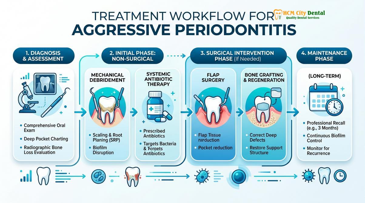
Gum flap surgery, specifically the Modified Widman Flap or apically positioned flap, is frequently utilized. The surgeon gently reflects the gum tissue away from the teeth, allowing for complete eradication of the deep-seated granulation tissue and calculus. In cases of localized aggressive periodontitis where vertical bone defects are present, Guided Tissue Regeneration (GTR) is highly effective. This advanced technique involves placing bone grafts (allografts or xenografts) into the bony defect and covering them with a biocompatible barrier membrane. The membrane prevents the fast-growing gum tissue from filling the hole, allowing the slower-growing bone cells time to regenerate and rebuild the lost support.
In modern clinical settings, laser gum treatment is also emerging as a powerful adjunctive tool. Specific dental lasers, such as Nd:YAG or Diode lasers, can be used to selectively ablate diseased epithelial lining and vaporize tissue-invasive bacteria within the pocket without the need for extensive scalpel incisions. The laser energy also promotes biostimulation, encouraging faster healing and reattachment of the connective tissue to the root surface.
Phase III: Rigorous Periodontal Maintenance
Aggressive periodontitis is never truly “cured”; it is managed. Patients who have been stabilized require a lifetime of rigorous periodontal maintenance. Standard six-month dental cleanings are insufficient. These patients must be placed on a strict 3-month recall schedule. During these visits, the clinician will re-probe all pocket depths, assess bleeding scores, and perform targeted subgingival debridement to disrupt any newly forming pathogenic biofilms before they can trigger a relapse of bone destruction. Adjunctive use of antimicrobial mouth rinses, such as chlorhexidine gluconate, and meticulous home care are non-negotiable components of long-term success.
When to See a Doctor (Clinical Considerations)
Early detection is the most critical factor in managing aggressive periodontitis. You should seek immediate evaluation from a qualified dental professional if you or your child experience any sudden changes in dental alignment, such as teeth shifting, flaring, or the development of new spaces between teeth. Additionally, if you notice increasing tooth mobility, persistent bad breath associated with gum disease, or if you observe dark or severely inflamed gums that bleed easily during brushing, do not delay care.
Given the strong genetic component, if a direct family member has been diagnosed with early-onset severe gum disease or has lost teeth at a young age, you should proactively schedule a comprehensive periodontal screening. At HCMC Dental Clinic, our clinical team utilizes advanced diagnostic probing and digital radiography to detect the earliest signs of bone loss. Treatment recommendations are always based on personalized clinical diagnostics, ensuring that therapeutic interventions are tailored to the specific microbiological and anatomical needs of each patient. While advanced therapies have a high success rate in halting disease progression, outcomes depend heavily on the stage of the disease at the time of intervention.
Frequently Asked Questions
Can young people get severe periodontal disease?
Yes, young people can develop severe periodontal disease, particularly a rapidly progressing form known clinically as aggressive periodontitis. This condition often affects adolescents and young adults who are otherwise completely healthy, driven by specific bacterial infections and genetic immune vulnerabilities rather than poor oral hygiene alone. Early diagnosis is critical to prevent irreversible bone loss.
Why is my bone loss progressing so fast?
Rapid bone loss is typically caused by highly virulent bacteria, such as Aggregatibacter actinomycetemcomitans, combined with a compromised local immune response. In aggressive periodontitis, the host’s white blood cells (neutrophils) may have chemotactic defects, meaning they cannot efficiently travel to the infection site to destroy bacteria, allowing the infection to rapidly destroy the alveolar bone.
Is aggressive periodontitis genetic?
Yes, there is a strong genetic predisposition associated with aggressive periodontitis. Familial aggregation is a hallmark of the disease, meaning it frequently runs in families. Genetic mutations can affect the immune system’s inflammatory response and neutrophil function, making certain individuals highly susceptible to rapid periodontal destruction even with minimal dental plaque.
What is the treatment for localized aggressive periodontitis?
Treatment for localized aggressive periodontitis typically involves a combination of mechanical scaling and root planing, systemic antibiotics (often Amoxicillin and Metronidazole), and strict home care. Because the bacteria invade the gum tissue, mechanical cleaning alone is insufficient. Advanced cases may also require periodontal flap surgery or laser therapy to regenerate lost bone and restore attachment.
Can aggressive periodontitis cause early tooth loss?
Without prompt and aggressive clinical intervention, aggressive periodontitis frequently leads to early tooth loss. The rapid destruction of the periodontal ligament and alveolar bone removes the foundational support for the teeth, leading to severe mobility and eventual exfoliation or the need for extraction, particularly affecting the first molars and incisors in young patients.
References
- Journal of Periodontology. Pathogenesis and microbiology of aggressive periodontitis. (2018).
- International Dental Journal. Systemic antibiotics in the treatment of aggressive periodontal diseases. (2020).
- Clinical Advances in Periodontics. Genetic and familial risk factors in early-onset periodontitis. (2019).
- Journal of Clinical Periodontology. Radiographic bone loss patterns in localized and generalized aggressive periodontitis. (2021).
- Periodontology 2000. Surgical management and regenerative therapy in severe periodontal defects. (2022).