The pinhole surgical technique is a minimally invasive periodontal procedure designed to treat gum recession without scalpels or sutures. By creating a tiny entry point, dentists gently reposition existing gum tissue over exposed roots, offering rapid recovery and immediate cosmetic improvement compared to traditional grafting methods.
Clinical Summary:
The pinhole surgical technique (PST) represents a significant paradigm shift in the management of marginal tissue recession. Unlike conventional connective tissue grafts that require harvesting donor tissue from the palate, this scalpel-free, suture-free approach utilizes specialized instruments to loosen and glide the patient’s existing gingival tissue coronally. Bio-absorbable collagen strips are subsequently inserted through the micro-incision to stabilize the new gum position and support cellular regeneration. Clinical studies demonstrate high root coverage predictability, particularly for Miller Class I and II recession defects. With significantly reduced postoperative pain, minimal bleeding, and rapid visual healing, PST offers a highly efficient, patient-friendly alternative to traditional periodontal plastic surgery, allowing multiple teeth to be treated in a single clinical session.
Key Takeaways:
- PST is a minimally invasive procedure requiring no scalpels, large incisions, or traditional sutures.
- The technique utilizes the patient’s existing local gum tissue rather than harvesting from the palate.
- Multiple adjacent teeth with recession can be treated simultaneously in a single appointment.
- Bio-absorbable collagen strips act as a biological scaffold to stabilize the repositioned gums.
- Recovery time and postoperative discomfort are drastically reduced compared to conventional grafting.
- Ideal candidates are those with Miller Class I or II recession and adequate attached gingiva.
What is the Pinhole Surgical Technique (PST)?
The Pinhole Surgical Technique (PST) is an advanced, scalpel-free periodontal procedure that corrects marginal gum recession by gently repositioning the patient’s existing gingival tissue through a tiny micro-puncture.
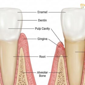
Marginal tissue recession, commonly known as gum recession, is a prevalent periodontal condition characterized by the apical migration of the gingival margin away from the cementoenamel junction (CEJ). This exposes the underlying root surface, leading to dentinal hypersensitivity, an increased risk of root caries, and significant aesthetic concerns. Historically, the gold standard for treating this condition involved complex grafting procedures. However, the introduction of the pinhole surgical technique has revolutionized comprehensive periodontal care by offering a highly predictable, minimally invasive alternative.
Developed to address the limitations and patient apprehension associated with traditional surgery, PST operates on the principle of tissue preservation rather than tissue replacement. By utilizing a specialized set of instruments, the clinician can access the subgingival structures through a single 2-3 millimeter entry point in the alveolar mucosa. This approach qualifies as a highly effective minimally invasive recession surgery, as it completely eliminates the need for broad flap elevation, releasing incisions, and complex suturing techniques[1].
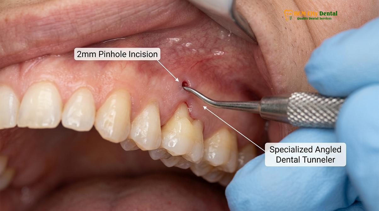
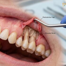
The biological rationale behind PST relies on the elasticity and vascularity of the mucogingival complex. When the periosteum is gently detached from the underlying bone, the entire tissue flap becomes mobile. This allows the clinician to advance the tissue coronally (downward over the upper teeth, or upward over the lower teeth) to cover the exposed root surfaces. Because the blood supply remains largely intact and the tissue is not severed from its primary vascular network, the healing process is accelerated, and the risk of tissue necrosis is significantly minimized.
How PST Rejuvenates Receded Gums Without Cuts
Instead of utilizing scalpels to cut and transfer tissue, PST employs specialized angled instruments inserted through a pinhole to perform a closed blunt dissection, freeing the gums to glide over the roots.
The mechanics of pinhole gum rejuvenation rely heavily on the precise manipulation of the mucogingival junction and the underlying connective tissue fibers. In a healthy periodontium, the gums are firmly attached to the underlying alveolar bone and tooth root by a dense network of collagenous fibers. To correct recession without cutting, these attachments must be gently released. The clinician creates a small access point—the “pinhole”—typically located high in the mucosal tissue above the affected teeth.
Through this micro-incision, custom-designed elevators are introduced. The clinician performs a supraperiosteal closed blunt dissection, carefully sweeping the instruments apically and laterally to separate the muscular and fibrous adhesions from the bone. This process extends to the interdental papillae, ensuring complete passive mobilization of the entire tissue complex. Once freed, the tissue can be easily draped over the exposed roots without any tension[2].
“The true elegance of the pinhole technique lies in its profound respect for the patient’s native vascular architecture. By avoiding releasing incisions, we maintain optimal blood flow to the repositioned tissue, which is the cornerstone of rapid, predictable periodontal healing.”
— Dr. Nguyen Van Cuong, Clinical Director
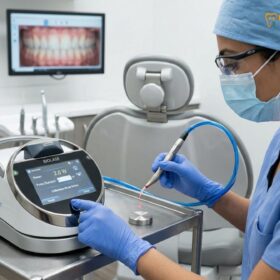
To maintain the tissue in its new coronal position without sutures, the technique utilizes bio-absorbable collagen strips. These strips are meticulously packed through the pinhole and into the interdental spaces. The collagen serves a dual purpose: mechanically, it creates “fullness” in the papillae that locks the tissue in place; biologically, it acts as a scaffold for fibroblasts and angiogenesis, promoting the formation of new tissue attachments. This innovative stabilization method is a key differentiator from other procedures, including advanced laser periodontal therapies, which focus primarily on disease eradication rather than physical tissue repositioning.
Pinhole Technique vs Traditional Gum Grafting
While traditional grafting requires harvesting donor tissue from the palate, the pinhole technique utilizes the patient’s local tissue, eliminating the painful secondary surgical site and the need for sutures.
When evaluating PST vs gum graft procedures, the most significant distinction lies in the patient experience and the surgical morbidity. Traditional connective tissue grafts (CTG) or free gingival grafts (FGG) have long been the standard for root coverage. These procedures involve creating a flap at the recession site, harvesting a piece of tissue from the roof of the patient’s mouth (the palate), transplanting it to the defect, and securing it with multiple sutures. While highly successful, the palatal donor site is notoriously uncomfortable during the healing phase.
The pinhole technique bypasses the need for a donor site entirely. By mobilizing the existing adjacent tissue, the procedure is confined to a single surgical area. This not only eliminates palatal pain but also drastically reduces the overall chair time. Furthermore, traditional grafting is often limited to treating one or two teeth per session due to the restricted amount of donor tissue available. In contrast, PST allows for the simultaneous treatment of an entire arch (10-14 teeth) in a single appointment, provided there is adequate local tissue volume[3].
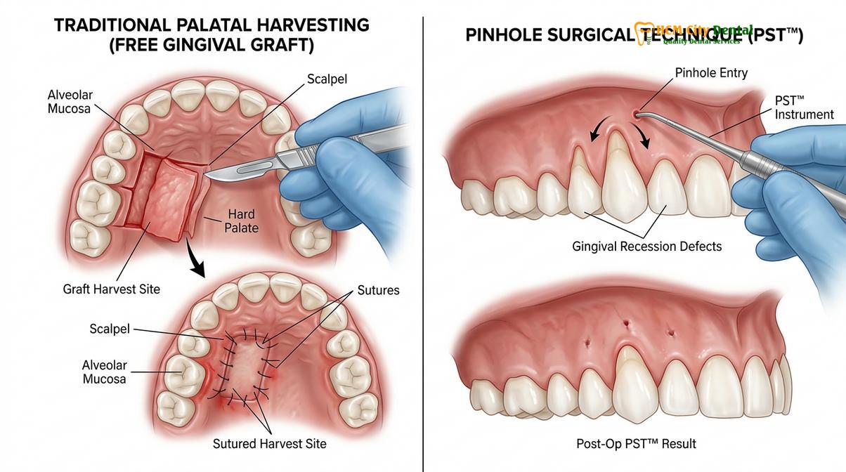
| Clinical Parameter | Pinhole Surgical Technique (PST) | Traditional Connective Tissue Graft |
|---|---|---|
| Incision Type | 2-3mm micro-puncture (no scalpels) | Broad flap elevation & palatal harvesting |
| Sutures Required | None (stabilized by collagen strips) | Extensive suturing at both sites |
| Donor Site Morbidity | None (uses local adjacent tissue) | High (palatal wound can be painful) |
| Teeth Treated per Session | Multiple (up to a full arch) | Limited (usually 1-3 teeth) |
| Visual Healing Time | 24 to 48 hours for the pinhole | 2 to 4 weeks for flap and palate |
Despite these advantages, it is crucial to understand that PST is not a universal replacement for traditional grafting. In cases where the patient has exceptionally thin biotypes or a complete lack of keratinized tissue, a traditional graft may still be required to thicken the gingiva and prevent future recession. A thorough clinical evaluation is necessary to determine the most appropriate approach, distinguishing between minimally invasive methods and traditional osseous surgical methods.
Step-by-Step Pinhole Gum Surgery Procedure
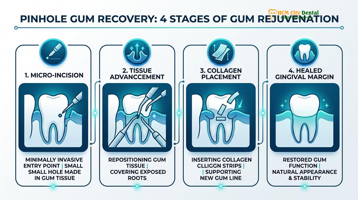
The procedure involves administering local anesthesia, creating a micro-puncture, loosening the gingival tissue with specialized elevators, advancing it coronally, and stabilizing the new gum line with collagen strips.
The execution of the pinhole surgical technique requires specialized training and a deep understanding of periodontal anatomy. The workflow is meticulously designed to maximize tissue mobilization while minimizing trauma. The procedure generally follows a strict sequence of clinical steps to ensure predictable root coverage and optimal aesthetic outcomes.
Step 1: Anesthesia and Site Preparation
The procedure begins with the administration of local infiltration anesthesia to ensure profound numbness in the treatment area. Because the technique is minimally invasive, the volume of anesthetic required is often less than that needed for extensive flap surgery. The exposed root surfaces are then thoroughly cleaned and prepared, often utilizing chemical root conditioning agents (like EDTA or citric acid) to remove the smear layer and facilitate cellular reattachment.
Step 2: The Pinhole Incision
Using a specialized needle or a micro-blade, the clinician creates a small 2-3 millimeter horizontal incision in the alveolar mucosa, typically positioned apical to the mucogingival junction of the centermost tooth in the treatment area. This single entry point provides access for multiple adjacent recession defects.
Step 3: Tissue Elevation and Mobilization
A series of custom-angled instruments are introduced through the pinhole. The clinician performs a closed blunt dissection, gently separating the periosteum from the bone. The instruments are swept laterally and apically to release all muscular and fibrous tension, particularly around the interdental papillae, until the entire tissue flap is completely passive and mobile[4].
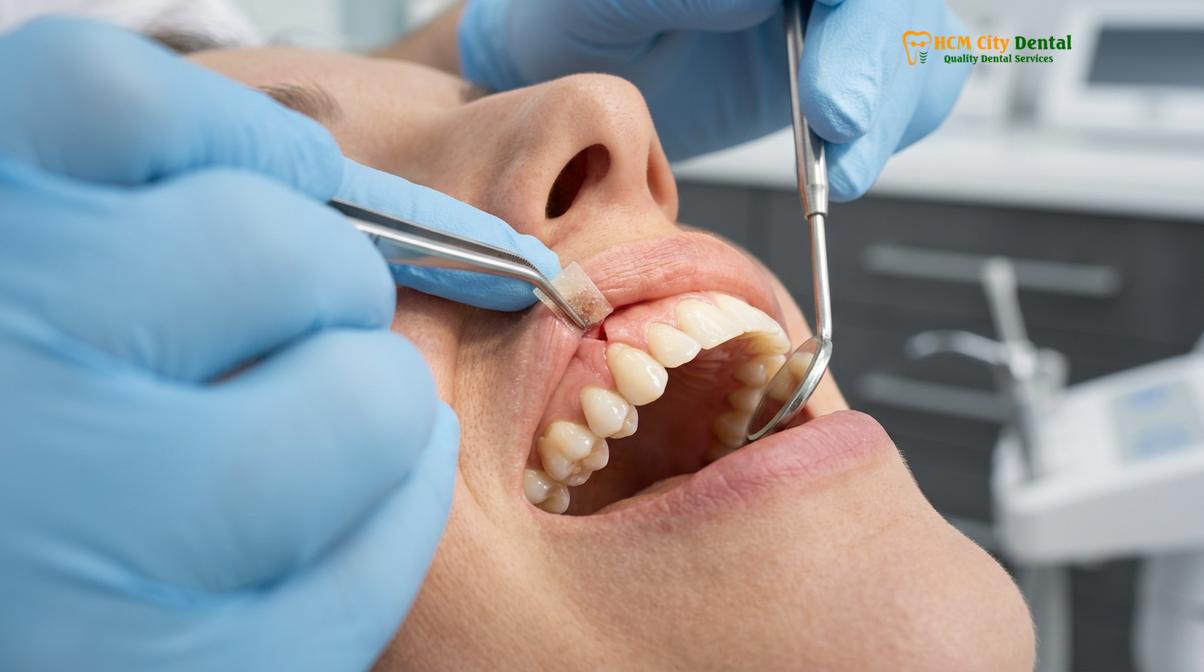
Step 4: Coronal Advancement
Once the tissue is fully released, it is gently guided coronally (downward for the maxilla, upward for the mandible) to cover the exposed root surfaces. The tissue should rest in its new position without any mechanical tension or pulling.
Step 5: Collagen Stabilization
To secure the tissue without sutures, bio-absorbable collagen membranes are cut into small strips. These strips are inserted through the pinhole and meticulously packed into the interdental spaces using specialized tampers. The collagen expands slightly upon contact with blood, creating a mechanical wedge that holds the tissue complex in its new coronal position while simultaneously providing a biological matrix for healing.
Clinical Case Review: Full-Arch Rejuvenation
A 38-year-old patient presented to HCMC Dental Clinic in Ho Chi Minh City complaining of severe root sensitivity and aesthetic concerns across six upper anterior teeth. Clinical examination revealed Miller Class I recession ranging from 2mm to 4mm. Dr. Nguyen Van Cuong performed the pinhole surgical technique under local anesthesia. The entire procedure was completed in under 90 minutes through two micro-incisions. At the 24-hour follow-up, the pinholes had closed, and the patient reported zero need for analgesic medication. At the 6-month evaluation, 100% root coverage was maintained with excellent tissue color match and complete resolution of dentinal hypersensitivity.
Recovery and Aftercare Following Pinhole Rejuvenation
Recovery from pinhole surgery is remarkably fast, requiring only mild analgesics, though patients must strictly adhere to brushing and dietary restrictions for several weeks to ensure tissue reattachment.
The postoperative phase of the pinhole surgical technique is characterized by rapid visual healing and minimal patient discomfort. Because there are no large incisions or sutures, the inflammatory response is significantly blunted. The 2-3mm access point typically seals itself within 24 to 48 hours. Most patients require nothing more than over-the-counter pain relievers (such as ibuprofen or paracetamol) for the first two to three days.
However, while the surface healing is swift, the underlying biological reattachment takes time. The newly positioned tissue is held in place solely by the collagen strips and the delicate fibrin clot that forms between the root surface and the gingiva. Any mechanical disruption during the first few weeks can dislodge the tissue and compromise the final result. Therefore, strict adherence to postoperative instructions is absolutely critical for success[5].
Important Postoperative Restrictions:
Patients must absolutely avoid brushing, flossing, or touching the surgical site for a minimum of 4 to 6 weeks. The use of electric toothbrushes and water flossers is strictly prohibited during the initial healing phase, as the mechanical force can easily detach the newly positioned gums.
During the non-brushing period, plaque control is maintained through the use of prescription antimicrobial mouthwashes (such as 0.12% Chlorhexidine gluconate). Patients are instructed to gently roll the liquid in their mouths rather than swishing vigorously. A soft diet is mandatory for the first week to prevent hard or crunchy foods from traumatizing the surgical area. Once the initial healing phase is complete, patients may be advised to undergo routine scaling and root planing in non-surgical areas to maintain overall periodontal health.
Am I a Good Candidate for Pinhole Surgery?
Ideal candidates for the pinhole technique have adequate attached gingiva and suffer from Miller Class I or II recession without severe underlying bone loss or active periodontal infections.
While the pinhole technique is highly versatile, careful patient selection is paramount to achieving predictable outcomes. The primary indication for PST is the treatment of Miller Class I and Class II marginal tissue recession. In these classifications, the recession does not extend past the mucogingival junction (Class I) or extends to/beyond it (Class II), but crucially, there is no loss of interdental bone or soft tissue. Because PST relies on the interdental papillae to hold the coronally advanced tissue, intact interproximal bone is a strict prerequisite.
Patients with Miller Class III or IV recession—where significant interdental bone loss has occurred—are generally not ideal candidates for this procedure, as the lack of papillary support makes tissue stabilization highly unpredictable. Additionally, patients must have a sufficient band of existing keratinized tissue to be mobilized. If the tissue is excessively thin or entirely absent, a traditional graft may be necessary to build tissue thickness.
“Patient selection is the ultimate determinant of success in minimally invasive periodontics. We must ensure the underlying bone architecture can support the repositioned tissue, and that all active inflammation is fully resolved prior to surgical intervention.”
— Dr. Nguyen Van Cuong
Furthermore, the periodontium must be healthy before surgery. Active periodontal disease, characterized by bleeding on probing, deep pocketing, or purulent exudate, is an absolute contraindication. Any active infection must be treated and stabilized first. Patients must also demonstrate excellent home care and be willing to correct the underlying causes of their recession, such as aggressive brushing habits or nocturnal bruxism. For international patients, the pinhole surgery cost is often a motivating factor, as specialized clinics in Vietnam offer this advanced procedure at a highly competitive rate while utilizing the exact same patented instruments and protocols as Western practices[6].
During the candidacy evaluation, the clinician will also assess other soft tissue parameters. If a patient presents with gingival pigmentation issues or halitosis linked to active gum infections, these conditions will be addressed as part of a comprehensive treatment plan prior to performing any root coverage procedures.
When to See a Doctor
Gum recession is a progressive condition that rarely halts without intervention. If you notice that your teeth appear longer than they used to, or if you experience sudden, sharp sensitivity when consuming hot, cold, or sweet foods, it is crucial to seek a professional periodontal evaluation. Exposed tooth roots are not protected by hard enamel; instead, they are covered by cementum, which is much softer and highly susceptible to rapid decay and toothbrush abrasion.
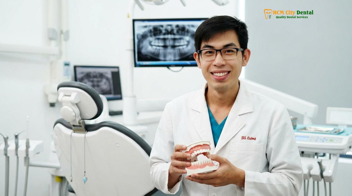
Do not wait until the recession causes significant pain or tooth mobility. Early intervention allows for more conservative, minimally invasive treatments like the pinhole technique. If you observe localized gingival swelling, persistent bleeding when brushing, or a visible notch near the gumline, schedule a consultation immediately. At HCMC Dental Clinic in Ho Chi Minh City, our periodontal specialists utilize advanced diagnostic imaging and precise clinical charting to determine the exact cause of your recession and develop a personalized, evidence-based treatment plan to restore your oral health and protect your smile.
Frequently Asked Questions
How does pinhole gum surgery work?
Pinhole gum surgery works by creating a tiny 2-3mm micro-incision in the alveolar mucosa, allowing specialized instruments to gently loosen and glide existing gum tissue over exposed tooth roots. Once the tissue is coronally advanced, bio-absorbable collagen strips are packed into the interdental spaces to stabilize the gums while natural healing and reattachment occur. This closed-flap approach preserves the blood supply, ensuring rapid cellular regeneration without the need for scalpels or sutures.
Is the pinhole surgical technique painful?
The pinhole surgical technique is generally associated with minimal postoperative discomfort, significantly less than traditional palatal grafting. Because there is no large incision or tissue harvesting from the roof of the mouth, most patients manage any mild soreness with standard over-the-counter analgesics for just a few days following the procedure. The micro-puncture itself typically heals and closes within 24 to 48 hours, making the recovery process highly tolerable.
How long does pinhole gum surgery last?
The clinical results of pinhole gum surgery are designed to be long-lasting, provided the patient maintains excellent oral hygiene and addresses underlying causes of recession. By eliminating aggressive brushing habits and controlling bruxism, the repositioned gingival tissue can remain stable and healthy for many years, effectively protecting the previously exposed roots. Regular periodontal maintenance and gentle brushing techniques are essential to preserve the surgical outcomes.
What is the cost of pinhole gum technique in Vietnam?
The pinhole surgery cost in Vietnam varies based on the number of teeth treated and the complexity of the recession defects. Generally, undergoing this advanced, minimally invasive procedure at a specialized center like HCMC Dental Clinic offers a highly cost-effective alternative compared to Western dental pricing, while maintaining strict international clinical standards. A personalized consultation is required to provide an accurate, case-specific financial estimate.
How fast is recovery from pinhole gum rejuvenation?
Recovery from pinhole gum rejuvenation is remarkably fast, with the initial micro-incision typically closing within 24 to 48 hours. While the visible healing is rapid and patients can resume normal daily activities almost immediately, the deeper tissue reattachment and collagen integration require several weeks of strict adherence to dietary and brushing restrictions. Patients must avoid mechanical trauma to the surgical site for up to six weeks to ensure permanent root coverage.
References
- International Journal of Periodontics & Restorative Dentistry. The Pinhole Surgical Technique for Treatment of Marginal Tissue Recession. (2012).
- Journal of Indian Society of Periodontology. Pinhole Surgical Technique for treatment of marginal tissue recession: A case series. (2018).
- Journal of Clinical Periodontology. Minimally invasive surgical techniques in periodontal regeneration. (2019).
- American Academy of Periodontology. Clinical guidelines on gingival augmentation and root coverage procedures. (2021).
- Journal of Periodontal Research. The role of collagen membranes in stabilizing coronally advanced flaps. (2020).
- Clinical Advances in Periodontics. Patient-reported outcomes following minimally invasive root coverage procedures. (2022).