Gum graft surgery is a specialized periodontal procedure designed to treat receding gums by adding healthy tissue to the affected area. This surgical intervention protects exposed tooth roots, reduces sensitivity, and restores the natural gumline to prevent further bone loss and decay.
Clinical Summary:
Gum graft surgery, or soft tissue grafting, is a highly effective periodontal treatment for gingival recession. By harvesting tissue from the palate or using donor matrices, periodontists can cover exposed roots, halt progressive tissue loss, and enhance aesthetic contours. Clinical success depends on precise surgical technique, proper patient selection, and strict adherence to postoperative care protocols. Modern advancements in microsurgery have significantly minimized discomfort, allowing for predictable root coverage and long-term periodontal stability.
Key Takeaways:
- Gum grafting restores lost tissue to protect exposed tooth roots from decay, structural damage, and hypersensitivity.
- The procedure utilizes either the patient’s own palatal tissue (autograft) or an acellular dermal matrix (allograft).
- Recovery typically spans one to two weeks, requiring a strict soft food diet and modified oral hygiene.
- Proper vascularization is critical; therefore, avoiding tobacco and mechanical trauma to the surgical site is mandatory.
- Early intervention prevents severe alveolar bone loss and potential tooth mobility or extraction.
What is Gum Graft Surgery?
Gum graft surgery involves transplanting healthy tissue to areas of gum recession to cover exposed roots and thicken the gingival margin. This prevents further tissue deterioration and protects the underlying jawbone.
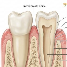
The periodontium is the specialized system of hard and soft tissues that supports your teeth and maintains them securely in the jaw. A critical component of this system is the attached gingiva—the thick, pink, keratinized tissue that tightly hugs the base of the teeth. When this tissue begins to wear away or pull back, a condition known as gingival recession occurs. This exposes the underlying tooth root, which lacks the protective enamel covering found on the crown of the tooth. Instead, the root is covered by cementum, a much softer substance that is highly susceptible to bacterial decay and mechanical abrasion.
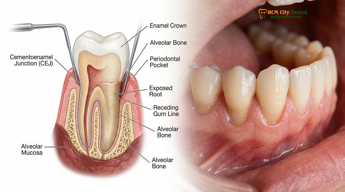
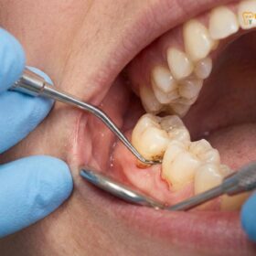
Gum graft surgery is the definitive clinical solution for this problem. By taking healthy tissue from another area—most commonly the roof of the mouth—and securing it over the exposed root, a periodontist can effectively rebuild the lost defense barrier. This procedure is not merely cosmetic; it is a vital structural repair. Without intervention, progressive recession can lead to the loss of the supporting alveolar bone, increased tooth mobility, and ultimately, tooth loss. The surgery aims to halt this progression, increase the width of the attached gingiva, and, when possible, achieve complete root coverage [1].
The etiology of gum recession is multifactorial. It can be triggered by aggressive toothbrushing with hard-bristled brushes, which physically wears away the delicate tissue. Periodontal disease (periodontitis) is another primary culprit, where bacterial toxins destroy the connective tissue and bone supporting the teeth. Additionally, genetic predispositions, such as naturally thin gingival biotypes, or orthodontic tooth movement that pushes teeth outside the bony housing, can exacerbate the condition. Identifying the root cause is a crucial step in the treatment planning phase, ensuring that the receding gums surgery provides a lasting, stable outcome rather than a temporary fix.
For patients exploring comprehensive oral health solutions, understanding the role of the gingiva is paramount. Procedures like grafting fall under the broader umbrella of General Dentistry and periodontics, focusing on establishing a healthy foundation before any restorative or cosmetic work is undertaken.
3 Main Types of Gum Grafts Explained
Periodontists primarily utilize connective tissue grafts, free gingival grafts, or pedicle grafts based on the patient’s specific anatomical needs, the severity of recession, and the desired aesthetic outcome.
Because every patient’s anatomical presentation is unique, there is no single approach to gingival graft surgery. The choice of technique depends heavily on the amount of existing tissue, the depth of the recession, and whether the primary goal is to cover an exposed root or simply to thicken the existing gum tissue to prevent future recession. The three primary autogenous (self-donated) grafting techniques are detailed below.
1. Connective Tissue Grafts
This is the most common and widely utilized method for treating root exposure. In this technique, the surgeon creates a small “trap door” or flap on the roof of the mouth (the palate). Instead of taking the surface tissue, the surgeon harvests the subepithelial connective tissue located underneath the flap. This harvested tissue is then carefully sutured to the recipient site, covering the exposed root. The palatal flap is then stitched back down. This method is highly favored because it provides excellent root coverage, blends seamlessly with the surrounding tissue color, and generally results in less postoperative discomfort at the donor site since the surface wound is closed [2].
2. Free Gingival Grafts
Unlike the connective tissue graft, a free gingival graft involves removing a small piece of tissue directly from the surface of the palate. This full-thickness graft includes both the surface epithelium and the underlying connective tissue. It is then attached directly to the area of recession. This technique is typically reserved for patients who have naturally thin gums and require an increase in the thickness and width of the keratinized tissue to halt further recession. While it is excellent for tissue augmentation, it is less commonly used for aesthetic root coverage because the grafted tissue often retains a slightly different color and texture (often described as a “tire patch” appearance) compared to the adjacent gums.
3. Pedicle Grafts (Lateral Sliding Flaps)
A pedicle graft is unique because it does not require harvesting tissue from the palate. Instead, the surgeon utilizes the healthy gum tissue immediately adjacent to the tooth with recession. A flap (pedicle) is partially cut away, leaving one edge attached to maintain its original blood supply. The flap is then stretched or rotated over the exposed root and sutured into place. This technique boasts an exceptionally high success rate because the blood vessels remain intact throughout the procedure. However, it can only be performed if the patient has an abundance of thick, healthy tissue adjacent to the recession site.
| Graft Type | Tissue Source | Primary Clinical Indication | Aesthetic Outcome |
|---|---|---|---|
| Connective Tissue | Subepithelial palatal tissue | Root coverage, treating sensitivity | Excellent color match and blending |
| Free Gingival | Surface palatal tissue | Thickening thin tissue, halting recession | Fair; may appear lighter or patch-like |
| Pedicle Graft | Adjacent gingival tissue | Root coverage when adjacent tissue is thick | Excellent; utilizes local tissue |
| Allograft (Matrix) | Acellular donor tissue bank | Treating multiple teeth, avoiding palatal wound | Very good; integrates well over time |
In modern periodontal practice, when patients require extensive grafting across multiple teeth, surgeons may opt for an acellular dermal matrix (allograft). This medically processed donor tissue eliminates the need for a palatal donor site entirely, significantly reducing surgical time and postoperative morbidity while providing an excellent scaffold for the patient’s own cells to repopulate and regenerate the gumline.
Why You Might Need a Connective Tissue Graft
A connective tissue graft is considered the gold standard for treating root exposure because it provides optimal blood supply and seamlessly blends with surrounding tissues for superior aesthetic and functional results.
When discussing a gum tissue graft procedure, the connective tissue graft frequently emerges as the treatment of choice, particularly for teeth in the aesthetic zone (the front of the mouth). The primary reason for its widespread application lies in its biological predictability and its ability to achieve dual objectives: covering the exposed, sensitive root surface and increasing the biotype (thickness) of the gingiva to prevent future relapse.
The biological mechanism behind the success of a connective tissue graft is fascinating. The tissue harvested from beneath the palatal surface is rich in fibroblasts—cells responsible for producing collagen and maintaining the structural integrity of connective tissues. When this tissue is placed over the exposed root and covered by the adjacent gum flap (a bilaminar technique), it receives a dual blood supply: one from the underlying periosteum (the membrane covering the bone) and one from the overlying mucosal flap. This rich vascular environment is crucial for the survival and integration of the graft during the initial healing phases of plasmatic imbibition and angiogenesis.
“The predictability of root coverage relies heavily on the vascular bed we create. A connective tissue graft, when properly stabilized, acts as a biological scaffold that not only protects the root but actively stimulates the regeneration of a robust, keratinized gingival margin.”
Patients often seek this specific treatment due to severe dentin hypersensitivity. When the root is exposed, the microscopic tubules leading to the tooth’s nerve center become open to the oral environment. Hot, cold, sweet, or acidic stimuli can cause sharp, sudden pain. By covering these tubules with a dense layer of connective tissue, the graft acts as a permanent biological bandage, effectively eliminating the sensitivity. Furthermore, restoring the gumline protects the root from cervical abrasion and root caries, which are notoriously difficult to treat and can compromise the longevity of the tooth.
If you are experiencing progressive recession, a thorough evaluation is necessary to determine if this approach is right for you. Comprehensive gum treatment protocols prioritize stabilizing the foundation of your smile before irreversible damage occurs to the supporting alveolar bone.
Step-by-Step Guide to the Grafting Procedure
The grafting procedure involves local anesthesia, meticulous site preparation, tissue harvesting, precise graft placement, and suturing to ensure optimal vascularization and seamless tissue integration.
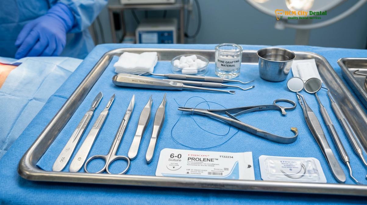
Understanding the clinical workflow of a gum graft can significantly alleviate patient anxiety. The procedure is performed using microsurgical techniques and specialized instruments to ensure precision and minimize tissue trauma. Here is a detailed, step-by-step breakdown of what occurs during the surgery.
Step 1: Diagnostic Preparation and Anesthesia
Before any incisions are made, the periodontist will thoroughly clean the affected area, removing any calculus (tartar) or bacterial plaque from the exposed root surface. This step, similar to deep dental scaling, ensures the root is biologically acceptable for the new tissue to attach. The surgical sites (both the recipient area and the palatal donor area) are then profoundly numbed using local anesthesia. Epinephrine is often included in the anesthetic to constrict blood vessels, minimizing bleeding and improving the surgeon’s visibility.
Step 2: Recipient Site Preparation
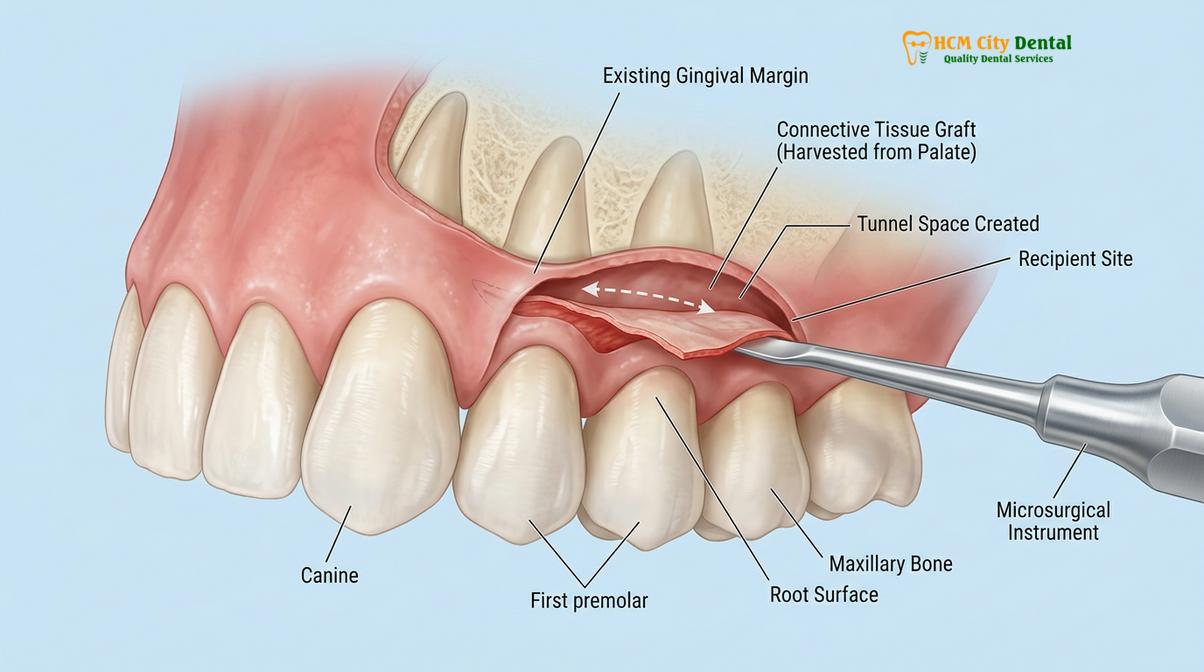
The surgeon carefully creates a split-thickness flap at the site of the recession. Using a micro-scalpel, the gum tissue is gently separated from the underlying bone and root, creating a small pouch or tunnel. The exposed root surface may be treated with chemical modifiers, such as EDTA or citric acid, to remove the smear layer and open the dentinal tubules, which encourages better cellular attachment of the graft [3].
Step 3: Tissue Harvesting
If an autograft is being used, the surgeon moves to the palate. For a connective tissue graft, a small, precise incision is made to lift the surface layer, and a measured section of the underlying connective tissue is carefully excised. The surface flap is then immediately sutured closed. If an allograft (donor matrix) is utilized, this step is entirely bypassed, and the pre-packaged matrix is hydrated and trimmed to the exact dimensions required.
Step 4: Graft Placement and Stabilization
The harvested tissue or matrix is carefully inserted into the prepared pouch or tunnel at the recipient site. Positioning is critical; the graft must completely cover the exposed root and extend slightly beyond the margins to ensure adequate blood supply. The graft is then immobilized using fine, often resorbable, sutures. Stabilization is the most critical factor for success; any micro-movement of the graft during the first few days can disrupt the fragile new blood vessels forming between the graft and the host bed.
Clinical Case Example: A 34-year-old patient visited HCMC Dental Clinic in Ho Chi Minh City presenting with severe root sensitivity and 4mm of recession on the lower canines due to aggressive brushing. Dr. Nguyen Van Cuong utilized a minimally invasive tunneling technique combined with a connective tissue graft. By avoiding vertical incisions, the blood supply was maximized. Within four weeks, the patient achieved 100% root coverage, complete resolution of sensitivity, and a highly aesthetic, scar-free result.
Step 5: Final Suturing and Protection
Once the graft is secured, the overlying flap is advanced coronally (pulled upward toward the crown of the tooth) to cover as much of the graft as possible and sutured into place. The surgeon may apply a periodontal dressing—a putty-like material—over the surgical site to protect it from mechanical trauma during eating and speaking in the initial days of healing.
Gum Graft Recovery Timeline and Diet Guidelines
Healing occurs in distinct biological phases, requiring a strict soft-food diet and modified oral hygiene practices for the first two weeks to prevent graft displacement and postoperative infection.
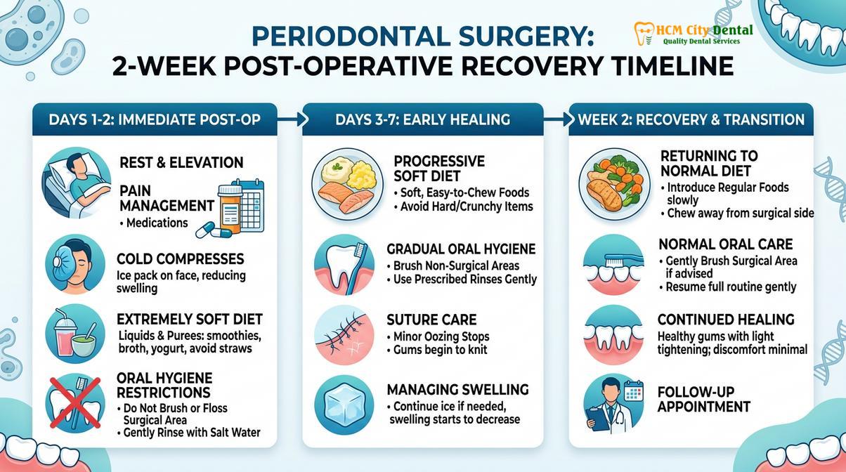
The success of a gum graft relies just as much on the patient’s postoperative compliance as it does on the surgeon’s skill. The healing process is delicate. During the first 24 to 72 hours, the graft survives solely on plasmatic imbibition—absorbing nutrients and oxygen directly from the surrounding fluid bed. It is not until days 3 to 7 that true angiogenesis (the formation of new blood vessels) begins to connect the graft to the host blood supply [4].
Days 1 to 3: The Critical Phase
Immediately following surgery, swelling and mild to moderate discomfort are expected, particularly at the palatal donor site. Patients are advised to apply ice packs to the outside of the face to minimize swelling. Bleeding should be minimal, though saliva may appear pink. During this phase, physical exertion must be strictly avoided to prevent an increase in blood pressure that could cause bleeding or graft displacement. Pain is typically managed effectively with over-the-counter NSAIDs (like Ibuprofen) or prescribed analgesics.
Days 4 to 14: Vascularization and Integration
Swelling generally peaks around day three and then begins to subside. The grafted area may look white, yellowish, or slightly sloughy—this is a normal part of the surface tissue turning over and does not necessarily indicate infection or failure. Sutures may begin to dissolve or loosen. It is imperative that patients do not pull on their lips or cheeks to inspect the graft, as this tension can tear the delicate healing tissues.
Dietary Restrictions
Nutrition plays a vital role in wound healing, but the mechanical action of eating poses a risk to the graft. A strict soft-food diet is mandatory for the first 10 to 14 days.
- Foods to Consume: Yogurt, scrambled eggs, mashed potatoes, lukewarm soups, smoothies (eaten with a spoon, never a straw), and flaky fish.
- Foods to Strictly Avoid: Hard, crunchy, or sharp foods (chips, nuts, crusty bread), spicy foods, acidic foods (citrus, tomatoes), and extremely hot foods or beverages that could dissolve the blood clot or irritate the wound.
Oral Hygiene Modifications
Traditional brushing and flossing at the surgical site are strictly prohibited until cleared by the periodontist. Mechanical trauma from toothbrush bristles will destroy the graft. Instead, patients are usually prescribed a chlorhexidine gluconate antimicrobial mouthrinse to gently bathe the area twice a day, preventing bacterial accumulation. The rest of the mouth can be brushed normally, taking great care to avoid the surgical zones.
Potential Risks and How to Ensure Graft Success
While highly successful, potential risks include graft failure, infection, or excessive bleeding, which can be mitigated by strictly following postoperative instructions and completely avoiding smoking.
Gum grafting is a highly predictable procedure with excellent long-term success rates when performed correctly and cared for properly. However, as with any surgical intervention, there are inherent risks. Understanding these risks and the biological requirements for healing empowers patients to take an active role in their recovery.
The most significant risk is graft failure, which occurs when the transplanted tissue fails to establish a blood supply (vascularize) and subsequently undergoes necrosis (dies). If a graft fails, the tissue will turn completely white and slough away, leaving the root exposed just as it was prior to surgery. The primary culprits for graft failure are mechanical trauma (brushing the area too soon, chewing hard foods, or pulling the lip to look at the site) and smoking.
Clinical Warning on Tobacco Use: Smoking or using any form of tobacco is an absolute contraindication for optimal healing. Nicotine is a potent vasoconstrictor, meaning it shrinks blood vessels and severely restricts the flow of oxygen and nutrients to the surgical site. Patients who smoke face a exponentially higher risk of graft necrosis, infection, and complete procedural failure. It is strongly advised to cease smoking for at least two weeks prior to surgery and four weeks post-operatively.
Infection is another potential complication, though relatively rare due to the excellent blood supply in the oral cavity and the routine use of antimicrobial rinses or prophylactic antibiotics. Signs of infection include increasing pain after the first few days, persistent swelling, purulent discharge (pus), and fever. If any of these symptoms occur, immediate clinical evaluation is required.
Bleeding from the palatal donor site can occasionally be a nuisance. The palate contains larger blood vessels, and if the protective stent or dressing is dislodged, bleeding may resume. Applying firm, continuous pressure with a moistened tea bag (which contains tannic acid, a natural coagulant) for 20-30 minutes usually resolves the issue [5].
To ensure maximum success, patients must adhere strictly to the postoperative protocol. This includes attending all scheduled follow-up appointments so the surgeon can monitor the integration of the tissue and safely remove any non-resorbable sutures. Long-term success also dictates a change in the habits that caused the recession initially—such as switching to an ultra-soft toothbrush and adopting a gentle, sweeping brushing technique.
When to See a Doctor
While minor swelling and mild discomfort are standard parts of the healing trajectory, certain symptoms warrant immediate professional attention. You should contact your dental provider if you experience uncontrolled bleeding that does not stop with applied pressure, a sudden spike in pain that is unresponsive to medication, or signs of systemic infection such as a fever or swollen lymph nodes.
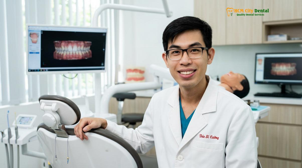
Furthermore, if you notice your gums beginning to recede, exposing the yellowish root surface, or if you experience sharp sensitivity when consuming cold beverages, it is time to schedule a clinical evaluation. Early diagnosis is critical. Waiting until the recession has caused significant bone loss limits the surgical options and decreases the predictability of complete root coverage.
“Proactive periodontal care is always preferable to reactive surgery. By addressing gingival recession early, we can utilize less invasive techniques, preserve the underlying bone, and ensure the long-term retention of the natural dentition.” — Dr. Nguyen Van Cuong.
Every patient’s periodontal health is unique, and treatment must be tailored to individual anatomical and biological factors. If you are concerned about receding gums or have been told you might need a graft, seeking a comprehensive assessment is the first step toward restoring your oral health. For expert evaluation and personalized treatment planning, patients are encouraged to consult with the experienced team at HCMC Dental Clinic in Ho Chi Minh City, where advanced diagnostics guide every clinical decision.
Frequently Asked Questions
How painful is gum graft surgery?
Gum graft surgery is performed under local anesthesia, ensuring you feel no pain during the procedure. Post-operative discomfort is typically mild to moderate and can be effectively managed with prescribed over-the-counter pain relievers for the first few days. The palatal donor site, if used, is usually the primary source of soreness, often described as feeling like a severe pizza burn, but this subsides within a week.
How long does it take for a gum graft to heal?
Initial soft tissue healing takes about one to two weeks, during which you must follow a soft diet. Complete maturation and integration of the graft with the surrounding gingival tissues can take up to several months. While you can return to normal daily activities and work within a day or two, the biological remodeling of the tissue continues beneath the surface long after the initial surgical wounds have closed.
Where do they take tissue from for a gum graft?
Tissue is most commonly harvested from the patient’s own palate (the roof of the mouth). Alternatively, periodontists may use an acellular dermal matrix (donor tissue) to avoid creating a second surgical site. The choice of tissue source depends on the specific type of graft required, the amount of tissue needed, and the patient’s anatomical suitability for palatal harvesting.
How much does gum graft surgery cost?
The cost varies depending on the severity of the recession, the number of teeth involved, and the type of graft material used. A personalized clinical consultation is required to provide an accurate and comprehensive treatment estimate. Currently, utilizing donor matrices may incur different costs compared to autogenous grafts due to the materials involved, making a professional assessment essential.
What happens if a gum graft fails?
If a graft fails to vascularize, the tissue may turn white and slough off, leaving the root exposed again. In such cases, the area is allowed to heal completely before a secondary grafting procedure is evaluated and planned. Failure is relatively rare but is most commonly associated with mechanical trauma to the surgical site during the early healing phase or the use of tobacco products.
References
- Journal of Periodontology. Clinical evaluation of connective tissue grafts for root coverage. (2021).
- International Journal of Periodontics & Restorative Dentistry. Healing dynamics of free gingival grafts. (2020).
- American Academy of Periodontology. Guidelines for the management of gingival recession. (2022).
- Clinical Oral Investigations. Postoperative morbidity associated with palatal donor sites. (2019).
- Journal of Clinical Periodontology. Long-term outcomes of periodontal plastic surgery. (2023).