To recover after wisdom tooth extraction, patients typically need 3 to 7 days for initial soft tissue healing, while complete bone regeneration takes several months. Following strict post-operative protocols—such as maintaining the blood clot, managing inflammation, and eating soft foods—helps support a smooth, complication-free healing trajectory.
Clinical Summary:
The biological process to recover after wisdom tooth extraction involves a highly orchestrated cascade of hemostasis, inflammation, proliferation, and remodeling. Immediate post-operative care focuses on stabilizing the fragile fibrin clot to prevent alveolar osteitis (dry socket). Over the subsequent 48 to 72 hours, localized edema and discomfort peak as part of the natural inflammatory response, requiring diligent management with prescribed analgesics and thermal therapy. By the end of the first week, epithelialization begins to seal the soft tissue, allowing patients to gradually resume normal dietary and physical activities. Advanced surgical techniques, such as the application of Platelet-Rich Fibrin (PRF) and minimally invasive ultrasonic instruments, can significantly accelerate this timeline. Adherence to clinical guidelines regarding oral hygiene, physical exertion, and avoiding negative intraoral pressure is paramount for optimal bone regeneration and preventing secondary infections.
Key Takeaways:
- The first 24 hours are critical for blood clot formation; avoid spitting, rinsing, or using straws.
- Swelling and discomfort naturally peak around day 2 or 3 before gradually subsiding.
- Soft tissue closes in 2 to 3 weeks, but underlying alveolar bone takes up to 6 months to remodel.
- Smoking and vaping must be strictly avoided for at least 72 hours to prevent dry socket.
- Advanced adjuncts like PRF (Platelet-Rich Fibrin) can significantly accelerate cellular healing.
Day-by-Day Wisdom Teeth Recovery Guide
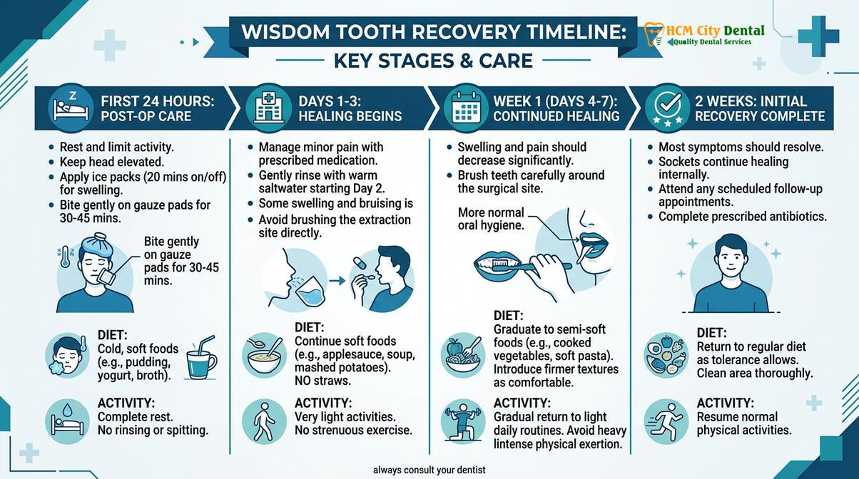
The wisdom teeth recovery timeline spans from immediate clot formation on day one to complete socket closure over several weeks, requiring specific care at each stage.
Understanding the physiological stages of healing is essential for patients preparing to undergo surgical removal of third molars. The human body heals extraction sites through a process known as secondary intention, where the wound remains open and gradually fills with granulation tissue from the base upward. This complex biological mechanism requires a stable environment free from mechanical disruption and bacterial overload.
Dr. Nguyen Van Cuong, a leading oral surgery specialist, emphasizes that patient compliance during the first week dictates the entire trajectory of the recovery. Dr. Cuong and his clinical team utilize advanced protocols to ensure minimal discomfort and optimal bone regeneration. The wisdom teeth recovery timeline is not merely a waiting period; it is an active phase where the patient’s dietary choices, oral hygiene practices, and physical activity levels directly influence cellular regeneration. Healing is generally divided into four distinct phases: hemostasis, inflammation, proliferation, and maturation[1]. These stages align closely with the standard protocols outlined by the Vietnam Ministry of Health (MOH) for post-operative oral care[6].
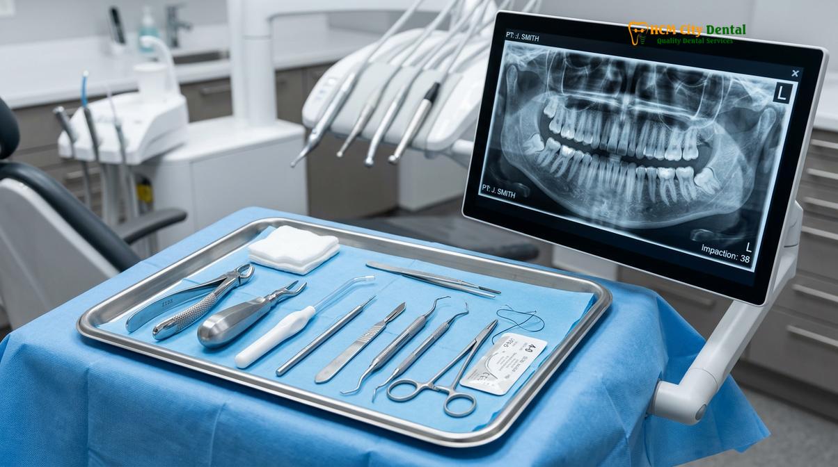
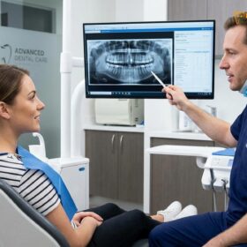
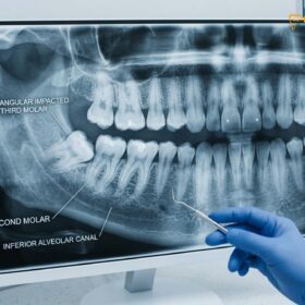
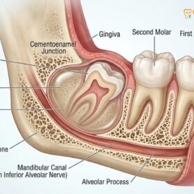
During the initial consultation, clinicians evaluate the anatomical position of the tooth, the density of the surrounding bone, and the proximity to vital structures such as the inferior alveolar nerve. These factors determine the surgical approach and, consequently, the expected duration of the recovery. Patients who undergo proactive extractions before the roots are fully formed often experience a more rapid and less complicated healing process compared to those who wait until acute symptoms arise.
Phase 1: The First 24 Hours (Clot Formation)
The first 24 hours focus strictly on hemostasis and stabilizing the fragile blood clot to prevent excessive bleeding and dry socket.
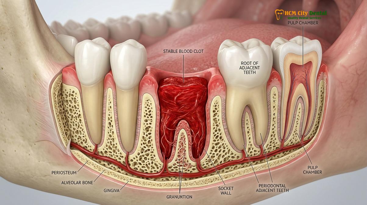
Immediately following the extraction, the primary biological objective is hemostasis—the cessation of bleeding. As the tooth is removed from the alveolar bone, severed blood vessels begin to bleed into the empty socket. The body responds by initiating the coagulation cascade. Platelets aggregate at the site, releasing chemical signals that convert fibrinogen into fibrin, creating a mesh-like network that traps red blood cells and forms a stable clot. This clot serves as a biological bandage, protecting the exposed bone and nerve endings from the oral environment[5].
To facilitate this process, patients are instructed to bite down firmly on sterile gauze placed over the surgical site for 30 to 45 minutes. The sustained mechanical pressure compresses the local blood vessels and provides a matrix for the clot to adhere to. If minor oozing continues, replacing the gauze and applying pressure for another 30 minutes is usually sufficient. It is entirely normal for saliva to be tinged pink with blood during this initial phase, which patients often mistake for active bleeding.
Clinical Warning: Preventing Alveolar Osteitis (Dry Socket)
The blood clot is extremely fragile during the first 24 hours. Any negative pressure in the mouth—created by using a straw, vigorous spitting, or aggressive rinsing—can dislodge the clot. If the clot is lost, the underlying bone and nerves are exposed, leading to a severely painful condition known as dry socket, which requires immediate clinical intervention.
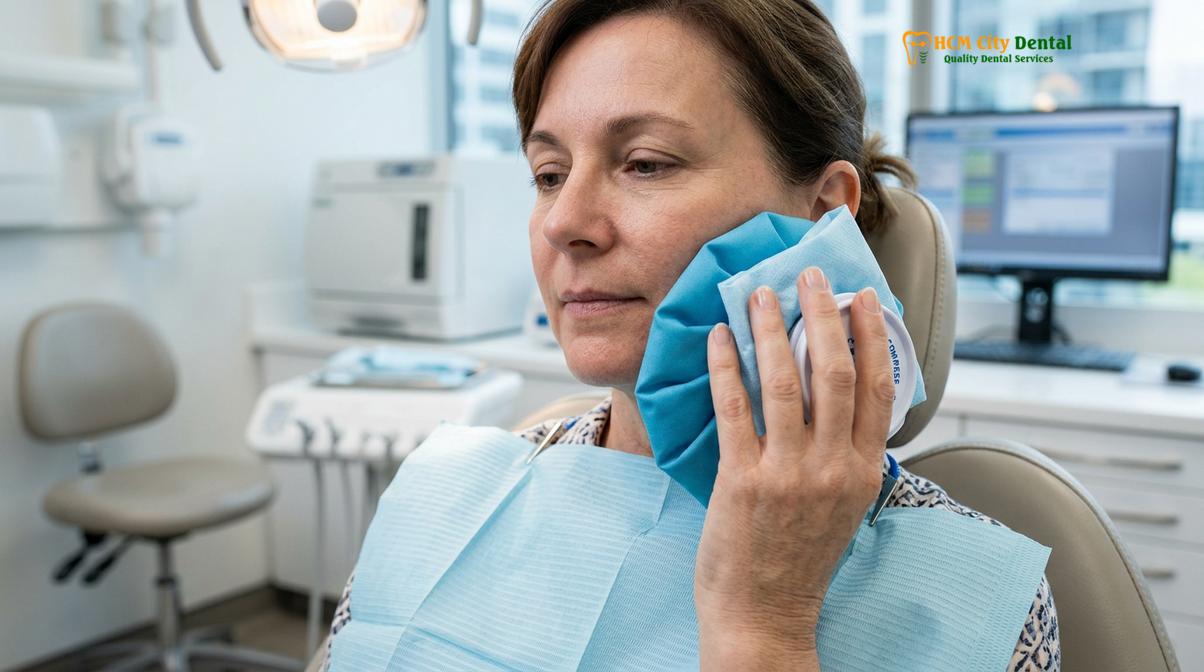
During this critical window, patients must avoid any actions that could disrupt the fibrin network. Hot liquids should be avoided, as heat induces vasodilation (widening of blood vessels), which can restart bleeding. Instead, patients should consume cold, soft foods like yogurt, applesauce, or smoothies. Furthermore, applying an ice pack to the outside of the face—20 minutes on, 20 minutes off—induces localized vasoconstriction, minimizing initial hematoma formation and setting the stage for manageable swelling in the coming days.
Phase 2: Day 2 to 3 (Peak Swelling & Discomfort)
Swelling and inflammatory discomfort typically peak between 48 and 72 hours post-surgery as the body increases blood flow to deliver healing cells.
As the body transitions from hemostasis to the inflammatory phase, patients often experience the peak of their post-operative symptoms. Inflammation is not a complication; it is a necessary biological response. The body releases histamines and prostaglandins, causing local blood vessels to dilate and become more permeable. This allows white blood cells (leukocytes) and nutrient-rich plasma to flood the surgical site, clearing away cellular debris and preventing bacterial colonization.
This influx of fluid manifests clinically as edema (swelling) in the cheeks and jawline. The swelling is often accompanied by trismus, a temporary stiffness or spasm of the masticatory muscles, making it difficult to open the mouth fully. Discomfort during this phase is typically managed through a combination of prescribed non-steroidal anti-inflammatory drugs (NSAIDs) like Ibuprofen, which specifically target the prostaglandin pathways, and acetaminophen for central pain relief. The use of advanced surgical tools, such as piezotome ultrasonic devices, has been shown to significantly reduce this localized trauma and subsequent swelling[2].
“Patients often feel alarmed when swelling increases on the second or third day, assuming an infection has occurred. In reality, this peak edema is a sign of a robust and healthy immune response delivering essential growth factors to the extraction site.”
By the third day, the protocol for thermal therapy shifts. While ice is used initially to restrict blood flow, patients are now advised to switch to warm, moist compresses. The gentle heat promotes vasodilation, enhancing local circulation to help the lymphatic system drain the accumulated fluid and reduce muscle stiffness. Dietary choices should remain soft, but patients can begin incorporating lukewarm, nutrient-dense options as outlined in post-operative dietary guidelines to support systemic healing.
Phase 3: Day 4 to 7 (Tissue Healing & Stitch Removal)
By the end of the first week, acute inflammation subsides, granulation tissue begins forming, and any non-dissolvable sutures are typically removed.
Entering the proliferative phase, the acute symptoms of swelling and throbbing pain should steadily decline. Inside the socket, fibroblasts are actively synthesizing collagen, creating a structural framework known as granulation tissue. Simultaneously, angiogenesis occurs, forming new microscopic blood vessels to supply oxygen and nutrients to the regenerating tissue. This is a critical milestone in the wisdom tooth healing stages, as the site transitions from a fragile clot to a more resilient, organized tissue matrix.
Oral hygiene becomes paramount during this phase. While patients must still avoid aggressive brushing directly over the extraction site, they should resume gentle brushing of the adjacent teeth. Warm saltwater rinses (saline) should be utilized after every meal. The mild osmotic gradient of the saline solution helps draw out localized fluid, reduces bacterial load, and soothes the healing gingiva without the harsh chemicals found in commercial mouthwashes.
If the oral surgeon utilized sutures to approximate the gum tissue, their management depends on the material used. Resorbable sutures (such as chromic gut or Vicryl) will begin to dissolve and fall out naturally between days 5 and 7. If non-resorbable sutures (like silk or nylon) were placed, the patient will return to the clinic for a brief, painless removal appointment. At this stage, patients generally feel comfortable enough to transition back to a relatively normal diet, though hard, crunchy, or heavily seeded foods should still be avoided to prevent mechanical trauma to the new tissue.
Phase 4: Week 2 and Beyond (Socket Closure & Bone Remodeling)
While soft tissue closes over the extraction site within two to three weeks, underlying alveolar bone remodeling continues for up to six months.
By the second week, the epithelial layer of the gums typically migrates across the surface of the granulation tissue, effectively sealing the socket from the oral cavity. However, patients often notice a slight indentation or “hole” where the tooth used to be. A common question during this phase is how long does it take for the socket hole to close? While the soft tissue surface may appear healed within 14 to 21 days, the space beneath is still undergoing profound changes.
The final phase of healing is maturation and remodeling. Osteoblasts (bone-forming cells) migrate into the collagen matrix and begin depositing calcium and phosphate, gradually converting the soft granulation tissue into woven bone, and eventually into mature lamellar bone. This osteogenic process is slow, often taking 3 to 6 months to achieve radiographic density comparable to the surrounding jawbone[4].
Clinical Case Review: Managing Socket Debris
A 24-year-old patient visited HCMC Dental Clinic in Ho Chi Minh City complaining of a foul taste and mild ache two weeks post-extraction. Clinical examination revealed no infection, but rather food debris trapped in the healing indentation. The patient was instructed on proper socket irrigation using a curved monoject syringe with warm saline. Regular flushing after meals immediately resolved the symptoms, allowing the soft tissue to close completely without further complication.
During this extended remodeling phase, patients can resume their normal diet and oral hygiene routines. The primary focus shifts from protecting a blood clot to maintaining a clean environment. Using a soft-bristled toothbrush and continuing gentle irrigation if a depression remains will prevent localized gingival inflammation as the bone solidifies.
Timeline for Resuming Activities (Exercise, Straws, Smoking/Vaping)
Patients must gradually reintroduce physical exertion and dietary habits based on clinical milestones to avoid disrupting the healing socket.
A successful recovery requires strict adherence to behavioral modifications. Prematurely resuming certain activities can induce mechanical stress or chemical irritation, leading to severe complications. The timeline for returning to normal life depends heavily on the physiological demands of the activity.
Physical exertion must be carefully managed. When patients ask how long off work wisdom teeth recovery requires, the answer depends on their occupation. Sedentary office workers may return in 2 to 3 days, but those in labor-intensive jobs should wait at least a week. Cardiovascular exercise elevates systemic blood pressure, which can cause the delicate capillaries in the healing socket to rupture, leading to secondary hemorrhage. Light walking is permissible after 48 hours, but heavy lifting, running, or contact sports should be postponed for 7 to 10 days.
The use of straws creates a vacuum inside the oral cavity. This negative hydrostatic pressure is strong enough to physically pull the blood clot out of the bony socket. Patients must drink directly from a cup for at least the first week. Similarly, questions regarding vaping after wisdom teeth recovery or when can i resume smoking after extraction are common. Both smoking and vaping are strictly contraindicated for a minimum of 72 hours, though 7 days is clinically preferred. Beyond the negative pressure of inhalation, the carbon monoxide and heat from smoke cause severe vasoconstriction, starving the healing tissues of oxygen and drastically increasing the risk of dry socket and infection.
| Activity Type | Restricted Period | Clinical Rationale |
|---|---|---|
| Using a Straw | Minimum 7 Days | Negative intraoral pressure can dislodge the fibrin clot. |
| Smoking / Vaping | 72 Hours to 7 Days | Vasoconstriction reduces oxygenation; suction risks dry socket. |
| Light Exercise (Walking) | 48 to 72 Hours | Allows initial hemostasis to stabilize before mild heart rate increases. |
| Heavy Lifting / Cardio | 7 to 10 Days | Prevents blood pressure spikes that cause secondary hemorrhage. |
| Eating Crunchy/Hard Foods | 2 to 3 Weeks | Prevents mechanical trauma to the newly formed epithelial tissue. |
Factors That Speed Up or Slow Down Recovery (PRF, Age, Case)
Individual recovery rates depend heavily on patient age, the anatomical complexity of the impaction, and the use of advanced surgical adjuncts like PRF.
No two extractions heal at the exact same rate. The biological timeline is influenced by a combination of patient-specific factors and the surgical techniques employed. Age is a primary determinant; younger patients (typically late teens to early twenties) possess highly vascularized bone and robust cellular turnover rates. As patients age, the surrounding bone becomes denser and less vascular, which can prolong the healing phase. This is why specialized protocols are often required for older adult patients undergoing third molar surgery.
The anatomical position of the tooth also dictates the surgical trauma required for removal. A fully erupted upper wisdom tooth may be extracted in minutes with minimal tissue disruption. Conversely, a horizontally impacted lower wisdom tooth requires surgical reflection of the gums and precise removal of overlying bone. Because the mandible (lower jaw) is denser than the maxilla (upper jaw), lower extractions inherently involve more swelling and a longer recovery period.
“The integration of Platelet-Rich Fibrin (PRF) has revolutionized post-operative healing. By centrifuging the patient’s own blood and placing the concentrated growth factors directly into the socket, we significantly accelerate soft tissue closure and drastically reduce the risk of dry socket.”
Modern surgical adjuncts play a massive role in recovery speed. The application of Platelet-Rich Fibrin (PRF) introduces a high concentration of leukocytes and growth factors directly into the surgical site, promoting rapid angiogenesis and tissue regeneration[3]. Additionally, utilizing painless extraction technologies such as ultrasonic bone surgery minimizes collateral tissue damage, leading to a much smoother post-operative experience.
When to Seek Immediate Clinical Attention
While mild discomfort and swelling are normal, certain symptoms indicate a disruption in the healing process that requires professional intervention.
Patients should closely monitor their symptoms during the first week. It is crucial to distinguish between normal inflammatory responses and signs of potential complications. You should contact your oral surgeon immediately if you experience any of the following warning signs:
- Uncontrolled Bleeding: If heavy bleeding continues despite applying firm pressure with gauze for over an hour.
- Severe, Worsening Pain: Pain that suddenly intensifies after the third day and is unresponsive to prescribed analgesics, which is a hallmark sign of dry socket.
- Fever or Chills: An elevated body temperature indicates a systemic response to a potential bacterial infection.
- Pus or Foul Discharge: A persistent bad taste or visible yellow/white discharge from the socket area.
- Prolonged Numbness: If the lips, tongue, or chin remain numb for more than 24 hours after the local anesthesia should have worn off, indicating potential nerve irritation.
Patients with underlying systemic conditions should be particularly vigilant. For instance, those managing diabetes or taking blood thinners may have altered healing timelines and require closer post-operative monitoring to prevent delayed wound closure or secondary hemorrhage.
If you are experiencing prolonged discomfort, suspect a complication, or need expert guidance on your recovery, contact HCMC Dental Clinic in Ho Chi Minh City for a comprehensive evaluation and personalized care plan.
References
- Journal of Oral and Maxillofacial Surgery. Recovery and healing after third molar extraction.
- International Journal of Oral and Maxillofacial Surgery. Piezotome versus rotary drill in wisdom teeth removal.
- British Dental Journal. Dry socket incidence and prevention with Platelet-Rich Fibrin.
- Journal of the American Dental Association. Mandibular third molar impaction classifications and surgical outcomes.
- Clinical Oral Investigations. Lingual and inferior alveolar nerve protection during surgical extraction.
- Vietnam Ministry of Health (MOH). National Guidelines on Oral and Maxillofacial Surgery and Post-Operative Care.