The surgical management of deeply impacted mandibular third molars (lower wisdom teeth) represents one of the most anatomically complex challenges in modern oral and maxillofacial surgery. For decades, the traditional paradigm dictated the complete removal of the entire tooth structure, regardless of its proximity to vital anatomical landmarks. However, when a lower wisdom tooth is intimately entangled with or compressing the inferior alveolar nerve (IAN), a standard full extraction carries a profound and unacceptable risk of iatrogenic nerve injury. This injury can manifest as temporary or, devastatingly, permanent paresthesia—a loss of sensation, tingling, or chronic neuropathic pain in the lower lip, chin, and gums. To mitigate this severe complication, advanced dental microsurgery has embraced a highly specialized, evidence-based alternative: the coronectomy. By evaluating the clinical efficacy of coronectomy wisdom tooth nerve damage prevention vs extraction, oral surgeons can offer a biologically sound, nerve-sparing protocol that intentionally retains the healthy, vital root fragments within the jawbone while completely eliminating the symptomatic, infection-prone dental crown.
At HCMC Dental Clinic in District 1, Ho Chi Minh City, our surgical philosophy is anchored in maximum anatomical preservation and uncompromising patient safety. Utilizing hospital-grade 0.075mm high-resolution 3D CBCT DICOM imaging, our clinical team meticulously maps the trajectory of the mandibular canal in three dimensions before any surgical intervention begins. If the radiographic evidence confirms a high-risk intersection between the tooth roots and the nerve bundle, we deploy a precise Piezosurgery protocol to perform an intentional partial odontectomy. This comprehensive clinical guide explores the biomechanical rationale, the strict diagnostic criteria, the step-by-step microsurgical execution, and the long-term biological outcomes of coronectomy, providing international patients and dental travelers with the authoritative knowledge required to make informed decisions about their oral surgical care in Vietnam.
Clinical Summary:
A coronectomy (intentional partial odontectomy) is an advanced, nerve-sparing oral surgery technique designed specifically for high-risk lower wisdom teeth whose roots are intimately associated with the inferior alveolar nerve. Instead of luxating and elevating the entire tooth—which could crush, stretch, or sever the nerve—the surgeon uses ultrasonic Piezosurgery to amputate the clinical crown 3 to 4 millimeters below the crestal bone, leaving the healthy, vital roots undisturbed in the socket. Supported by 3D CBCT diagnostics, this protocol virtually eliminates the risk of permanent lip and chin numbness (paresthesia) while effectively resolving pericoronitis and preventing damage to the adjacent second molar. The retained roots remain biologically vital, eventually becoming encased in newly formed autogenous bone.
Key Takeaways:
- Primary Indication: Coronectomy is strictly indicated when 3D CBCT imaging confirms that the wisdom tooth roots are wrapping, grooving, or compressing the inferior alveolar nerve canal.
- Nerve Safety Efficacy: Clinical studies demonstrate that coronectomy reduces the incidence of inferior alveolar nerve injury to near 0%, compared to a 5% to 19% risk during full extraction of high-risk teeth.
- Biological Vitality: The procedure is only successful if the retained roots are healthy, vital, and completely immobile; infected or carious roots are strict contraindications.
- Root Migration Phenomenon: In 15% to 30% of cases, the retained roots may slowly migrate coronally (upward) over 1 to 3 years, safely moving away from the nerve canal.
- Advanced Technology: The procedure relies on 0.075mm 3D CBCT nerve mapping and 24–36 kHz Piezosurgery to ensure micrometric precision without thermal necrosis to the surrounding bone.
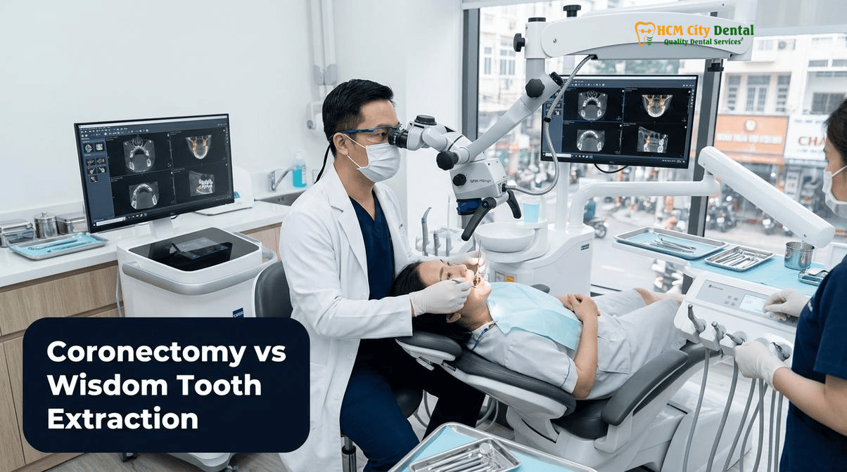
When Full Lower Wisdom Tooth Extraction Risks Permanent Lip and Chin Paresthesia (Inferior Alveolar Nerve Injury)
The inferior alveolar nerve (IAN) is a major sensory branch of the mandibular nerve, which itself is the third division of the trigeminal nerve (Cranial Nerve V). The IAN travels through the mandibular canal within the lower jawbone, providing critical sensory innervation to the lower teeth, the gums, the lower lip, and the skin of the chin. Because the human jaw has evolutionary shortened over millennia, the third molars (wisdom teeth) often lack sufficient space to erupt properly. Consequently, they become impacted deep within the posterior mandible, frequently developing roots that extend downward into dangerous proximity with the mandibular canal. When a patient presents with recurrent pericoronitis (infection of the gum tissue overlying the tooth), cystic lesions, or damage to the adjacent second molar, surgical intervention becomes mandatory. However, the biomechanics of traditional tooth extraction pose a severe threat to the IAN in these specific anatomical scenarios.
Traditional full extraction requires the application of mechanical force using elevators and forceps to luxate (loosen) the tooth within its socket. This process expands the surrounding alveolar bone and tears the periodontal ligament. If the roots of the wisdom tooth are merely resting against the roof of the mandibular canal, this pressure might cause temporary neuropraxia—a mild nerve contusion resulting in transient numbness that resolves in a few weeks. However, if the roots are hooked around the nerve, or if the nerve actually passes between fused roots (a phenomenon known as inter-radicular grooving), the mechanical elevation of the tooth will physically drag, stretch, crush, or completely sever the nerve bundle. This catastrophic trauma leads to axonotmesis or neurotmesis, resulting in permanent paresthesia (loss of sensation), dysesthesia (painful, burning, or abnormal sensations), or anesthesia (complete numbness) of the lower lip and chin. This complication profoundly impacts a patient’s quality of life, affecting speech, eating, and psychological well-being[1].
To prevent this irreversible iatrogenic trauma, the dental profession recognized the need for a paradigm shift. If the pathology associated with an impacted wisdom tooth is primarily localized to the clinical crown (such as plaque accumulation, pericoronitis, or food impaction against the second molar), and the roots themselves are healthy and vital, there is no biological imperative to remove the roots at the expense of the nerve. By evaluating the clinical efficacy of coronectomy wisdom tooth nerve damage prevention vs extraction, surgeons can eliminate the source of the infection (the crown) while leaving the high-risk anatomical relationship (the root-nerve interface) completely undisturbed.
“The primary directive in oral and maxillofacial surgery is to do no harm. When a 3D CBCT scan reveals that a wisdom tooth root is intimately wrapped around the inferior alveolar nerve, attempting a heroic full extraction is a violation of biological safety. Coronectomy is not a compromise; it is a highly sophisticated, evidence-based microsurgical protocol that definitively cures the patient’s symptoms while guaranteeing the preservation of their sensory nerve function.”
— Dr. Nguyen Van Cuong, DDS (Clinical Director, HCMC Dental Clinic)
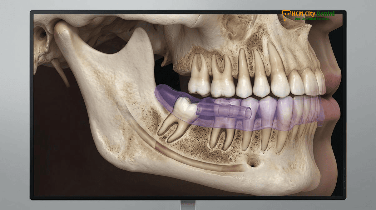
3D CBCT Diagnostic Criteria: Canal Cortication Loss, Inter-Radicular Nerve Grooving, and Juxtacanal Root Hook
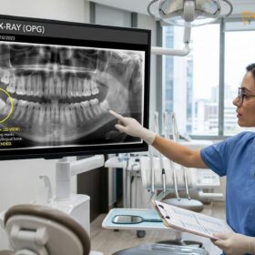
The decision to perform a coronectomy rather than a standard extraction cannot be made based on clinical intuition or outdated 2D imaging. Traditional panoramic radiographs (OPG) provide a flattened, two-dimensional representation of a three-dimensional anatomical structure. On a 2D X-ray, the roots of a wisdom tooth may appear to overlap or intersect the mandibular canal due to superimposition, even if they are safely separated by several millimeters of bone in the buccal-lingual (side-to-side) dimension. Relying solely on 2D imaging leads to both false positives (performing unnecessary coronectomies) and false negatives (causing unexpected nerve damage during extraction). Therefore, the definitive diagnosis relies on the identification of specific radiographic risk markers, originally described by Rood and Shehab, which must then be verified using advanced 3D volumetric imaging.
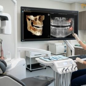
The Rood and Shehab radiographic nerve risk signs are classic indicators observed on panoramic films that suggest a high probability of IAN involvement. These signs include: darkening of the tooth root where it crosses the canal (indicating a loss of bone density), deflection or sudden deviation of the canal’s path, narrowing of the root apex, dark and bifid apices, and an interruption in the white radiopaque lines that define the borders of the mandibular canal (loss of canal cortication). When any of these high-risk signs are detected, the standard of care mandates a transition to three-dimensional diagnostics. At HCMC Dental Clinic, we utilize a 0.075mm high-resolution 3D CBCT (Cone Beam Computed Tomography) DICOM scan to provide a flawless, cross-sectional view of the mandible[2].
The 3D CBCT scan allows the oral surgeon to slice through the jawbone virtually, examining the exact spatial relationship between the root and the nerve from the axial, coronal, and sagittal planes. We specifically look for definitive wisdom tooth touching mandibular nerve CBCT signs. The most critical findings that mandate a coronectomy include:
- Canal Cortication Loss: The CBCT reveals that the dense bony roof of the mandibular canal has been completely resorbed by the encroaching tooth root, meaning the root is in direct physical contact with the nerve sheath.
- Inter-Radicular Nerve Grooving: The scan shows that the nerve canal is actually compressed or indented by the root, or in severe cases, the nerve passes directly through a space between multiple roots.
- Juxtacanal Root Hook: The apical portion of the root curves sharply (dilaceration) and hooks directly under or around the nerve bundle, making safe elevation physically impossible.
By utilizing this rigorous diagnostic protocol, we ensure that every patient receives the safest and most appropriate surgical intervention. For patients seeking the Best Oral Surgeon for Wisdom Tooth Extraction in Saigon | HCMC Dental, our reliance on 3D CBCT nerve mapping is the cornerstone of our zero-nerve-damage safety record.
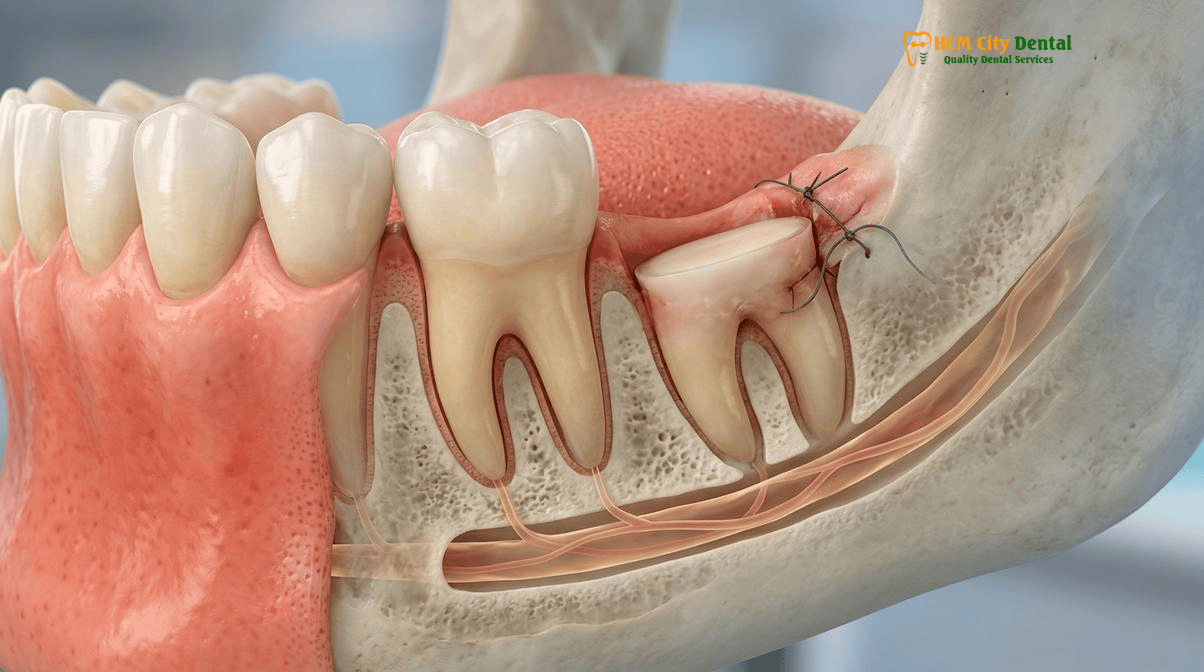
What Is a Coronectomy (Intentional Partial Odontectomy) and Why Can Healthy Vital Root Fragments Safely Remain in Bone?
A coronectomy, formally known in the medical literature as an intentional partial odontectomy, is a surgical procedure where the clinical crown of a tooth is deliberately amputated and removed, while the root complex is intentionally left undisturbed within the alveolar bone. To patients unfamiliar with the procedure, the concept of leaving a “broken” root behind may seem counterintuitive or alarming. A common misconception is that any retained root will inevitably rot, become infected, or be rejected by the body as a foreign object. However, this assumption fundamentally misunderstands the biology of a healthy tooth and the principles of intentional root retention inferior alveolar nerve protocols.
The success of a coronectomy relies entirely on the biological vitality of the retained root. A tooth is not a dead piece of stone; it is a living organ. The inner chamber of the root contains the dental pulp, a complex matrix of nerves, blood vessels, and connective tissue that enters the tooth through the apical foramen (the opening at the tip of the root). The outer surface of the root is covered in cementum, which is attached to the surrounding jawbone by the periodontal ligament (PDL). When a coronectomy is performed on a healthy, vital tooth, the blood supply to the root remains completely intact. Because the root is not mobilized or luxated during the surgery, the delicate apical blood vessels are not severed, and the PDL remains firmly attached to the bone[3].
Following the amputation of the crown, the exposed pulp tissue at the sectioned surface undergoes a predictable and highly successful healing process. The vital pulp tissue reacts to the surgical trauma by initiating a localized inflammatory response, followed by the rapid deposition of secondary dentin (reparative dentin) by odontoblasts. This calcified barrier effectively seals the pulp chamber from the oral environment. Simultaneously, a blood clot forms over the sectioned root surface within the bony socket. Through the process of osteogenesis, this blood clot organizes into granulation tissue, which is eventually replaced by woven bone and, ultimately, mature lamellar bone. Within a few months, the retained root becomes completely encased in healthy, native jawbone. The body does not reject the root because it recognizes the vital pulp, the cementum, and the PDL as its own living tissue.
Clinical Case Study: Expat Patient in District 1, Ho Chi Minh City
Patient Profile: A 28-year-old Australian expatriate working in District 1 presented to HCMC Dental Clinic with severe, recurrent pericoronitis associated with a horizontally impacted lower right wisdom tooth (Tooth 48). The patient reported significant pain, trismus (difficulty opening the mouth), and localized swelling.
Diagnostic Findings: A 2D panoramic X-ray suggested a high risk of nerve involvement. A subsequent 0.075mm 3D CBCT scan revealed severe inter-radicular nerve grooving; the roots of Tooth 48 were completely wrapped around the inferior alveolar nerve, with total loss of canal cortication.
Surgical Intervention: Dr. Nguyen Van Cuong, DDS, performed a 3D CBCT-guided Piezosurgery coronectomy. The crown was sectioned and removed without mobilizing the roots. The roots were reduced 3mm below the crestal bone, and the site was closed primarily.
Clinical Outcome: The patient experienced zero postoperative numbness or paresthesia. At the 6-month follow-up, a new CBCT scan confirmed complete bone fill over the retained roots, with the roots remaining stable and asymptomatic. The patient successfully avoided a high probability of permanent nerve damage.
Step-by-Step Microsurgical Protocol: Piezosurgery Crown Sectioning 3–4mm Below the Alveolar Crest Without Mobilizing the Roots
The execution of a coronectomy requires a significantly higher level of surgical precision than a standard extraction. The procedure must be performed under strict aseptic conditions, utilizing advanced microsurgical instrumentation to ensure that the crown is cleanly amputated without transmitting any luxating forces to the root complex. At HCMC Dental Clinic, our protocol integrates 3D CBCT guidance with ultrasonic Piezosurgery to achieve unparalleled safety and predictability.
Step 1: 3D CBCT Planning and Anesthesia
The procedure begins with a thorough review of the 3D CBCT DICOM data to map the exact angulation of the tooth and the depth of the nerve. Profound local anesthesia is administered using an inferior alveolar nerve block, supplemented by buccal infiltration, ensuring complete patient comfort throughout the surgery.
Step 2: Flap Design and Bone Exposure
A full-thickness mucoperiosteal flap is carefully elevated to expose the surgical site. The flap design must provide adequate visualization of the clinical crown and the surrounding alveolar crest while preserving the integrity of the lingual nerve, which runs along the inner aspect of the mandible.
Step 3: Piezosurgery Bone Osteotomy
To access the crown of a deeply impacted tooth, some overlying bone must often be removed. Instead of using traditional high-speed rotary burs, which can cause thermal necrosis and soft tissue trauma, we utilize advanced Piezosurgery. This technology uses ultrasonic micro-vibrations (24–36 kHz) to perform selective bone osteotomy. Piezosurgery cuts mineralized bone with extreme precision but bounces off soft tissues, providing an additional layer of safety for the inferior alveolar nerve and the adjacent gingival tissues.
Step 4: Crown Sectioning and Amputation
The most critical phase of the procedure is the sectioning of the crown. Using a specialized surgical fissure bur or a piezotome insert, the surgeon makes a deep cut into the tooth structure, typically at a 45-degree angle, aiming just below the cementoenamel junction (CEJ). It is imperative that the cut extends almost entirely through the tooth. The surgeon then uses a straight elevator to gently fracture the remaining sliver of enamel and remove the crown. Crucially, the surgeon must never use the root as a fulcrum. The roots must remain absolutely immobile during this process.
Step 5: Root Reduction and Profiling
Once the crown is removed, the exposed surface of the retained roots must be modified. Using a long-shank round bur under copious sterile saline irrigation, the surgeon grinds the root surface down until it sits at least 3 to 4 millimeters below the crest of the surrounding alveolar bone. This subcrestal reduction is essential to provide sufficient space for a robust blood clot to form and for new bone to regenerate over the top of the root, preventing future exposure to the oral cavity[4].
Step 6: Irrigation and Primary Closure
The surgical site is thoroughly irrigated with sterile saline to remove any dentin debris or bone dust. The mucoperiosteal flap is then repositioned and secured with watertight, tension-free sutures (typically 4-0 or 5-0 resorbable sutures) to achieve primary closure. This seals the socket, protecting the vital root and facilitating rapid osteogenesis.
For a deeper understanding of the safety protocols surrounding this procedure, patients can review our comprehensive Coronectomy Wisdom Teeth: Clinical Guide & Nerve Safety | HCMC Dental.
Strict Contraindications for Coronectomy (Non-Vital Infected Pulps, Carious Roots, and Mobilized Root Fragments) & Root Migration Follow-Up
While coronectomy is a highly effective nerve-sparing technique, it is not a universal solution for all impacted wisdom teeth. The procedure is governed by strict biological prerequisites, and attempting a coronectomy when contraindications are present will inevitably lead to surgical failure, chronic infection, and the need for a complex secondary surgery. The fundamental rule of coronectomy is that the retained root must be healthy, vital, and immobile.
Strict Clinical Contraindications:
- Non-Vital or Infected Pulps: If the wisdom tooth has a history of deep dental caries that has penetrated the pulp chamber, causing irreversible pulpitis or pulpal necrosis, a coronectomy cannot be performed. A necrotic root acts as a reservoir for bacteria and will cause a severe periapical abscess if left in the bone.
- Periapical Pathology: The presence of a periapical radiolucency (a dark spot at the tip of the root on an X-ray, indicating a cyst or granuloma) is an absolute contraindication. The entire tooth, including the infected root, must be extracted.
- Horizontal Impactions Along the Nerve: In some cases of deep horizontal impaction, it is physically impossible to section the crown without the surgical bur directly cutting into the underlying nerve canal. In these rare scenarios, alternative techniques, such as orthodontic extraction (using braces to slowly pull the tooth away from the nerve over several months), may be considered.
- Mobilized Root Fragments: This is the most critical intraoperative complication. If, during the process of sectioning and removing the crown, the roots become loose or luxated within the socket, the coronectomy has failed. A mobilized root has had its apical blood supply severed; it is no longer vital and has become a necrotic foreign body. The surgeon must immediately convert the procedure to a full extraction, carefully removing the loosened roots.
The Phenomenon of Root Migration:
A well-documented biological response following a successful coronectomy is root migration. In approximately 15% to 30% of cases, the retained roots will slowly migrate coronally (upward) over a period of 1 to 3 years. This is actually a highly favorable outcome. As the root moves upward, it travels safely away from the inferior alveolar nerve. In most cases, the migration stops within the bone, and the root remains permanently encased and asymptomatic. However, in a small percentage of cases, the root may eventually erupt through the gum tissue. If this occurs, the root can be easily and safely extracted in a minor, minimally invasive procedure, as it is no longer anywhere near the nerve canal. Patients must commit to routine radiographic follow-up (typically at 6, 12, and 24 months) to monitor the position of the retained roots.
Clinical Precautions & Post-Operative Care
Following a coronectomy, patients must adhere strictly to the prescribed pharmacological protocol, which includes broad-spectrum antibiotics to prevent surgical site infection, corticosteroids to manage localized edema (swelling), and NSAIDs for analgesia. Patients must maintain impeccable oral hygiene using a prescribed chlorhexidine mouthwash. If you experience severe, throbbing pain that worsens after 72 hours, foul-tasting discharge, or a sudden fever, you must contact Dr. Nguyen Van Cuong, DDS, immediately, as these may be signs of a postoperative infection requiring prompt clinical intervention. For international patients planning their return journey, please consult our guidelines on Flying After Wisdom Tooth Extraction: Safe Timelines & Risks.
2026 3D CBCT Nerve Mapping, Piezosurgery Coronectomy & Impacted Wisdom Tooth Fee Schedule at HCMC Dental Clinic (District 1)
At HCMC Dental Clinic, we believe that advanced, nerve-sparing oral surgery should be accessible and transparently priced. Under the clinical leadership of Dr. Nguyen Van Cuong, DDS, our facility operates strictly according to international hospital-grade sterilization and surgical protocols. We utilize the exact same 3D CBCT imaging, Piezosurgery units, and biomaterials found in top-tier clinics in Australia, the United States, and Europe, but at a significantly reduced cost due to the economic efficiencies of operating in Vietnam.
“Transparency in clinical capabilities and pricing is the foundation of trust for our international patients. We do not compromise on diagnostic imaging or surgical technology. Our 2026 fee schedule reflects our commitment to providing world-class, nerve-safe wisdom tooth management and comprehensive implantology at a fair and accessible price point.”
— Dr. Nguyen Van Cuong, DDS (Vietnam MOH License 0003265/BL-CCHN)
The table below outlines our official 2026 fee schedule, highlighting the -40% online booking rates for our comprehensive range of specialist dental services, including our advanced Piezosurgery wisdom tooth extraction and coronectomy protocols. For a complete overview of our clinical pricing, please visit our Official 2026 Dental Price List & -40% Online Booking Fee Schedule.
| Specialist Dental Procedure (HCMC Dental Clinic, District 1) | 2026 Online Booking Fee (-40% USD) | 2026 Online Booking Fee (VND) | Walk-In / US / Australian Benchmark |
|---|---|---|---|
| 3D CBCT Scan (0.075mm High-Resolution Nerve Mapping) | $18 USD | 480,000 VND | $150 – $300 USD |
| Surgical Wisdom Tooth Extraction / Coronectomy (Piezotome) | $58 – $115 USD / tooth | 1,500,000 – 3,000,000 VND | $400 – $800 USD |
| All 4 Wisdom Teeth Surgical Package | $231 – $308 USD | 6,000,000 – 8,000,000 VND | $1,500 – $3,000 USD |
| Single Korean Implant (Dentis / Neo Biotech / Dentium) | $462 – $654 USD | 12,000,000 – 17,000,000 VND | $769 – $1,090 walk-in |
| Single European / Swiss Implant (Straumann BLT-SLA / Active) | $962 – $1,346 USD | 25,000,000 – 35,000,000 VND | $1,603 – $2,244 walk-in |
| All-on-4 Full-Arch Implants (Complete Zirconia Package) | From $6,346 USD / arch | 165,000,000 VND | $15,000 – $25,000 USD |
| IPS E.max Porcelain Veneer | $185 USD / tooth | 4,800,000 VND | $308 walk-in |
| Full Zirconia Crown (Cercon Standard / HT Multilayer) | $154 – $192 USD / tooth | 4,000,000 – 5,000,000 VND | $256 – $321 walk-in |
| Invisalign Comprehensive (Unlimited aligners) | $2,538 USD | 66,000,000 VND | $4,231 walk-in |
| Microscopic Endodontic Root Canal Therapy (Complex Molar) | $115 – $154 USD / tooth | 3,000,000 – 4,000,000 VND | $192 – $256 walk-in |
To learn more about our clinical director’s credentials and expertise in advanced oral surgery, please read about Dr. Nguyen Van Cuong, DDS (Clinical Director — MOH License 0003265/BL-CCHN). For comprehensive information on preparing for your surgical journey to Vietnam, explore our International Patient Medical Safety & Post-Op Recovery Guides.
Frequently Asked Questions
Will the wisdom tooth roots left behind during a Coronectomy become infected or rejected by my body?
No, provided the tooth was vital and healthy prior to the procedure. Because the intentional root retention inferior alveolar nerve protocol relies on the biological vitality of the tooth, the retained root fragments maintain their blood supply via the apical foramen. The pulp tissue heals, secondary dentin forms over the exposed pulp chamber, and the surrounding periodontal ligament (PDL) remains intact. The body recognizes the root as native tissue, not a foreign object, allowing healthy bone to fill the surgical site over the root.
What happens if the retained wisdom tooth roots slowly migrate upward away from the nerve over 1 to 3 years?
Root migration after coronectomy wisdom tooth surgery is a well-documented and generally favorable biological response occurring in approximately 15% to 30% of cases. As the bone heals, the retained root may slowly move coronally (upward) and away from the inferior alveolar nerve. If the root eventually erupts through the gum tissue and causes mild irritation, it can be easily and safely extracted in a minor secondary procedure, as it is no longer in dangerous proximity to the mandibular canal.
What happens if the root accidentally loosens during a planned Coronectomy?
A strict rule of the coronectomy procedure is that the retained roots must remain absolutely immobile. If the roots are accidentally mobilized or luxated during the crown sectioning phase, they lose their vital blood supply and become a necrotic foreign body risk. In this scenario, the surgeon must immediately convert the procedure to a full extraction, carefully removing the loosened roots while employing advanced microsurgical techniques to minimize trauma to the adjacent nerve.
How does a 0.075mm 3D CBCT scan prove whether I need a Coronectomy or a standard Piezosurgery extraction?
A standard 2D panoramic X-ray can show superimposition, making it look like the tooth root intersects the nerve when they are actually separated in 3D space. A 0.075mm high-resolution 3D CBCT scan provides a volumetric, cross-sectional view of the mandible. It allows the oral surgeon to definitively identify high-risk signs, such as wisdom tooth touching mandibular nerve CBCT signs, canal cortication loss, or a juxtacanal root hook. If the CBCT proves the root is intimately wrapped around the nerve, a coronectomy is indicated [5].
How much does a 3D CBCT-guided Coronectomy cost at HCMC Dental Clinic in District 1?
At HCMC Dental Clinic in District 1, Ho Chi Minh City, a high-resolution 3D CBCT Scan costs just $18 USD online (480,000 VND). The surgical procedure itself, utilizing advanced Piezosurgery technology, ranges from $58 to $115 USD per tooth online (1,500,000 – 3,000,000 VND), depending on the complexity of the impaction and the sectioning required. This represents a highly transparent, hospital-grade surgical protocol at a fraction of the cost found in Australia, the US, or the UK.
References
- Pogrel, M. A., Lee, J. S., & Muff, D. F. (2004). Coronectomy: a technique to protect the inferior alveolar nerve. Journal of Oral and Maxillofacial Surgery, 62(12), 1447-1452.
- Rood, J. P., & Shehab, B. A. (1990). The radiological prediction of inferior alveolar nerve injury during third molar surgery. British Journal of Oral and Maxillofacial Surgery, 28(1), 20-25.
- Leung, Y. Y., & Cheung, L. K. (2009). Safety of coronectomy versus excision of wisdom teeth: a randomized controlled trial. Oral Surgery, Oral Medicine, Oral Pathology, Oral Radiology, and Endodontology, 108(6), 821-827.
- Patel, V., Moore, S., & Sproat, C. (2010). Coronectomy – oral surgery’s answer to modern day conservative dentistry. British Dental Journal, 209(3), 111-114.
- Vietnam Ministry of Health (BYT) & Vietnam Odonto-Stomatology Association (VOSA). National Clinical Guidelines on Specialist Dental Care, Biomaterial Traceability & EN 13060 Class B Infection Control. (2024).