For international patients and dental travelers experiencing persistent, localized pain in a previously treated tooth, achieving an accurate diagnosis is the most critical step in preventing catastrophic jawbone loss. When evaluating vertical root fracture symptoms, CBCT diagnosis vs. root canal retreatment becomes the definitive clinical crossroads. A misdiagnosis can lead to months of unnecessary, painful, and expensive endodontic retreatments on a tooth that is fundamentally non-savable. Conversely, extracting a tooth that merely suffers from a treatable coronal crack deprives the patient of their natural dentition. At HCMC Dental Clinic in District 1, Ho Chi Minh City, the integration of 0.075mm high-resolution 3D Cone Beam Computed Tomography (CBCT) and 25x Carl Zeiss Extaro 300 surgical microscopy has revolutionized the management of longitudinal tooth fractures. Under the clinical direction of Dr. Nguyen Van Cuong, DDS (Clinical Director — MOH License 0003265/BL-CCHN), our specialist team employs evidence-based biomimetic and endodontic microsurgery protocols to definitively differentiate between savable cracked teeth and terminal vertical root fractures, ensuring that every patient receives the most predictable, conservative, and biologically sound treatment available in Vietnam.
Clinical Summary:
Differentiating between Cracked Tooth Syndrome and a Vertical Root Fracture (VRF) requires advanced diagnostic imaging and microscopic visualization. While 2D periapical X-rays miss up to 75% of early VRFs due to anatomical superimposition, a 0.075mm endo-mode 3D CBCT scan can reveal pathognomonic signs such as the J-shaped periradicular halo lesion and buccal cortical plate fenestration. A savable cracked tooth can often be preserved using Cracked Tooth Root Canal: Clinical Guide | HCMC Dental protocols and biomimetic onlay splinting. However, a confirmed VRF represents a terminal prognosis; the tooth must be atraumatically extracted using Piezosurgery to halt rapid bone destruction, followed by immediate dental implant placement and Sticky Bone grafting to restore function and aesthetics.
Key Takeaways:
- Diagnostic Superiority: 3D CBCT imaging is essential for detecting VRFs, as traditional 2D X-rays cannot visualize fractures running parallel to the X-ray beam.
- Clinical Hallmarks: A deep, isolated, narrow periodontal pocket combined with a J-shaped radiolucency is highly indicative of a terminal root fracture.
- Microscopic Verification: 25x Carl Zeiss microscopy, combined with 1% methylene blue dye and fiber-optic transillumination, provides direct visual confirmation of crack propagation.
- Biomimetic Preservation: Savable cracked teeth are treated using Immediate Dentin Sealing (IDS) and Deep Margin Elevation (DME) under strict Rubber Dam Isolation in Biomimetic Dentistry & Root Canal Therapy: Safety & Clinical Protocol in Vietnam.
- Immediate Implantology: True VRFs require immediate, atraumatic extraction and immediate implant placement to preserve the buccal bone plate and prevent severe osteolysis.
AAE Classification of Longitudinal Tooth Fractures: Craze Lines, Fractured Cusp, Cracked Tooth, Split Tooth, and Vertical Root Fracture (VRF)
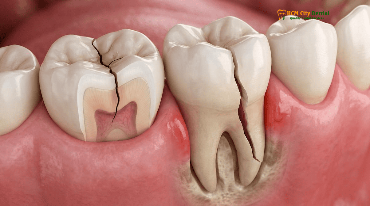
To accurately diagnose and manage structural compromises in the dentition, clinicians rely on the American Association of Endodontists (AAE) classification of longitudinal tooth fractures. This standardized framework categorizes fractures into five distinct types based on their origin, direction of propagation, and clinical severity. Understanding these distinctions is paramount when evaluating cracked tooth syndrome vs. vertical root fracture, as the prognosis and treatment modalities differ drastically.
1. Craze Lines: These are micro-fractures confined entirely to the enamel. They are highly common in adult teeth, asymptomatic, and require no treatment other than aesthetic management if desired. They do not cross the dentinoenamel junction (DEJ) and pose no threat to the structural integrity of the tooth.
2. Fractured Cusp: This occurs when a crack originates in the crown, extends into the dentin, and terminates in the cervical region, often breaking off a cusp. Because the fracture does not typically involve the pulp or extend subgingivally into the root, the tooth is highly savable. Treatment usually involves removing the weakened cusp and restoring the tooth with a biomimetic onlay or full-coverage crown.
3. Cracked Tooth: A cracked tooth features an incomplete fracture that originates in the crown and extends apically (downward) toward the root. The crack may or may not reach the pulp chamber. If the pulp is inflamed or necrotic, Microscopic Endodontic Root Canal Therapy at HCMC Dental Clinic is required, followed by a cuspal-coverage restoration to splint the crack and prevent further propagation. The prognosis is generally favorable if the crack does not extend deep below the alveolar bone crest.
4. Split Tooth: This is the end-stage evolution of an untreated cracked tooth. The incomplete crack has propagated completely through the tooth, separating it into two distinct, mobile segments. A split tooth is fundamentally non-savable in its entirety, though in rare cases involving multi-rooted molars, hemisection or root amputation might salvage a portion of the tooth. In most instances, extraction is indicated.
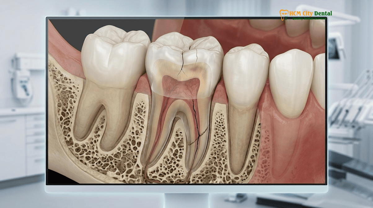
5. Vertical Root Fracture (VRF): Unlike a cracked tooth, a VRF originates in the root itself and propagates coronally (upward) toward the crown. VRFs almost exclusively occur in previously endodontically treated teeth, often due to excessive condensation forces during obturation, the placement of oversized, non-passive metal posts, or the loss of critical pericervical dentin. Because the fracture creates a direct, unsealable communication between the root canal space and the periodontium, bacteria rapidly colonize the fracture gap. This leads to severe, localized bone destruction. A tooth with a true VRF is non-savable and must be extracted[1].
Pathognomonic Clinical Signs of Vertical Root Fracture: The Isolated Narrow Deep Periodontal Probing Defect and Dual Sinus Tracts
Diagnosing a VRF clinically can be exceptionally challenging because the symptoms often mimic those of a failed root canal treatment or primary periodontal disease. Patients frequently present with a history of mild, intermittent pain upon chewing, vague discomfort, or a recurrent “gum boil” (sinus tract) adjacent to a tooth that has already undergone endodontic therapy. However, careful clinical examination by a specialist can reveal specific pathognomonic signs that strongly suggest a root fracture.
The most definitive clinical indicator is the presence of a deep, isolated, narrow periodontal pocket indicating a cracked root. When Dr. Nguyen Van Cuong, DDS performs a circumferential periodontal probing of the suspected tooth, the surrounding gingival attachment may be completely healthy with normal probing depths of 2–3mm. However, at the exact site of the fracture line (typically on the direct buccal or direct lingual aspect), the periodontal probe will suddenly drop into a narrow, deep defect, often measuring 7–10mm or extending all the way to the root apex. This isolated defect occurs because the fracture line acts as a conduit for bacteria, causing rapid, localized destruction of the periodontal ligament (PDL) and adjacent alveolar bone.
Another strong clinical indicator is the presence of dual sinus tracts. While a standard endodontic infection typically produces a single sinus tract near the root apex, a VRF can produce multiple sinus tracts along the lateral aspect of the root, corresponding to the extent of the fracture line. Furthermore, the tooth may exhibit mild mobility and sensitivity to percussion, though these signs are non-specific. Because these clinical signs are highly suggestive but not entirely definitive on their own, advanced 3D imaging is required to confirm the diagnosis and rule out other potential causes of endodontic failure, such as missed anatomy or Calcified Root Canals (Pulp Canal Obliteration): 3D CBCT Guided Endodontics & 25x Microscope Protocol[2].
“The tragedy of the vertical root fracture is that it often masquerades as a standard endodontic failure. Without meticulous periodontal probing and high-resolution 3D CBCT imaging, clinicians may subject the patient to futile, expensive retreatments. Identifying that isolated, narrow probing defect is the first critical step in halting the cycle of misdiagnosis and preventing catastrophic bone loss.”
— Dr. Nguyen Van Cuong, DDS (Clinical Director, HCMC Dental Clinic)
Radiographic & 0.075mm 3D CBCT Hallmarks: The ‘J-Shaped’ Periradicular Halo Lesion and Buccal Cortical Plate Fenestration
The limitations of traditional 2D periapical radiography in detecting longitudinal root fractures cannot be overstated. Because a 2D X-ray compresses three-dimensional anatomy into a single flat plane, a fracture line will only be visible if the X-ray beam passes exactly parallel to the fracture gap. Since the vast majority of VRFs run in a bucco-lingual direction, they are perpendicular to the standard X-ray beam and remain completely invisible on 2D films. Furthermore, the superimposition of the dense buccal and lingual cortical bone plates, as well as radiopaque root filling materials (gutta-percha) and metal posts, easily obscures the microscopic fracture gap.
This is why the implementation of high-resolution, small field-of-view (FOV) 3D Cone Beam Computed Tomography (CBCT) is the gold standard for endodontic diagnosis at HCMC Dental Clinic. Utilizing an endo-mode CBCT with a voxel size of 0.075mm, Dr. Nguyen Van Cuong, DDS can evaluate the tooth in axial, coronal, and sagittal planes, completely eliminating anatomical superimposition.
While the fracture line itself may sometimes be visible on a CBCT scan as a distinct radiolucent line separating the root segments, the most reliable diagnostic indicators are the secondary signs of bone destruction caused by the fracture. The classic radiographic hallmark is the J-shaped halo bone loss on a radiograph indicating a VRF. Unlike a typical endodontic lesion, which presents as a round, well-circumscribed radiolucency confined to the root apex, a VRF lesion begins at the apex and extends coronally along the lateral surface of the root, creating a distinct “J” or “halo” shape that envelops the root structure.
Additionally, 3D CBCT imaging frequently reveals buccal cortical plate fenestration or dehiscence. As the localized infection spreads outward from the fracture line, it rapidly destroys the thin buccal bone plate. This specific pattern of osteolysis, clearly visible on axial and coronal CBCT slices, is highly predictive of a vertical root fracture and dictates the necessity for immediate extraction and comprehensive bone grafting prior to implant placement[3].
Clinical Case Study: Expat Patient from Australia
Patient Profile: A 52-year-old Australian expatriate living in Thao Dien, District 2, presented to HCMC Dental Clinic with persistent, dull pain and a recurrent sinus tract adjacent to a mandibular first molar (Tooth #46). The tooth had undergone root canal therapy and crown placement in Sydney five years prior.
Clinical Findings: A standard 2D periapical X-ray taken at another clinic showed a seemingly acceptable root canal filling with no obvious signs of fracture. However, clinical examination by Dr. Nguyen Van Cuong, DDS revealed an isolated 9mm probing defect on the mid-buccal aspect of the mesial root.
CBCT Diagnosis & Treatment: A 0.075mm 3D CBCT scan was immediately performed. The axial slices clearly demonstrated a bucco-lingual fracture line in the mesial root, accompanied by a classic J-shaped halo radiolucency and complete loss of the buccal cortical plate. The diagnosis of a terminal VRF was confirmed. The tooth was atraumatically extracted using Piezosurgery, the infected granulation tissue was meticulously debrided, and a Sticky Bone graft (PRF + allograft) was placed to regenerate the lost buccal bone. Four months later, a premium Straumann BLT-SLA implant was successfully placed, restoring full function and aesthetics.
Direct Visual Confirmation Under 25x Carl Zeiss Microscopy: Fiber-Optic Transillumination and 1% Methylene Blue Dye
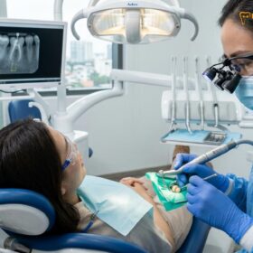
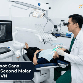
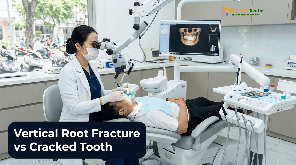
While 3D CBCT imaging provides unparalleled insight into the osseous changes surrounding a fractured root, direct visual confirmation of the crack remains the ultimate diagnostic standard. At HCMC Dental Clinic, this is achieved through the rigorous application of Wave 3 microscopic endodontic protocols. Utilizing the advanced optics and powerful illumination of the 25x Carl Zeiss Extaro 300 operating microscope, Dr. Nguyen Van Cuong, DDS can identify microscopic structural defects that are entirely invisible to the naked eye or standard dental loupes.
When evaluating vertical root fracture symptoms in a tooth, or when attempting to determine the apical extent of a coronal crack, the integration of methylene blue transillumination under a 25x Zeiss microscope is indispensable. The diagnostic protocol involves several precise steps:
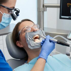
First, the tooth is isolated under a 100% latex-free nitrile rubber dam to ensure a sterile, moisture-free operating field. Existing restorations, posts, and core materials are carefully removed using ultrasonic instruments to expose the underlying dentin and the floor of the pulp chamber. Next, a 1% methylene blue dye is applied to the internal surfaces of the tooth. Methylene blue is a highly penetrative biological stain that wicks into microscopic cracks and fracture lines. After the excess dye is rinsed away, the fracture line remains stained, appearing as a distinct dark blue line against the lighter dentin.
To further verify the depth and propagation of the crack, fiber-optic transillumination is employed. A high-intensity fiber-optic light wand is placed against the external surface of the tooth. In a healthy, intact tooth, the light transmits uniformly through the crystalline structure of the enamel and dentin. However, when a crack is present, the fracture plane disrupts the transmission of light, causing the segment of the tooth beyond the crack to appear dark or shadowed. This dramatic visual contrast, magnified 25 times under the Carl Zeiss microscope, allows the clinician to precisely map the extent of the fracture.
If the crack is confined to the coronal structure and does not extend significantly below the alveolar crest, the tooth can often be saved. In these cases, Dr. Cuong employs advanced biomimetic restorative protocols, including Immediate Dentin Sealing (IDS) with a low-viscosity flowable resin biobase (increasing microtensile dentin bond strength to 45–55 MPa) and Deep Margin Elevation (DME). The tooth is then restored with a hand-layered refractory Feldspathic porcelain or IPS E.max lithium disilicate onlay to splint the crack and distribute occlusal forces evenly. However, if the methylene blue dye and transillumination reveal that the crack extends deep into the root structure, or if a true VRF is identified originating from the apical region, the diagnosis shifts from preservation to extraction[4].
For patients comparing international treatment options, understanding the value of this microscopic precision is crucial. You can explore a detailed comparison in our Root Canal Cost Vietnam vs Korea: Expat Guide | HCMC Dental.

Why True Vertical Root Fractures Must Be Extracted Immediately to Prevent Catastrophic Bone Loss Before Implant Placement
The definitive diagnosis of a Vertical Root Fracture carries a grim prognosis for the affected tooth. Unlike an endodontic infection caused by necrotic pulp tissue—which can be predictably resolved through thorough chemomechanical debridement and 3D obturation—a VRF represents a fundamental mechanical failure of the root structure. The fracture gap creates an open, unsealable highway for oral bacteria to continuously invade the periodontal ligament and surrounding alveolar bone.
Attempting to retreat a tooth with a confirmed VRF is not only futile but clinically negligent. No amount of sodium hypochlorite irrigation, calcium hydroxide medication, or advanced bioceramic obturation can fuse a fractured root back together or seal the microscopic gap against bacterial microleakage. As long as the fractured root remains in the jaw, the localized infection will persist, driving a rapid and aggressive osteolytic process. This chronic inflammation destroys the critical buccal bone plate and interproximal bone peaks, severely compromising the site for future restorative options.
Therefore, the standard of care dictates that clinicians must extract and place an immediate implant after a vertical root fracture, or perform a delayed implant placement following comprehensive site development. At HCMC Dental Clinic, the extraction of a fractured root is performed with meticulous care to preserve the remaining bone architecture. Dr. Nguyen Van Cuong, DDS utilizes ultrasonic Piezosurgery—a technology that cuts tooth structure and bone using micro-vibrations while completely sparing soft tissue. This allows for the atraumatic sectioning and removal of the fractured root segments without applying excessive luxation forces that could fracture the fragile buccal bone plate.
Once the root is extracted, the socket is thoroughly curetted to remove all infected granulation tissue. If the 3D CBCT scan indicates sufficient apical bone beyond the extraction socket to achieve primary stability (typically >35 Ncm of torque), an immediate dental implant (such as a premium Straumann BLT-SLA or Dentium fixture) can be placed during the same surgical appointment. To address the bone voids caused by the VRF osteolysis and the jumping gap between the implant and the socket walls, a Sticky Bone graft is utilized. Sticky Bone is created by mixing autologous Platelet-Rich Fibrin (PRF)—derived from the patient’s own blood—with particulate allograft or xenograft material. This bioactive matrix accelerates angiogenesis, promotes rapid osseointegration, and ensures the regeneration of a robust, healthy bone foundation for the final Zirconia implant crown[5].
Clinical Precaution: When to Contact Dr. Cuong Immediately
If you experience localized swelling, a recurring “pimple” on your gums, or sharp pain when releasing bite pressure on a tooth that has previously had a root canal and crown, do not ignore these symptoms. Delaying diagnosis of a potential Vertical Root Fracture allows the infection to rapidly destroy your jawbone, which can make future dental implant placement significantly more complex and expensive. Contact HCMC Dental Clinic via WhatsApp at +84 853 020 003 to schedule an immediate 3D CBCT and Microscopic Diagnostic Evaluation with Dr. Nguyen Van Cuong, DDS.
2026 Microscopic Crack Diagnosis, Biomimetic Onlay Splinting & Immediate Implant Fee Schedule at HCMC Dental Clinic (District 1)
At HCMC Dental Clinic, we are committed to providing world-class, evidence-based endodontic and implantology care with absolute financial transparency. By utilizing our Official 2026 Dental Price List & -40% Online Booking Fee Schedule, international patients and expatriates can access elite-level diagnostics and surgical interventions at a fraction of the cost compared to clinics in Australia, the US, or Europe. Below is the detailed 2026 fee schedule for diagnostic, endodontic, and implant procedures.
| Diagnostic / Surgical Procedure | 2026 Online Booking Fee (-40% USD) | 2026 Online Booking Fee (VND) | Standard Walk-In / US Benchmark |
|---|---|---|---|
| 3D CBCT Scan (0.075mm High-Resolution Endo-Mode) | $18 USD | 480,000 VND | $30 Walk-in / $250+ US |
| Microscopic Endodontic Root Canal Therapy (Anterior/Premolar) Includes 25x Carl Zeiss Microscope & Nitrile Dam | $77 – $115 USD | 2,000,000 – 3,000,000 VND | $128 – $192 Walk-in / $900+ US |
| Microscopic Endodontic Root Canal Therapy (Complex Molar) | $115 – $154 USD | 3,000,000 – 4,000,000 VND | $192 – $256 Walk-in / $1,200+ US |
| Microscopic Endodontic Retreatment / Separated File Bypass | $154 – $231 USD | 4,000,000 – 6,000,000 VND | $256 – $385 Walk-in / $1,500+ US |
| IPS E.max All-Ceramic Crown / Inlay / Onlay / Overlay Biomimetic splinting for savable cracked teeth | $231 USD | 6,000,000 VND | $385 Walk-in / $1,200+ US |
| Surgical Wisdom Tooth / Fractured Root Extraction (Piezotome) | $58 – $115 USD | 1,500,000 – 3,000,000 VND | $96 – $192 Walk-in / $400+ US |
| Single Korean Implant (Dentis / Neo Biotech / Dentium) Includes titanium fixture + abutment | $462 – $654 USD | 12,000,000 – 17,000,000 VND | $769 – $1,090 Walk-in / $2,500+ US |
| Single European / Swiss Implant (Straumann BLT-SLA / Active) | $962 – $1,346 USD | 25,000,000 – 35,000,000 VND | $1,603 – $2,244 Walk-in / $4,500+ US |
| Implant Zirconia HT Crown + Custom Abutment | $154 USD | 4,000,000 VND | $256 Walk-in / $1,500+ US |
“Our commitment at HCMC Dental Clinic is to provide uncompromising clinical excellence. By integrating 3D CBCT diagnostics and 25x Carl Zeiss microscopy, we eliminate the guesswork from endodontic diagnosis. Whether we are preserving a cracked tooth with biomimetic protocols or replacing a fractured root with a premium Straumann implant, our patients receive the highest standard of care at transparent, accessible prices.”
— Dr. Nguyen Van Cuong, DDS (Clinical Director, HCMC Dental Clinic)
To learn more about our advanced diagnostic capabilities and to see how we manage complex cases, please review our comprehensive Microscope Root Canal Vietnam: Clinical Guide | HCMC Dental.
Frequently Asked Questions
What is the difference between a savable Cracked Tooth (coronal crack) and a non-savable Vertical Root Fracture (VRF)?
A savable cracked tooth typically involves an incomplete fracture originating in the crown and extending apically, which can often be managed with biomimetic onlay splinting or microscopic root canal therapy if the pulp is involved. Conversely, a Vertical Root Fracture (VRF) originates in the root itself (often in previously endodontically treated teeth) and extends coronally. Because VRFs create an unsealable pathway for bacterial ingress directly into the periodontium, they result in rapid, catastrophic bone loss and render the tooth non-savable, necessitating immediate extraction.
Why do 2D periapical X-rays miss 75% of early Vertical Root Fractures that a 0.075mm endo-mode 3D CBCT detects?
Standard 2D periapical X-rays compress three-dimensional anatomy into a flat image. For a fracture line to be visible on a 2D radiograph, the X-ray beam must pass perfectly parallel to the fracture plane. Since most VRFs run bucco-lingually, they are perpendicular to the beam and remain invisible. A 0.075mm high-resolution endo-mode 3D CBCT eliminates anatomical superimposition, allowing the endodontist to view the root in axial, coronal, and sagittal slices, revealing the fracture line, cortical plate fenestrations, and the characteristic J-shaped halo bone loss.
Why should a tooth with a confirmed Vertical Root Fracture be extracted gently with Piezosurgery rather than retreated?
Attempting to retreat a tooth with a true VRF is futile because the fracture gap cannot be biologically sealed; bacteria will continue to leak into the surrounding bone, causing progressive osteolysis. Extracting the tooth atraumatically using ultrasonic Piezosurgery preserves the delicate buccal bone plate. This minimally invasive approach is critical for maintaining the necessary bone volume and soft tissue architecture required for successful immediate or delayed dental implant placement.
Can a dental implant and Sticky Bone graft be placed on the same day a fractured root is extracted?
Yes, provided there is sufficient apical bone beyond the extraction socket to achieve primary stability (typically >35 Ncm of torque) and the acute infection is thoroughly debrided. At HCMC Dental Clinic, Dr. Nguyen Van Cuong, DDS utilizes 3D CBCT-guided surgery to extract the fractured root, thoroughly curette the granulation tissue, place a premium titanium implant (such as Straumann or Dentium), and fill any jumping gaps with a Sticky Bone graft (autologous PRF mixed with particulate allograft) in a single appointment.
How much does a 3D CBCT + 25x Microscopic Crack Diagnostic Evaluation cost at HCMC Dental Clinic in District 1?
At HCMC Dental Clinic in District 1, Ho Chi Minh City, a comprehensive diagnostic evaluation utilizing a high-resolution 3D CBCT scan costs just $18 USD (480,000 VND) when booked online. If microscopic endodontic intervention is required, Microscopic Endodontic Root Canal Therapy utilizing a 25x Carl Zeiss microscope and nitrile rubber dam ranges from $77 to $154 USD per tooth, depending on anatomical complexity. This represents a transparent, -40% online booking savings compared to walk-in rates.
References
- American Association of Endodontists (AAE). (2022). Endodontic Diagnosis and the Management of Longitudinal Tooth Fractures: Clinical Guidelines and CBCT Efficacy.
- Journal of Endodontics. (2023). Diagnostic Accuracy of High-Resolution Cone-Beam Computed Tomography in Detecting Vertical Root Fractures: A Systematic Review and Meta-Analysis.
- International Endodontic Journal. (2021). The Pathognomonic Signs of Vertical Root Fractures: J-Shaped Lesions and Isolated Periodontal Defects in Endodontically Treated Teeth.
- Journal of Prosthetic Dentistry. (2024). Biomimetic Restorative Protocols and Immediate Dentin Sealing in the Prevention of Coronal Cracks and Root Fractures.
- Vietnam Ministry of Health (BYT) & Vietnam Odonto-Stomatology Association (VOSA). National Clinical Guidelines on Specialist Dental Care, Biomaterial Traceability & EN 13060 Class B Infection Control. (2024).