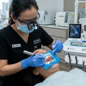
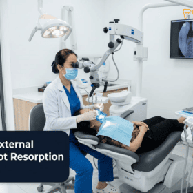
The successful endodontic management of the mandibular second molar represents one of the most formidable technical challenges in modern clinical dentistry, particularly when treating patients of East and Southeast Asian descent. Unlike standard molars that typically feature two distinct roots with three or four predictable, tubular canals, a significant percentage of Asian mandibular second molars exhibit a highly complex anatomical variation known as the C-shaped root canal system. This configuration is characterized by fused roots and a continuous, ribbon-like internal web that connects the individual canals, creating a labyrinthine network of fins, isthmuses, and microscopic irregularities. Attempting to clean, shape, and seal this intricate anatomy using traditional tactile-driven, blind endodontic techniques frequently results in catastrophic clinical failures, including missed anatomy, persistent apical periodontitis, and iatrogenic strip perforations. At HCMC Dental Clinic in District 1, Ho Chi Minh City, the management of c-shaped root canal anatomy mandibular second molar microscope Vietnam protocols is elevated to the highest international standards. Under the clinical direction of Dr. Nguyen Van Cuong (Dr. Cuong), DDS, our specialized endodontic team utilizes high-resolution 3D Cone Beam Computed Tomography (CBCT) and 25x Carl Zeiss Extaro operating microscopes to navigate, debride, and hermetically seal these complex ribbon-shaped systems, ensuring long-term tooth retention for our international patients, expatriates, and dental travelers.
Clinical Summary:
The C-shaped root canal configuration is a complex anatomical variation found in up to 40% of mandibular second molars in Asian populations. Characterized by a ribbon-like cross-section and fused roots, this anatomy poses severe risks for strip perforation in the mesiolingual “danger zone” (where dentin thickness is often <0.6mm) and harbors necrotic tissue in microscopic fins that standard rotary files cannot reach. At HCMC Dental Clinic, Dr. Nguyen Van Cuong, DDS, employs a strict, evidence-based protocol utilizing 0.075mm 3D CBCT imaging to map the Fan classification (C1, C2, C3) prior to access. Treatment is executed under a 25x Carl Zeiss microscope with sealed nitrile rubber dam isolation. The protocol mandates passive ultrasonic irrigation (PUI) with closed-end side-vented 30G needles to safely dissolve biofilm within the C-shaped webs without apical extrusion, followed by 3D thermoplasticized warm vertical compaction and bioceramic sealer obturation. This advanced micro-endodontic approach maximizes the success rate of primary treatments and complex retreatments while strictly adhering to international safety standards.
Key Takeaways:
- High Ethnic Prevalence: Up to 44% of East and Southeast Asian mandibular second molars feature a C-shaped root canal configuration, compared to less than 8% in Caucasian populations.
- The Anatomical Danger Zone: The mesiolingual longitudinal groove creates a paper-thin dentinal wall (often <0.6mm thick), making the tooth highly susceptible to iatrogenic strip perforation if aggressive rotary files are used.
- Mandatory 3D CBCT Diagnostics: Standard 2D X-rays cannot accurately depict the internal ribbon shape; a high-resolution 3D CBCT scan is essential to classify the canal (Fan’s C1, C2, C3) and measure dentin thickness.
- Advanced Irrigation Protocol: Because solid NiTi files cannot clean the ribbon-shaped webs, 100% prevention of NaOCl apical extrusion is achieved via closed-end side-vented 30G irrigation needles kept ≥2mm short of the electronic working length, combined with passive ultrasonic agitation.
- Microscopic Precision: A 25x Carl Zeiss operating microscope is required to safely de-roof the isthmus, locate hidden orifices, and perform 3D thermoplasticized warm vertical compaction with bioceramic sealers.
Why Up to 40% of Asian & Viet Kieu Lower Second Molars Feature Fused ‘C-Shaped’ Roots Instead of Two Separate Roots
To understand the clinical complexity of the c-shaped root canal mandibular second molar, one must first examine the embryological development of the human tooth. During odontogenesis, the shape of the tooth root is determined by Hertwig’s epithelial root sheath (HERS). In a typical mandibular molar, HERS invaginates from both the buccal and lingual aspects, eventually fusing in the center to create a distinct furcation that separates the mesial and distal roots. However, in a significant portion of the global population, a genetic and embryological variation occurs where HERS fails to fuse on either the buccal or lingual side. This failure of invagination results in the formation of a single, conical, or fused root structure that houses a continuous, ribbon-like, or C-shaped internal canal system rather than separate, tubular canals.
Extensive endodontic literature and anthropological studies have established that this anatomical variation exhibits a profound ethnic predilection. While the prevalence of C-shaped canals in mandibular second molars is relatively low in Caucasian and African populations (typically ranging from 2% to 8%), it is remarkably high in East Asian, Southeast Asian, and Native American cohorts. Clinical studies indicate that between 35% and 44% of Chinese, Korean, Vietnamese, and broader Asian populations possess this complex anatomy. For expatriates living in Vietnam, Viet Kieu (Overseas Vietnamese) returning for dental care, and international dental travelers seeking an 2026 Clinical Dental Research & Statistical Reports Hub, understanding this high prevalence is critical. When an international patient presents to an asian molar root canal anatomy endodontist Vietnam, the clinician must maintain a high index of suspicion for a C-shaped configuration before initiating any endodontic access.
Furthermore, this genetic clustering of complex root morphologies in Asian populations is not limited to the second molar. It is closely related to another significant anatomical variant: the Radix Entomolaris. The Radix Entomolaris is a supernumerary (extra) distolingual third root found in 15% to 30% of Asian mandibular first molars. According to the De Moor classification, these extra roots often exhibit a Type I, II, or III configuration, frequently featuring a severe 60° to 90° hidden buccolingual curvature. Successfully negotiating a Radix Entomolaris requires a modified trapezoidal access cavity preparation under 25x magnification to locate the hidden distolingual orifice without gouging the pulp chamber floor. Both the C-shaped second molar and the Radix Entomolaris first molar underscore the absolute necessity of advanced diagnostic imaging and high-level magnification when performing endodontics in Vietnam. Attempting to treat these teeth with standard 2D radiography and the naked eye is a primary cause of endodontic failure, leading to persistent infection and the eventual need for complex retreatment or extraction.[1]
Fan’s 3D CBCT & Cross-Sectional Classification (C1 Continuous Ribbon, C2 Semicolon, and C3 Separate Canals Linked by Fin Isthmuses)
The internal anatomy of a C-shaped root canal is not uniform; it is highly variable and changes dramatically from the pulp chamber orifice down to the apical foramen. In 1991, Melton et al. first proposed a classification system based on the cross-sectional shape of these canals. However, with the advent of high-resolution micro-computed tomography and clinical 3D CBCT, Dr. Bing Fan and colleagues introduced a more precise, modified classification system that has become the international gold standard for endodontists evaluating the fan classification c1 c2 c3 c-shaped canal.
Fan’s classification categorizes the cross-sectional morphology of the C-shaped canal into five distinct types, which can vary at different vertical levels of the same root:
- Category C1 (Continuous C-shape): The canal presents as an uninterrupted, continuous ribbon-like “C” shape from the mesial to the distal aspect, with no separation of dentin. This is often seen at the orifice level but rarely persists all the way to the apex.
- Category C2 (Semicolon shape): The dentin separates the main C-shaped ribbon into two distinct canals, resembling a semicolon (;). Typically, there is a larger, curved canal (often encompassing the mesial and buccal aspects) and a smaller, distinct round canal (usually the distal or mesiolingual).
- Category C3 (Two or three separate canals): The continuous C-shape is divided by thicker bridges of dentin into two or three distinct, separate canals. However, these canals are often still connected by microscopic, paper-thin fins or isthmuses that harbor necrotic tissue.
- Category C4 (Single round or oval canal): The complex ribbon merges into a single, distinct round or oval canal, typically found in the extreme apical third of the root near the foramen.
- Category C5 (No canal lumen): The canal space is completely obliterated or absent, usually seen at the very apex just before the apical foramen exits the root surface.
“The greatest clinical deception of the C-shaped mandibular second molar is that its anatomy is a moving target. A canal that appears as a continuous C1 ribbon at the pulp chamber floor may divide into a C2 semicolon in the middle third, and further split into a C3 configuration with hidden isthmuses in the apical third. Without a high-resolution 0.075mm 3D CBCT scan to map this exact morphological shifting before we even pick up a handpiece, we are essentially operating blindfolded. 3D imaging is not an optional luxury in these cases; it is the fundamental blueprint for safe and predictable endodontic success.”
— Dr. Nguyen Van Cuong, DDS (Clinical Director, HCMC Dental Clinic)
At HCMC Dental Clinic, the utilization of a small-field-of-view, high-resolution 3D CBCT scan is a mandatory prerequisite for the treatment of any suspected C-shaped molar. Standard 2D periapical radiographs suffer from anatomical superimposition; the dense cortical bone of the mandible and the fused nature of the C-shaped root often mask the true internal complexity. A 2D X-ray might show what appears to be two standard roots, or a single conical root with a blurry, indistinct canal space. Only through the multi-planar slicing of a 3D CBCT scan can Dr. Cuong accurately classify the canal according to Fan’s criteria, trace the pathway of the ribbon from orifice to apex, and identify the exact location of the apical foramina. This precise digital mapping is the cornerstone of our Microscope Root Canal Vietnam: Clinical Guide | HCMC Dental protocol, ensuring that no hidden anatomy is missed during the debridement phase.[2]
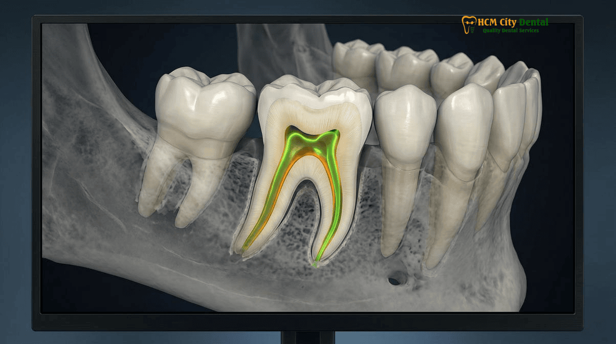
The Anatomical ‘Danger Zone’: Why the Lingual/Mesial Groove Wall Is Often Thinner Than 0.6mm and Prone to Strip Perforation
While the complex internal web of the C-shaped canal presents a significant challenge for cleaning and disinfection, the external morphology of the root presents an equally severe mechanical hazard. The defining external feature of a C-shaped mandibular second molar is the presence of a deep longitudinal developmental groove, most commonly located on the lingual or mesiolingual aspect of the fused root. This deep invagination creates a severe concavity in the root structure, resulting in an area where the dentinal wall separating the internal root canal space from the external periodontal ligament is alarmingly thin.
In endodontic literature, this specific area is universally referred to as the “danger zone.” In a standard molar, the dentin thickness in the furcation area might be 1.0mm to 1.5mm. However, in the strip perforation danger zone c-shaped molar, the dentin thickness along the invaginated groove is frequently less than 0.6mm, and in some cases, as thin as 0.2mm to 0.3mm. This paper-thin wall poses a monumental risk during the shaping phase of root canal therapy.
Clinical Warning: The Risk of Iatrogenic Strip Perforation
The use of aggressive, large-tapered rotary Nickel-Titanium (NiTi) files (such as .06 or .08 taper) or traditional Gates Glidden drills in the mesiolingual aspect of a C-shaped canal is strictly contraindicated. Because rotary files inherently attempt to center themselves within the canal space, they will aggressively cut into the inner curve of the C-shape. If the clinician is unaware of the danger zone, the file will rapidly strip away the remaining <0.6mm of dentin, causing a lateral “strip perforation” into the periodontal ligament. This iatrogenic error causes massive bleeding, severe postoperative pain, rapid bone loss, and often renders the tooth completely non-restorable, necessitating immediate extraction and implant replacement.
To safely navigate this danger zone, Dr. Cuong employs a highly conservative, biologically driven shaping protocol. Instead of using large, stiff rotary files, the shaping of the C-shaped canal is performed using heat-treated, controlled-memory (CM) NiTi files with minimal tapers (e.g., .04 taper) and flexible martensitic alloy properties. These files are used in a brushing motion directed strictly away from the danger zone and towards the bulky, safe outer walls of the root (the buccal and distal aspects). Furthermore, the preparation size is kept deliberately small (often no larger than an ISO size 25 or 30) to preserve maximum peri-cervical dentin.
The restorative implications of this thin dentinal wall are equally critical. Following the completion of the root canal, a C-shaped molar is structurally compromised. The placement of rigid prefabricated metal posts or custom cast posts inside a C-shaped canal is an absolute contraindication. The wedging forces of a metal post against the paper-thin lingual wall will inevitably cause a catastrophic vertical root fracture. Instead, at HCMC Dental Clinic, these teeth are restored using biomimetic principles, utilizing fiber-reinforced composite core buildups and cuspal-coverage restorations (such as IPS E.max overlays or full Zirconia crowns) that distribute occlusal forces evenly without relying on deep intraradicular post retention.[3]
25x Carl Zeiss Microscopic Isthmus De-Roofing & Passive Ultrasonic Irrigation (PUI / XP-endo Finisher) Protocol
Because the mechanical shaping of a C-shaped canal must be highly conservative to prevent strip perforation, solid titanium or NiTi rotary files cannot physically contact all the internal walls of the ribbon-shaped webs, fins, and isthmuses. Studies show that up to 60% of the canal walls in a C-shaped molar remain untouched by rotary instruments. If these uninstrumented areas are left filled with necrotic pulp tissue and bacterial biofilm, the root canal will inevitably fail, leading to a complex failed root canal lower second molar retreatment.
To overcome this limitation, the endodontic protocol at HCMC Dental Clinic shifts the paradigm from mechanical cutting to advanced chemical disinfection and fluid dynamics, executed under extreme magnification. The procedure begins with the application of a sealed nitrile rubber dam. This is not merely for patient comfort; it is an absolute, non-negotiable safety requirement to maintain a sterile operating field and protect the patient’s airway from strong chemical irrigants. For more details on our safety standards, review our Rubber Dam Isolation in Biomimetic Dentistry & Root Canal Therapy: Safety & Clinical Protocol in Vietnam.
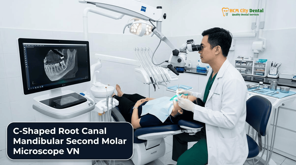
Operating under a 25x Carl Zeiss Extaro 300 microscope, Dr. Cuong first identifies the complex C-shaped orifice. Using specialized ultrasonic micro-tips (such as Start-X or Munce Discovery burs), the calcified roof of the pulp chamber is carefully troughed and de-roofed to expose the full extent of the ribbon-like anatomy. The microscope’s coaxial LED illumination penetrates deep into the root, allowing the clinician to visually confirm the removal of tissue from the isthmuses that connect the main canals.
Clinical Case Scenario: Bypassing a Separated File in a C-Shaped Molar
An expatriate patient presented to HCMC Dental Clinic with severe pain and swelling in the lower left jaw. A 3D CBCT scan revealed a failing root canal in the mandibular second molar (tooth 37), which exhibited a Fan C2 (semicolon) C-shaped configuration. The previous dentist, operating without a microscope, had fractured a rotary file deep inside the narrow mesiolingual fin and failed to locate the distal aspect of the ribbon. Under 25x Zeiss magnification, Dr. Cuong utilized ultrasonic micro-tips to carefully trough the dentin around the separated file, successfully bypassing and retrieving the fragment without perforating the <0.6mm danger zone. The hidden distal web was then located, thoroughly disinfected using passive ultrasonic irrigation, and sealed with bioceramic sealer, saving the tooth from extraction.
Once the canals are conservatively shaped, the critical phase of ultrasonic irrigation activation warm gutta percha preparation begins. The primary irrigant is 5.25% Sodium Hypochlorite (NaOCl), a powerful antimicrobial and tissue-dissolving agent. However, NaOCl is highly toxic if extruded past the apex into the periapical bone. To ensure 100% prevention of NaOCl apical extrusion, Dr. Cuong strictly adheres to the following safety rules:
- Safe Delivery: Irrigants are delivered using closed-end, side-vented 30-gauge irrigation needles. The needle is carefully measured and kept at least 2mm short of the electronic working length, ensuring the fluid flows laterally and coronally, never apically.
- Passive Ultrasonic Irrigation (PUI): A smooth, non-cutting ultrasonic wire (e.g., Irrisafe) is introduced into the canal and activated. The ultrasonic energy creates acoustic streaming and microscopic cavitation bubbles within the NaOCl. When these bubbles implode, they generate powerful shear forces that physically tear bacterial biofilm off the walls of the uninstrumented fins and dissolve the necrotic tissue hidden deep within the C-shaped web.
- Mechanical Agitation (XP-endo Finisher): In highly complex ribbon anatomies, a specialized NiTi instrument called the XP-endo Finisher may be used. At body temperature, this highly flexible file expands and whips around inside the canal, mechanically agitating the irrigant and scrubbing the irregular walls of the C-shape without cutting dentin.
This synergistic combination of microscopic visualization, conservative shaping, and advanced ultrasonic fluid dynamics ensures that the entire C-shaped labyrinth is rendered surgically clean and ready for obturation.[4]
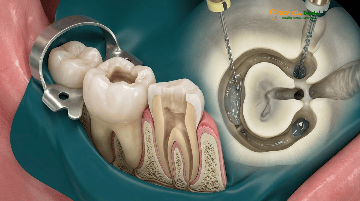
3D Thermoplasticized Warm Vertical Compaction & Bioceramic Sealer Obturation of Ribbon-Shaped Canal Webs
The final, critical step in the endodontic management of a C-shaped mandibular second molar is obturation—the complete, hermetic sealing of the disinfected root canal system. The goal of obturation is to entomb any remaining microscopic bacteria and prevent the ingress of tissue fluids from the periapical tissues or coronal leakage from the oral cavity. In a standard, round root canal, traditional cold lateral condensation (where a master gutta-percha cone is placed and smaller accessory cones are wedged in beside it) may be sufficient. However, in a C-shaped canal, cold lateral condensation is a recipe for failure.
Because the C-shaped canal is a continuous ribbon with irregular fins and webs, cold, solid gutta-percha cones cannot physically deform to fill these microscopic voids. Attempting to force cold cones into a C-shaped web will leave massive empty spaces (voids) within the isthmuses, which serve as breeding grounds for recurrent bacterial infection. Furthermore, excessive lateral pressure applied with a metal spreader during cold condensation can easily cause a vertical root fracture in the fragile, thin walls of the danger zone.
“You cannot seal a 3D anatomical labyrinth with a 2D cold technique. The ribbon-shaped webs of a C-shaped molar demand a fluid, hydraulic seal. By utilizing 3D thermoplasticized warm vertical compaction combined with advanced calcium silicate bioceramic sealers, we can literally flow the filling material into every microscopic fin, lateral canal, and apical delta that the microscope reveals. This is the only way to guarantee a hermetic seal and long-term clinical success in these highly complex Asian molar anatomies.”
— Dr. Nguyen Van Cuong, DDS (Clinical Director, HCMC Dental Clinic)
To achieve a flawless 3D seal, Dr. Cuong utilizes the Continuous Wave of Condensation technique (warm vertical compaction). The protocol involves the following precise steps:
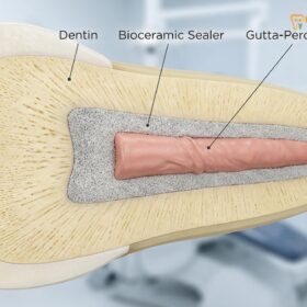
- Bioceramic Sealer Application: A state-of-the-art, biocompatible calcium silicate-based sealer (such as BioRoot RCS or TotalFill BC Sealer) is introduced into the canal. Unlike traditional resin sealers that shrink upon setting, bioceramic sealers are hydrophilic; they utilize the natural moisture within the dentinal tubules to initiate a setting reaction, expanding slightly to create a chemical bond with the root dentin and producing hydroxyapatite, which promotes periapical bone healing.
- Downpack Phase: A master gutta-percha cone, precisely matched to the conservative taper of the prepared canal, is coated in bioceramic sealer and seated to the working length. An electrically heated plugger (set to a precise temperature, typically 200°C) is driven down into the canal, severing the coronal portion of the gutta-percha and instantly plasticizing (melting) the apical plug. The softened gutta-percha is then vertically compacted, hydraulically forcing the bioceramic sealer deep into the microscopic fins, isthmuses, and lateral canals of the C-shaped web.
- Backfill Phase: The remaining coronal portion of the ribbon-shaped canal is then backfilled using a motorized extruder that injects molten, thermoplasticized gutta-percha. This warm material flows seamlessly into the irregular C-shaped cross-section, adapting perfectly to the complex internal anatomy before cooling and solidifying into a dense, void-free 3D mass.
Following obturation, the pulp chamber is immediately sealed with a bonded composite resin to prevent any coronal microleakage, and the patient is scheduled for a definitive biomimetic restoration (such as an overlay or crown) to protect the structurally compromised tooth from occlusal fracture. For patients interested in learning more about Dr. Cuong’s credentials and expertise in complex restorative dentistry, please visit Dr. Nguyen Van Cuong, DDS (Clinical Director — MOH License 0003265/BL-CCHN).[5]
2026 Microscopic Molar Endodontics & C-Shaped Canal Retreatment Fee Schedule at HCMC Dental Clinic (District 1)
For international patients, expatriates, and dental travelers, navigating the costs of complex specialist endodontics can be daunting. In countries like Australia, the United States, or the UK, a microscopic root canal treatment on a complex molar performed by a specialist endodontist can easily exceed $1,500 to $2,500 USD, not including the cost of the 3D CBCT scan or the final crown. At HCMC Dental Clinic, we believe in absolute financial transparency and providing world-class, technology-driven specialist care at highly accessible price points.
By utilizing our Official 2026 Dental Price List & -40% Online Booking Fee Schedule, international patients can access the exact same 25x Carl Zeiss microscopic protocols, 3D CBCT diagnostics, and bioceramic obturation techniques used in top-tier Western clinics at a fraction of the cost. Below is the official 2026 fee schedule for microscopic endodontics and associated diagnostic procedures at our District 1 facility.
| Specialist Endodontic Procedure (2026) | Walk-In / Standard Price (USD) | -40% Online Booking Price (USD) | Online Booking Price (VND) |
|---|---|---|---|
| 3D CBCT Scan (Mandatory for C-Shaped Molars) | $30 USD | $18 USD | 480,000 VND |
| Microscopic Root Canal (Anterior / Premolar) (25x Carl Zeiss + Nitrile Dam) | $128 – $192 USD | $77 – $115 USD / tooth | 2,000,000 – 3,000,000 VND |
| Microscopic Root Canal (Complex Molar / C-Shaped) (25x Carl Zeiss + Nitrile Dam + PUI) | $192 – $256 USD | $115 – $154 USD / tooth | 3,000,000 – 4,000,000 VND |
| Microscopic Endodontic Retreatment (Includes Separated File Bypass / C-Shaped Failure) | $256 – $385 USD | $154 – $231 USD / tooth | 4,000,000 – 6,000,000 VND |
| Bioactive Perforation Repair (ProRoot MTA / Septodont Biodentine) | $96 – $160 USD | $58 – $96 USD | 1,500,000 – 2,500,000 VND |
| Biomimetic Quartz/Glass Fiber Post & Core Buildup | $63 – $96 USD | $38 – $58 USD / tooth | 1,000,000 – 1,500,000 VND |
| Full Zirconia Crown (Cercon Standard / HT Multilayer) (Definitive Restoration post-Endo) | $256 – $321 USD | $154 – $192 USD / tooth | 4,000,000 – 5,000,000 VND |
Patients traveling to Ho Chi Minh City for complex endodontic care can rest assured that there are no hidden fees. The online booking price reflects a transparent, all-inclusive approach to specialist dental care. To secure these exclusive 2026 online rates and schedule a comprehensive 3D CBCT consultation with Dr. Nguyen Van Cuong, DDS, patients are encouraged to contact our international coordination team via WhatsApp at +84 853 020 003 prior to their arrival in Vietnam.

Frequently Asked Questions
Why do standard root canals performed without a microscope frequently fail on lower second molars with C-shaped roots?
Standard root canals performed without high-level magnification frequently fail on C-shaped molars because the naked eye cannot visualize the microscopic ribbon-shaped isthmuses and fins that connect the main canals. Without a 25x Carl Zeiss microscope, these hidden webs harbor residual necrotic pulp tissue and bacterial biofilm, leading to persistent apical periodontitis and the eventual need for a failed root canal lower second molar retreatment.
Can a regular 2D dental X-ray clearly show the internal ribbon shape of a C-shaped root canal without a 3D CBCT scan?
No, a standard 2D periapical or panoramic X-ray cannot definitively show the internal cross-sectional ribbon shape of a C-shaped canal. Because 2D imaging compresses complex 3D anatomy into a flat picture, the thin webs and fused roots often superimpose over one another. A high-resolution 3D CBCT scan is absolutely mandatory to map the Fan classification (C1, C2, C3) and measure the exact dentin thickness of the mesiolingual danger zone prior to treatment.
Why must aggressive large tapered rotary files and metal posts never be placed inside the thin mesiolingual wall of a C-shaped molar?
The mesiolingual wall of a C-shaped mandibular second molar features a deep anatomical invagination (the longitudinal groove), making the dentin in this ‘danger zone’ extremely thin—often less than 0.6mm. Using aggressive large tapered rotary files or attempting to place rigid metal posts in this area will almost certainly result in a catastrophic strip perforation or vertical root fracture, rendering the tooth non-restorable and requiring extraction.
How does ultrasonic activation of sodium hypochlorite clean the paper-thin web connecting the canals?
Ultrasonic activation utilizes a smooth, non-cutting oscillating wire inside the canal to generate acoustic streaming and cavitation within the sodium hypochlorite (NaOCl) irrigant. This powerful fluid dynamics effect shears bacterial biofilm and dissolves necrotic tissue deep inside the paper-thin webs and fins of a C-shaped canal—areas that solid titanium rotary files physically cannot reach without destroying the fragile root structure.
How much does 25x microscopic root canal treatment for a complex C-shaped molar cost at HCMC Dental Clinic in District 1?
At HCMC Dental Clinic in District 1, Ho Chi Minh City, the fee for Microscopic Endodontic Root Canal Therapy (utilizing a 25x Carl Zeiss microscope and Nitrile Dam isolation) for a complex molar is $115 – $154 USD per tooth when booked online (3,000,000 – 4,000,000 VND). If the tooth requires microscopic endodontic retreatment to bypass separated files or fix a previous failure, the online fee is $154 – $231 USD per tooth.
References
- Fan B, Cheung GS, Fan M, Gutmann JL, Bian Z. (2004). C-Shaped Canal System in Mandibular Second Molars: Part I—Anatomical Features. Journal of Endodontics, 30(12), 899-903.
- Jafarzadeh H, Wu YN. (2007). The C-shaped Root Canal Configuration: A Review. Journal of Endodontics, 33(5), 517-523.
- Melton DC, Krell KV, Fuller MW. (1991). Anatomical and histological features of C-shaped canals in mandibular second molars. Journal of Endodontics, 17(8), 384-388.
- Cooke HG, Cox FL. (1979). C-shaped canal configurations in mandibular molars. Journal of the American Dental Association, 99(5), 836-839.
- Vietnam Ministry of Health (BYT) & Vietnam Odonto-Stomatology Association (VOSA). National Clinical Guidelines on Specialist Dental Care, Biomaterial Traceability & EN 13060 Class B Infection Control. (2024).