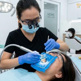
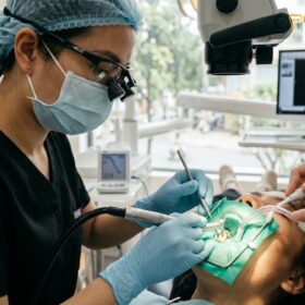
For international patients, expatriates, and dental travelers seeking advanced endodontic care in Vietnam, the diagnosis of a “calcified root canal” often brings anxiety and frustration. When a tooth undergoes severe trauma, chronic irritation, or natural aging, the internal pulp chamber can shrink and fill with hard tertiary dentin—a condition known as Pulp Canal Obliteration (PCO). In these complex scenarios, traditional freehand root canal therapy frequently fails, leading to catastrophic root perforations, untreated hidden infections, and ultimately, unnecessary tooth extraction. However, the landscape of endodontic salvage has been revolutionized by digital dentistry. At HCMC Dental Clinic (District 1, Ho Chi Minh City), Dr. Nguyen Van Cuong (Dr. Cuong), DDS (Vietnam MOH License 0003265/BL-CCHN) employs a hospital-grade protocol combining calcified root canal treatment using 3D guided endodontics and 25x Carl Zeiss optical magnification. This micro-surgical approach allows for the millimeter-precise location and negotiation of obliterated canals, preserving the natural tooth structure and ensuring long-term biomechanical stability.
Clinical Summary:
Pulp Canal Obliteration (PCO) presents one of the most formidable challenges in modern endodontics. Attempting to locate a microscopic, calcified canal using conventional 2D X-rays and freehand drilling carries a high risk of iatrogenic root perforation. HCMC Dental Clinic mitigates this risk entirely through a dual-technology protocol: 3D Static Guided Endodontics (merging 0.075mm voxel CBCT data with intraoral STL scans to 3D-print a custom drill-targeting stent) and 25x Carl Zeiss Microscopic Endodontics. Led by Dr. Nguyen Van Cuong, DDS, this evidence-based workflow ensures absolute precision, allowing for the safe negotiation of blocked canals using ultrasonic micro-troughing, C-Pilot files, and bioceramic obturation, all performed under strict 100% latex-free Nitrile Rubber Dam isolation.
Key Takeaways:
- The Pathology of PCO: Dental trauma, chronic bruxism, and aging trigger odontoblasts to rapidly deposit tertiary dentin, shrinking the pulp space until it becomes radiographically invisible.
- The Danger of Freehand Drilling: Without 3D guidance, attempting to drill into a calcified root carries a 40%+ risk of lateral root perforation, often condemning the tooth to extraction.
- 3D Guided Precision: By superimposing a high-resolution CBCT scan with a digital surface scan, a 3D-printed stent is fabricated to guide a micro-bur exactly to the calcified canal orifice with sub-millimeter accuracy.
- Microscopic Verification: A 25x Carl Zeiss Extaro 300 microscope allows the specialist to read the “dentin color map,” distinguishing between the darker floor of the pulp chamber and the lighter, calcified tertiary dentin.
- Transparent 2026 Pricing: HCMC Dental Clinic offers world-class microscopic endodontics starting at just $77 – $115 USD per anterior tooth online, representing a -40% savings compared to walk-in rates and a fraction of Western dental costs.

What Causes Pulp Canal Obliteration (Calcified Root Canals)? Dental Trauma, Chronic Bruxism, and Aging Secondary Dentin
To understand the complexity of treating a calcified tooth, one must first understand the biological mechanism of Pulp Canal Obliteration (PCO), also known as calcific metamorphosis. The dental pulp is a highly vascularized and innervated tissue housed within the rigid walls of the root canal system. When the tooth is subjected to severe stress, the pulp’s primary defense mechanism is to lay down additional layers of dentin to insulate itself from the irritant. This process is driven by specialized cells called odontoblasts.
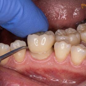
The most common catalyst for rapid PCO is acute dental trauma, such as a luxation injury (where the tooth is displaced) or a concussion injury (a blunt impact without displacement) sustained during childhood sports, falls, or accidents. Following the trauma, the blood supply to the pulp is temporarily disrupted but often re-establishes itself. In response to the shock, the surviving odontoblasts go into overdrive, rapidly depositing irregular, disorganized hard tissue known as reparative tertiary dentin. Over several years, this relentless deposition shrinks the pulp chamber and the root canal space until it appears completely obliterated on a standard 2D dental X-ray. It is highly common for a patient to present with a yellow, darkened front tooth after trauma becomes calcified, completely unaware that the internal canal has vanished[1].
Beyond acute trauma, chronic, low-grade irritants can also cause gradual calcification. Severe bruxism (teeth grinding), chronic occlusal trauma, deep carious lesions, and the natural physiological aging process all stimulate the slow, continuous deposition of secondary dentin. In elderly patients, it is not uncommon to find molars where the pulp horns have completely receded and the canals are hair-thin. While a calcified tooth may remain asymptomatic for decades, the microscopic remnants of pulpal tissue can eventually undergo aseptic necrosis (tissue death). Because the canal is blocked, the necrotic debris and bacteria cannot escape, leading to a chronic apical periodontitis—a silent infection at the tip of the root that slowly destroys the surrounding jawbone.
Why Conventional Freehand Drilling in Calcified Teeth Leads to 40%+ Root Perforation and Unnecessary Tooth Loss
When a calcified tooth develops an apical infection, endodontic intervention becomes mandatory to save the tooth. However, a blocked root canal where the dentist cannot find the canal presents a terrifying scenario for a general practitioner relying on traditional freehand techniques and 2D periapical radiographs.
In a normal tooth, the clinician drills an access cavity and relies on tactile feedback—the distinct “drop” into the hollow pulp chamber—to locate the canals. In a tooth with PCO, there is no hollow chamber. The entire internal space is a solid mass of dentin. The clinician is forced to drill blindly down the long axis of the root, hoping to intersect the microscopic remnant of the canal. Because the human hand cannot maintain a perfectly straight trajectory without visual landmarks, the dental bur inevitably deviates. The bur tends to follow the path of least resistance, cutting into the softer lateral dentin walls rather than the harder calcified core.
Clinical studies have shown that attempting to negotiate severely calcified canals using freehand drilling without magnification or 3D guidance results in catastrophic iatrogenic errors in a staggering percentage of cases. The most common complication is a lateral root perforation—where the drill breaches the side of the root and enters the periodontal ligament and surrounding bone. Once a perforation occurs deep within the root, the prognosis of the tooth plummets, and extraction often becomes the only viable option. Furthermore, aggressive freehand drilling removes excessive amounts of healthy cervical dentin, severely weakening the tooth and predisposing it to vertical root fractures. This is why root perforation prevention in a calcified molar or incisor requires a paradigm shift away from tactile guesswork and toward digital, image-guided micro-surgery[2].
“The era of ‘drilling and hoping’ in endodontics is over. When a patient presents with a severely calcified canal, attempting freehand access is a gamble with the patient’s natural tooth. By integrating 3D CBCT imaging with static guided stents and 25x optical magnification, we remove the guesswork entirely. We can target a canal that is less than 0.1 millimeters wide with absolute mathematical precision, preserving the structural integrity of the root and saving teeth that would otherwise be condemned to the extraction forceps.”
— Dr. Nguyen Van Cuong, DDS (Clinical Director, HCMC Dental Clinic)
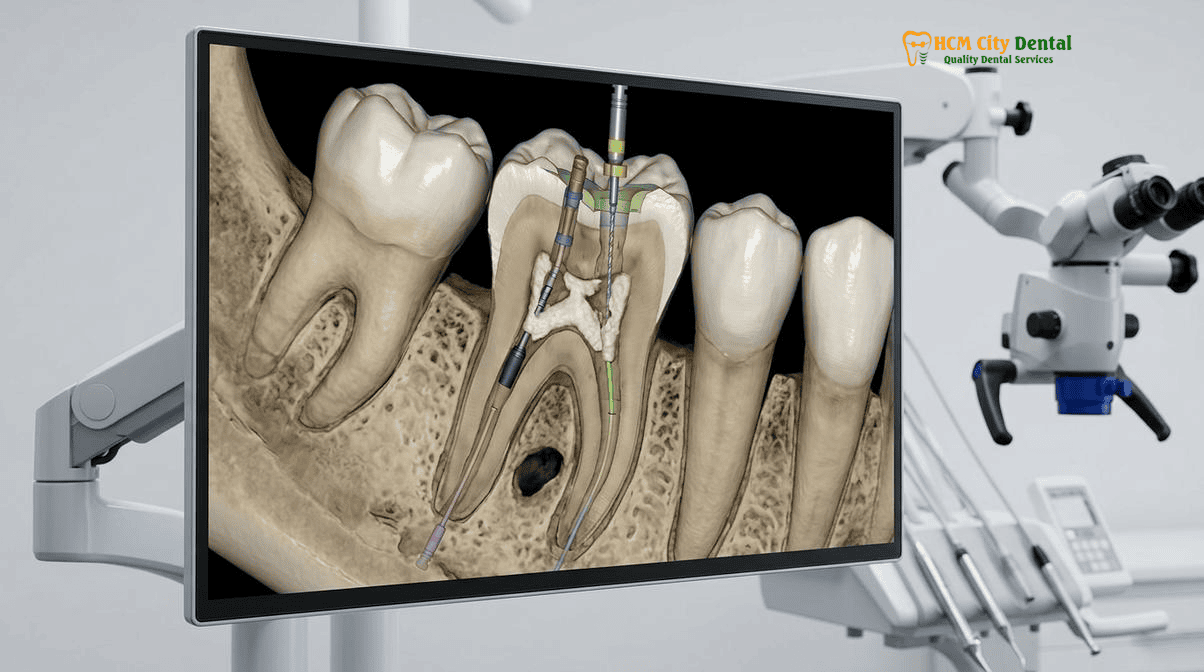
25x Carl Zeiss Microscope & Ultrasonic Diamond Micro-Troughing: Following the Dentin Color Map (Dark Floor vs. White Tertiary Dentin)
The first pillar of advanced PCO management at HCMC Dental Clinic is the utilization of hospital-grade optical magnification. Performing calcified root canal treatment using a microscope is not a luxury; it is an absolute clinical necessity. Dr. Nguyen Van Cuong, DDS utilizes the industry-leading 25x Carl Zeiss Extaro 300 dental operating microscope. This optical powerhouse provides brilliant, shadow-free coaxial LED illumination that penetrates deep into the root canal system, revealing anatomical details that are invisible to the naked eye or standard dental loupes.
Under 25x magnification, the clinician can read the “dentin color map.” The natural floor of the pulp chamber is typically darker and greyish, while the calcified tertiary dentin that blocks the canal orifices is usually lighter, whitish, or yellowish. By carefully distinguishing these subtle color variations, Dr. Cuong can identify the exact location where the original canal once existed. For more insights into our microscopic protocols, patients can review our Microscope Root Canal Vietnam: Clinical Guide | HCMC Dental.
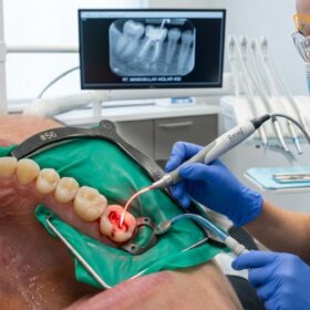
Once the calcified orifice is visually identified, traditional high-speed rotary burs are abandoned. They are too bulky, obscure the line of sight, and remove dentin too aggressively. Instead, Dr. Cuong employs Satelec/Acteon Titanium Ultrasonic Micro-Tips (such as the ET20 or ET25) coated with fine diamond grit. Ultrasonic troughing for a calcified root canal involves using these microscopic vibrating tips to gently brush away the calcified tissue in increments of fractions of a millimeter. Because the ultrasonic tip is incredibly slender, it does not block the light from the microscope, allowing Dr. Cuong to maintain continuous visual control as he slowly uncovers the hidden canal pathway. This conservative, visually guided approach preserves maximum peri-cervical dentin, which is critical for the long-term fracture resistance of the tooth[3].
3D Static Guided Endodontics: Merging High-Resolution CBCT (0.075mm Voxel) with Intraoral STL Scans to 3D-Print Micro-Access Stents
While the operating microscope is invaluable for visual navigation, some canals are calcified so deeply into the apical third of the root that even ultrasonic troughing becomes risky without a definitive roadmap. This is where the second pillar of our protocol—3D Static Guided Endodontics—becomes the ultimate safety net.
The workflow for 3D guided endodontics for pulp canal obliteration begins with a high-resolution Cone Beam Computed Tomography (CBCT) scan. At HCMC Dental Clinic, we utilize a state-of-the-art CBCT unit capable of capturing images at an ultra-fine 0.075mm voxel resolution. This 3D scan slices the tooth into microscopic layers, allowing Dr. Cuong to visualize the exact depth, angulation, and curvature of the remaining patent (open) canal space, even if it is located millimeters below the gumline.
Simultaneously, an intraoral optical scanner is used to capture a highly accurate 3D surface map of the patient’s dental arches, generating an STL (Stereolithography) file. Using advanced implant planning software (such as BlueSkyBio or Sicat Endo), the DICOM data from the CBCT scan is superimposed over the STL surface scan. In this virtual 3D environment, Dr. Cuong designs a precise drill trajectory. He plots a straight line from the biting surface of the tooth directly into the microscopic target of the calcified canal, ensuring the path stays perfectly centered within the root to avoid perforation.
Once the virtual plan is perfected, the software designs a custom surgical stent (template) that fits snugly over the patient’s adjacent teeth. This design is sent to an in-house high-resolution 3D resin printer. A metal guide sleeve is inserted into the printed stent at the exact angle and position dictated by the software. During the clinical procedure, the stent is seated in the patient’s mouth. A specialized, long-shank micro-bur is inserted through the metal sleeve. The sleeve physically restricts the bur, allowing it to move only along the pre-planned 3D trajectory and stopping it at the exact pre-planned depth. This technology transforms a highly unpredictable, high-risk procedure into a safe, mathematically precise, and minimally invasive operation[4].
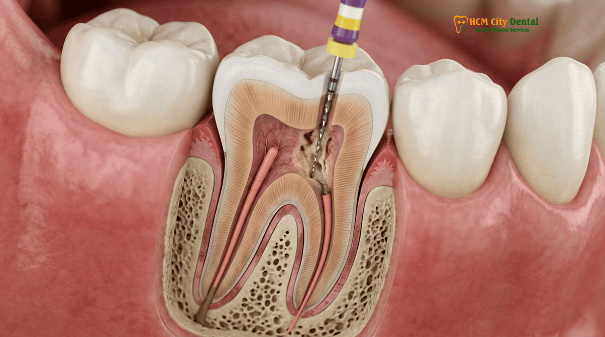
Negotiating Obliterated Canals with #06/#08 Carbon-Steel C-Pilot Files, 17% EDTA Chelation, and Bioceramic Obturation
Once the 3D guided bur or ultrasonic tip has successfully bypassed the calcified blockage and reached the patent portion of the canal, the next challenge is negotiating the remaining canal space to the root apex. Because the canal has been subjected to chronic calcification, it is often incredibly narrow, tortuous, and packed with dense collagenous debris.
Standard nickel-titanium (NiTi) rotary files cannot be used initially, as they will instantly unwind and separate (break) in such tight, calcified spaces. Instead, Dr. Cuong relies on specialized manual hand files, specifically #06, #08, and #10 Carbon-Steel C-Pilot files or D-Finders. Unlike standard stainless steel files, these instruments have a stiffer alloy and a specialized flute design that allows them to penetrate dense calcifications without buckling or bending. The negotiation process is performed with extreme patience, using a “watch-winding” motion to slowly glide the file down the canal.
To assist in breaking down the calcifications, copious chemical irrigation is employed. A 17% EDTA (Ethylenediaminetetraacetic acid) solution is used as a chelating agent. EDTA chemically binds to the calcium ions in the calcified dentin, softening the canal walls and lubricating the passage for the micro-files. This is alternated with heated 5.25% Sodium Hypochlorite (NaOCl) to dissolve any remaining necrotic organic tissue and eradicate bacterial biofilms. All of this is performed under strict 100% latex-free heavy-gauge Nitrile Rubber Dam isolation, ensuring absolute sterility and patient safety. For more details on our isolation standards, please see Rubber Dam Isolation in Biomimetic Dentistry & Root Canal Therapy: Safety & Clinical Protocol in Vietnam.
Once the canal is fully negotiated, cleaned, and shaped, it must be sealed to prevent reinfection. For calcified cases, HCMC Dental Clinic utilizes advanced Bioceramic (calcium silicate-based) sealers (such as TotalFill or BioRoot RCS). Unlike traditional resin sealers that shrink upon setting, bioceramics are highly biocompatible, hydrophilic (they utilize the natural moisture in the dentinal tubules to set), and actually expand slightly to create a hermetic, gap-free seal. Furthermore, they release calcium hydroxide during the setting phase, providing a highly alkaline, antibacterial environment that promotes the healing of apical bone lesions[5].
Internal Walking Bleach + Microscopic Endodontic Pricing for Traumatized Dark Front Teeth at HCMC Dental Clinic
One of the most distressing aesthetic consequences of pulp canal obliteration in anterior (front) teeth is severe discoloration. As the traumatized pulp slowly necroses and calcifies, red blood cells rupture, releasing iron-rich hemoglobin into the dentinal tubules. This hemoglobin breaks down into dark pigments (hemosiderin), causing the tooth to turn a noticeable yellow, grey, or even dark brown. Patients often assume that the only way to fix a dark, calcified front tooth is to grind it down for a porcelain crown.
However, following successful calcified root canal treatment using 3D guided endodontics, Dr. Cuong can perform a highly conservative “Internal Walking Bleach” procedure. Because the access cavity is already open on the back of the tooth, a specialized whitening paste (typically sodium perborate mixed with distilled water) is placed directly inside the pulp chamber. The cervical area of the root is carefully sealed with a glass ionomer barrier to prevent the bleaching agent from leaking into the root and causing external cervical resorption. The tooth is temporarily sealed, and the patient “walks” with the bleach inside for 3 to 7 days. The peroxide safely oxidizes the dark hemosiderin pigments from the inside out. Once the tooth matches the adjacent natural teeth, the bleach is removed, and the access cavity is permanently restored with a biomimetic composite resin, completely avoiding the need for an invasive crown.
At HCMC Dental Clinic, we believe that advanced, hospital-grade endodontic care should be accessible and transparent. By booking online, international patients and expats can access our official 2026 fee schedule, which offers a -40% discount compared to walk-in rates. For a comprehensive overview of all our clinical data and outcomes, visit our 2026 Clinical Dental Research & Statistical Reports Hub.
| Advanced Endodontic & Diagnostic Procedure | 2026 Online Booking Price (-40% USD) | 2026 Walk-In Price (USD) | VND Equivalent (Online) |
|---|---|---|---|
| High-Resolution 3D CBCT Scan (0.075mm Voxel) | $18 USD | $30 USD | 480,000 VND |
| Microscopic Endodontics (Anterior/Premolar) – 25x Carl Zeiss + Nitrile Dam | $77 – $115 USD | $128 – $192 USD | 2,000,000 – 3,000,000 VND |
| Microscopic Endodontics (Complex Molar) – 25x Carl Zeiss + Nitrile Dam | $115 – $154 USD | $192 – $256 USD | 3,000,000 – 4,000,000 VND |
| Microscopic Endodontic Retreatment / Separated File Bypass | $154 – $231 USD | $256 – $385 USD | 4,000,000 – 6,000,000 VND |
| Bioactive ProRoot MTA / Septodont Biodentine Perforation Repair | $58 – $96 USD | $96 – $160 USD | 1,500,000 – 2,500,000 VND |
| Biomimetic Quartz/Glass Fiber Post & Core Buildup | $38 – $58 USD | $63 – $96 USD | 1,000,000 – 1,500,000 VND |
To view our complete, transparent pricing for all procedures, including implants and aesthetics, please visit our Official 2026 Dental Price List & -40% Online Booking Fee Schedule. To learn more about the credentials of our clinical director, visit Dr. Nguyen Van Cuong, DDS (Clinical Director — MOH License 0003265/BL-CCHN).
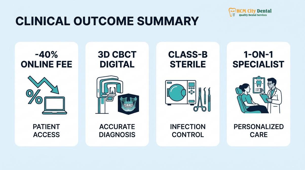
Clinical Case Study: Salvaging a Calcified Maxillary Central Incisor
Patient Profile: A 34-year-old Australian expatriate presented to HCMC Dental Clinic in District 1, Ho Chi Minh City, complaining of a dark yellow upper right central incisor and a recurring “pimple” on the gums (a sinus tract indicating chronic infection). The patient reported a skateboarding accident at age 14. A previous dentist in Sydney had attempted a root canal but abandoned the procedure, stating the canal was “completely blocked” and recommended extraction and a dental implant.
Clinical Intervention: Dr. Nguyen Van Cuong, DDS performed a high-resolution 3D CBCT scan, which revealed severe Pulp Canal Obliteration in the coronal two-thirds of the root, but a patent canal in the apical third surrounded by a 5mm periapical radiolucency (bone infection). An intraoral STL scan was taken, and a 3D static guided endodontic stent was designed and printed in-house. Under 25x Carl Zeiss microscopic magnification and Nitrile Rubber Dam isolation, the guided micro-bur safely bypassed 12mm of solid calcified dentin without deviating. The apical canal was negotiated using #08 C-Pilot files and 17% EDTA, disinfected with ultrasonic activation, and obturated with bioceramic sealer. An internal walking bleach procedure was subsequently performed over 5 days, restoring the tooth to its natural bright shade.
Outcome: The natural tooth was saved, the infection resolved completely within 3 months, and the patient avoided the cost and surgical trauma of a dental implant, all for under $250 USD total.
Clinical Precautions & When to Contact Dr. Cuong
If you have been told by a general dentist that your root canal is “calcified,” “blocked,” or “impossible to find,” do not consent to immediate extraction. Attempting to force a drill into a calcified tooth without 3D CBCT guidance and a high-powered operating microscope carries an unacceptably high risk of root perforation. Stop the procedure and seek a specialist endodontic evaluation immediately. Contact HCMC Dental Clinic via WhatsApp at +84 853 020 003 for a direct 5-minute X-ray or CBCT triage with Dr. Nguyen Van Cuong, DDS. We can evaluate your scans remotely and determine if 3D guided endodontics can save your natural tooth.
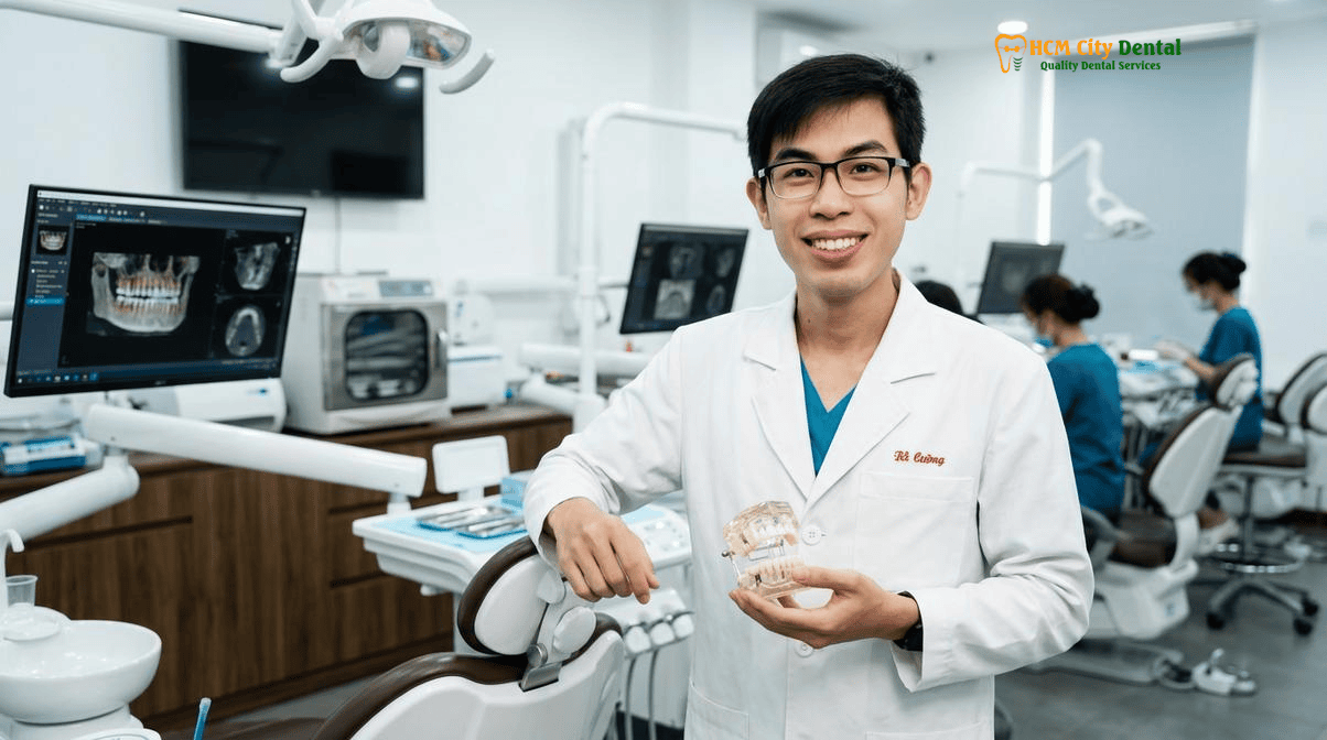
“The preservation of the natural dentition is the ultimate goal of biomimetic dentistry. A calcified root canal is not a death sentence for a tooth; it is simply a mechanical puzzle that requires the right digital keys to unlock. With 3D guided stents and 25x microscopy, we have the keys to safely navigate these complex anatomies and restore health to teeth that were previously considered hopeless.”
— Dr. Nguyen Van Cuong, DDS
Frequently Asked Questions
My front tooth turned yellow-grey years after a childhood fall, and my dentist said the canal is completely calcified—does it need a root canal?
Yes, if the tooth exhibits signs of apical periodontitis (infection at the root tip) on a 3D CBCT scan, or if you wish to restore its natural color through internal bleaching. A yellow, darkened front tooth after trauma becomes calcified as the pulp lays down tertiary dentin in response to the injury. Eventually, the pulp may necrose (die) silently, leading to a chronic hidden infection. At HCMC Dental Clinic, Dr. Nguyen Van Cuong, DDS utilizes 3D guided endodontics to safely access these obliterated canals without compromising the remaining tooth structure.
How does a 3D-printed Guided Endodontic template locate a calcified canal down to the millimeter without perforating the root?
3D static guided endodontics merges a high-resolution CBCT scan (which maps the internal root anatomy) with an intraoral STL optical scan (which maps the surface of your teeth). Using specialized software, Dr. Nguyen Van Cuong, DDS designs a virtual drill path that targets the exact coordinates of the calcified canal. A custom resin stent is then 3D-printed with a metal guide sleeve. During the procedure, a specialized micro-bur passes through this sleeve, restricting its movement to the pre-planned trajectory, ensuring absolute root perforation prevention in a calcified molar or anterior tooth.
Can a severely calcified molar with a periapical abscess still be saved without extraction?
Absolutely. While a blocked root canal where the dentist cannot find the canal using traditional freehand methods often leads to extraction recommendations, advanced microscopic and guided techniques change the prognosis entirely. By combining a 25x Carl Zeiss operating microscope, ultrasonic troughing for a calcified root canal, and 3D CBCT imaging, we can locate and negotiate the microscopic remnants of the canal space, disinfect the abscess, and save the natural molar.
Once the calcified root canal is treated on a dark front tooth, can internal bleaching restore its natural white color without a crown?
Yes. Following successful calcified root canal treatment using 3D guided endodontics, the access cavity can be utilized for an ‘internal walking bleach’ procedure. A specialized whitening agent (sodium perborate) is sealed inside the tooth for several days. This breaks down the dark blood breakdown products (hemosiderin) trapped in the dentinal tubules. Once the desired shade is achieved, the tooth is permanently sealed with a biomimetic composite resin, often eliminating the need for an invasive porcelain crown.
What is the cost of 3D CBCT imaging and microscopic calcified root canal therapy at HCMC Dental Clinic in District 1?
Under our official 2026 fee schedule, a high-resolution 3D CBCT Scan is just $18 USD online (480,000 VND). Microscopic Endodontic Root Canal Therapy (using a 25x Carl Zeiss microscope and Nitrile Dam) ranges from $77 – $115 USD per tooth online for anteriors/premolars, and $115 – $154 USD per tooth online for complex molars. This represents a transparent -40% online booking discount compared to walk-in rates, providing world-class specialist care at a fraction of Western prices.
References
- Krastl, G., Zehnder, M. S., Connert, T., Weiger, R., & Kühl, S. (2016). Guided Endodontics: a novel treatment approach for teeth with pulp canal obliteration and apical periodontitis. Clinical Oral Investigations, 20(3), 739-744.
- Connert, T., Zehnder, M. S., Weiger, R., Kühl, S., & Krastl, G. (2017). Microguided Endodontics: Accuracy of a Miniaturized Technique for Apically Extended Access Cavity Preparation in Teeth with Pulp Canal Obliteration. Journal of Endodontics, 43(5), 787-790.
- Buchgreitz, J., Buchgreitz, M., Mortensen, D., & Bjørndal, L. (2016). Guided access cavity preparation using cone-beam computed tomography and optical surface scans – an ex vivo study. International Endodontic Journal, 49(8), 790-795.
- Zehnder, M. S., Connert, T., Weiger, R., Krastl, G., & Kühl, S. (2016). Guided endodontics: accuracy of a novel method for guided access cavity preparation and root canal location. International Endodontic Journal, 49(10), 966-972.
- Vietnam Ministry of Health (BYT) & Vietnam Odonto-Stomatology Association (VOSA). National Clinical Guidelines on Specialist Dental Care, Biomaterial Traceability & EN 13060 Class B Infection Control. (2024).