Tooth sensitivity treatment involves clinical interventions designed to block exposed dentinal tubules or desensitize hyperactive nerve endings. Effective management ranges from topical fluoride varnishes and specialized desensitizing agents to restorative procedures like composite fillings or root canal therapy, depending on the underlying cause of the dentin hypersensitivity.
Clinical Summary:
Dentin hypersensitivity manifests as sharp, transient pain in response to thermal, tactile, osmotic, or chemical stimuli. Clinical treatment follows a tiered, evidence-based approach, beginning with non-invasive topical desensitizers and high-concentration fluoride applications to physically occlude dentinal tubules or alter nerve excitability. If structural damage such as severe enamel erosion, abfraction, deep decay, or significant gingival recession is present, restorative treatments—including dental sealants, composite bonding, periodontal grafting, or endodontic therapy—are required. Accurate clinical diagnosis by a dental professional ensures targeted, effective relief, prevents irreversible pulpal damage, and restores optimal oral function.
Key Takeaways:
- Dentin hypersensitivity occurs when protective enamel wears away or gums recede, exposing microscopic tubules that lead to the dental nerve.
- First-line clinical treatments include potassium nitrate to depolarize nerves and high-concentration fluoride varnishes to physically block tubule openings.
- Restorative options, such as composite resin fillings or dental sealants, are utilized to repair structural defects like non-carious cervical lesions.
- Severe, lingering sensitivity that does not respond to conservative therapy may indicate irreversible pulpitis, necessitating root canal treatment.
- Consistent preventive care, including the use of soft-bristled brushes and non-abrasive toothpaste, is critical to preventing further enamel erosion.
The Pathophysiology of Dentin Hypersensitivity
Dentin hypersensitivity is primarily explained by the hydrodynamic theory, where external stimuli cause rapid fluid movement within exposed dentinal tubules, triggering sharp nerve pain.
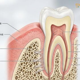
To fully comprehend the rationale behind any tooth sensitivity treatment, one must first understand the intricate anatomy of the human tooth and the physiological mechanisms that trigger pain. A healthy tooth is composed of three primary layers: the outer protective enamel (or cementum on the root surface), the underlying dentin, and the innermost dental pulp, which houses the nerves and blood vessels. Dentin is not a solid mass; rather, it is a porous tissue containing thousands of microscopic channels known as dentinal tubules. These tubules radiate outward from the pulp to the junction of the enamel or cementum.
When the protective layer of enamel is compromised or the gingival tissue recedes, these dentinal tubules become exposed to the oral environment. According to Brännström’s hydrodynamic theory—the most widely accepted scientific explanation for dentin hypersensitivity—external stimuli such as cold air, hot liquids, sweet foods, or tactile pressure cause the fluid inside these tubules to expand, contract, or shift rapidly. This sudden fluid movement stimulates the mechanoreceptors on the A-delta nerve fibers located at the pulp-dentin interface, resulting in the transmission of a sharp, localized, and transient pain signal to the brain [1].
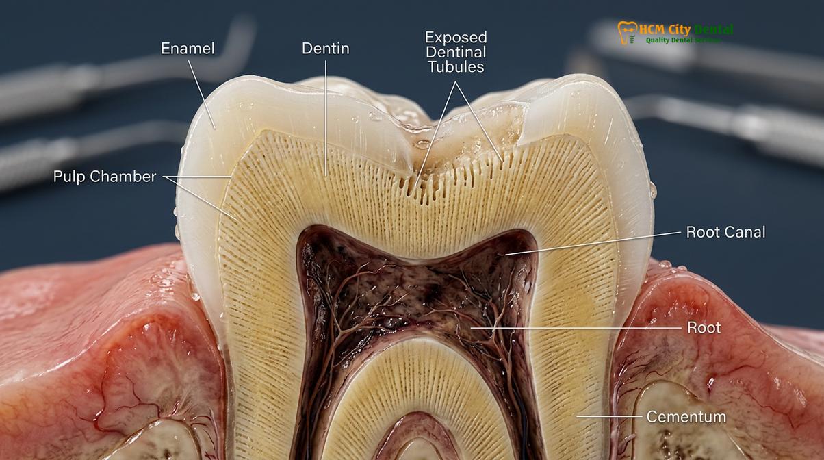
The etiology of this exposure is multifactorial and often requires a comprehensive evaluation within the scope of General Dentistry to determine the exact cause. Common culprits include:
- Enamel Erosion: The chemical dissolution of tooth structure caused by intrinsic acids (such as gastric acid from reflux or bulimia) or extrinsic acids (frequent consumption of citrus fruits, sodas, and sports drinks).
- Abrasion: The mechanical wear of the tooth surface, most commonly resulting from aggressive toothbrushing techniques or the use of highly abrasive whitening toothpastes.
- Abfraction: V-shaped notches that form at the cervical margin (gumline) of the tooth, theorized to be caused by biomechanical loading forces from bruxism (teeth grinding) or malocclusion, which cause the enamel to flex and micro-fracture.
- Gingival Recession: The apical migration of the gum margin, exposing the root surface. The root is covered by cementum, which is significantly softer and thinner than enamel, making it highly susceptible to rapid wear and tubule exposure.
“Dentin hypersensitivity is not merely a symptom of discomfort; it is a clinical indicator of compromised tooth structure or periodontal attachment loss that requires prompt diagnostic evaluation to prevent progressive pulpal pathology.”
First-Line Clinical Interventions: Non-Invasive Therapies
First-line treatments for sensitive teeth utilize topical agents like fluoride varnishes, potassium nitrate, and advanced calcium phosphate technologies to physically occlude tubules or chemically depolarize nerve endings.
When a patient presents with mild to moderate dentin hypersensitivity without significant structural loss, the standard of care dictates a conservative, non-invasive approach. The primary goals of these therapies are either to block the transmission of the nerve impulse or to physically plug the exposed dentinal tubules, thereby preventing the hydrodynamic fluid movement that triggers pain.
Nerve Depolarization Agents
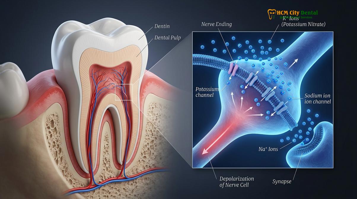
Potassium-based compounds, most notably potassium nitrate, are the most common active ingredients in over-the-counter desensitizing toothpastes. When applied consistently, the potassium ions penetrate the dentinal tubules and accumulate around the pulpal nerve endings. These ions alter the extracellular potassium concentration, effectively preventing the nerve fibers from repolarizing after an initial stimulus. By keeping the nerve in a depolarized state, the transmission of pain signals to the brain is blocked. Dr. Nguyen Van Cuong emphasizes that while potassium nitrate provides excellent symptomatic relief, it requires continuous, twice-daily use over several weeks to build and maintain its efficacy; if the patient discontinues use, the sensitivity will likely return.
Tubule Occluding Agents
For more immediate and robust relief, dental professionals utilize agents designed to physically seal the open tubules. These treatments are often applied in-office and include:
- Fluoride Varnishes: High-concentration sodium fluoride (typically 5%) is painted onto the sensitive root surfaces. The fluoride reacts with the calcium and phosphate in saliva to form calcium fluoride crystals, which precipitate inside the tubules, effectively plugging them and reducing permeability [2].
- Stannous Fluoride: Unlike sodium fluoride, stannous fluoride not only provides remineralization benefits but also creates a robust, acid-resistant smear layer over the exposed dentin, offering superior protection against erosive stimuli.
- Arginine and Calcium Carbonate: This technology mimics the natural biological process of saliva. The positively charged arginine binds to the negatively charged dentin surface, drawing in calcium carbonate to form a plug that seals the tubule.
- Glutaraldehyde and HEMA: In severe cases of localized sensitivity, dentists may apply a liquid desensitizer containing glutaraldehyde and hydroxyethyl methacrylate (HEMA). Glutaraldehyde coagulates the plasma proteins within the dentinal fluid, while HEMA acts as a wetting agent to draw the solution deep into the tubule, creating a deep, durable seal.
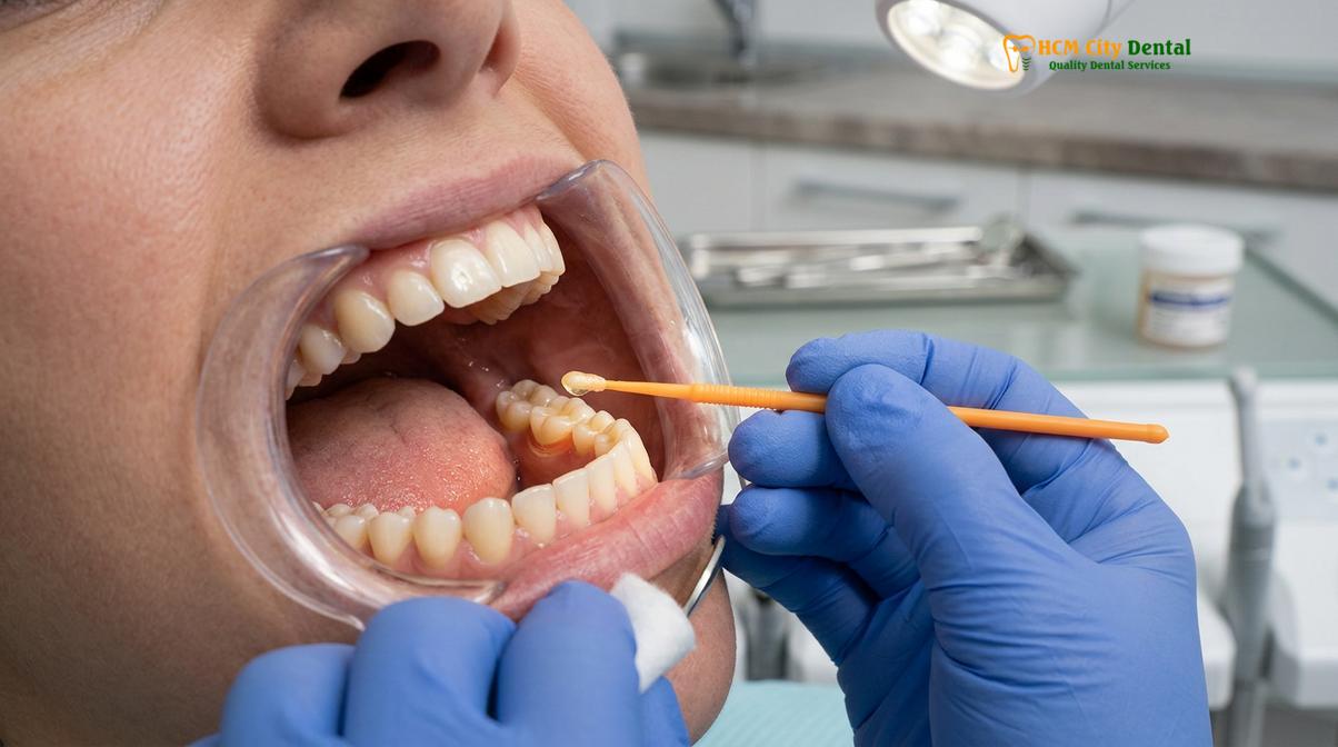
While these non-invasive treatments are highly effective, they are most successful when combined with proper oral hygiene instruction. For instance, utilizing gentle cleaning methods, such as those detailed in a Jet Dental: Clinical Air Polishing Guide, can help maintain periodontal health without causing further abrasive damage to the vulnerable root surfaces.
Restorative Procedures for Structural Damage
When sensitivity stems from significant enamel loss, deep cavities, or severe abfraction lesions, restorative treatments such as composite fillings or dental sealants are necessary to rebuild the protective barrier.
Topical desensitizers are highly effective for microscopic tubule exposure, but they are insufficient when macroscopic structural damage has occurred. If a patient suffers from non-carious cervical lesions (NCCLs), deep dental caries, or fractured enamel, the physical barrier protecting the dentin must be artificially reconstructed. Restorative dentistry provides several modalities to achieve this, restoring both function and aesthetics while eliminating hypersensitivity.
Composite Resin Restorations
For V-shaped abfraction lesions or areas of severe toothbrush abrasion at the gumline, composite resin bonding is the treatment of choice. The clinical workflow involves isolating the tooth, mildly etching the exposed dentin, and applying a dentin bonding agent. This bonding agent infiltrates the dentinal tubules, creating a hybrid layer that completely seals the dentin. A tooth-colored composite resin is then layered, cured with a specialized curing light, and polished to match the natural contour of the tooth. This not only provides immediate and long-lasting relief from sensitivity but also protects the tooth from further structural wear [3].
Dental Sealants and Glass Ionomer Cements
In cases where the structural loss is minimal but the sensitivity is persistent, dental sealants or flowable resins may be applied over the exposed root surfaces. Additionally, Glass Ionomer Cements (GIC) are frequently utilized for cervical restorations. GICs have the unique advantage of chemically bonding to the tooth structure without the need for extensive etching, and they continuously release fluoride into the surrounding dentin, providing an ongoing desensitizing and anti-cariogenic effect.
Maintaining a complete and structurally sound dentition is vital for distributing occlusal forces evenly. As explored in clinical resources regarding How Many Teeth Do Adults Have: Clinical Guide, missing teeth can lead to altered bite mechanics, placing excessive stress on remaining teeth, which accelerates abfraction and subsequent hypersensitivity.
| Treatment Modality | Primary Indication | Mechanism of Action | Expected Clinical Outcome |
|---|---|---|---|
| Potassium Nitrate Toothpaste | Generalized, mild sensitivity | Nerve depolarization | Gradual relief; requires continuous daily use |
| Fluoride Varnish (5% NaF) | Localized, moderate sensitivity | Tubule occlusion via calcium fluoride | Immediate relief; may require periodic reapplication |
| Composite Resin Bonding | Abfraction, abrasion, structural loss | Physical barrier and hybrid layer formation | Long-term relief and structural restoration |
| Periodontal Gum Grafting | Severe gingival recession | Soft tissue coverage of exposed roots | Permanent root coverage and aesthetic improvement |
| Root Canal Therapy | Irreversible pulpitis, severe lingering pain | Removal of inflamed pulpal tissue | Definitive elimination of nerve pain |
Periodontal and Endodontic Management
Advanced cases of tooth sensitivity may require periodontal grafting to cover exposed roots or root canal therapy to remove irreversibly damaged nerve tissue.
When conservative and restorative measures are insufficient, or when the etiology of the sensitivity lies in severe anatomical defects or pulpal pathology, advanced surgical or endodontic interventions become necessary.
Periodontal Plastic Surgery (Gum Grafting)
Gingival recession is a leading cause of dentin hypersensitivity. When the gums recede due to periodontal disease, aggressive brushing, or anatomical predispositions (such as a thin periodontal biotype), the root surface is exposed. While composite fillings can cover these roots, they do not restore the natural gingival architecture and can sometimes create plaque-retentive margins. In such cases, periodontal plastic surgery, specifically connective tissue grafting, is highly recommended.
During this procedure, a periodontist harvests a small piece of connective tissue, typically from the patient’s palate, and sutures it over the exposed root surface. As the graft heals and integrates, it thickens the gingival biotype, covers the exposed dentinal tubules, and provides a permanent, biological solution to root sensitivity while simultaneously enhancing the aesthetic appearance of the smile.
Endodontic Therapy (Root Canal Treatment)
It is crucial to differentiate between reversible pulpitis (typical tooth sensitivity) and irreversible pulpitis. Reversible pulpitis is characterized by a sharp, fleeting pain that dissipates immediately upon removal of the stimulus. However, if the tooth has sustained deep trauma, extensive decay, or repeated restorative procedures, the pulp may become irreversibly inflamed. Symptoms of irreversible pulpitis include spontaneous pain, pain that wakes the patient at night, or sensitivity to hot and cold that lingers for more than 30 seconds after the stimulus is removed [4].
Clinical Warning: Tooth sensitivity that transitions from a short, sharp sensation to a dull, throbbing, or lingering ache is a strong indicator of irreversible nerve damage. Ignoring these symptoms can lead to pulpal necrosis, dental abscesses, and systemic complications. Immediate endodontic evaluation is required.
In cases of irreversible pulpitis, topical desensitizers and fillings will fail to provide relief. The definitive treatment is root canal therapy. This endodontic procedure involves accessing the pulp chamber, meticulously cleaning and shaping the root canal system to remove all infected and inflamed nerve tissue, and sealing the space with a biocompatible material called gutta-percha. By completely removing the sensory nerve of the tooth, the sensitivity is permanently eliminated. The tooth is subsequently restored with a dental crown to ensure long-term structural integrity.
Understanding the systemic implications of untreated dental infections is critical. Chronic oral inflammation and untreated abscesses can have far-reaching effects, a concept thoroughly examined in literature discussing The Link Between Dental Health and Heart Attacks.
Preventive Strategies and Long-Term Care
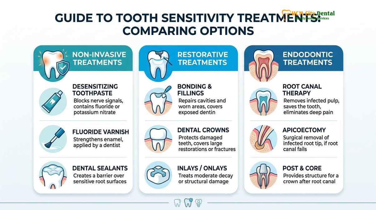
Preventing the recurrence of tooth sensitivity requires adopting proper oral hygiene techniques, using soft-bristled brushes, and minimizing the consumption of highly acidic dietary triggers.
Successful management of dentin hypersensitivity extends beyond the dental chair; it requires active patient compliance and the implementation of robust preventive strategies. Once a patient’s sensitivity has been clinically managed, preventing its recurrence is paramount.
Dietary Modifications
Diet plays a significant role in enamel erosion. Frequent consumption of acidic foods and beverages lowers the pH of the oral cavity, leading to the demineralization of enamel and the opening of dentinal tubules. Patients are advised to limit their intake of citrus fruits, carbonated sodas, sports drinks, and wine. When acidic beverages are consumed, using a straw can help bypass the teeth, minimizing direct contact. Furthermore, Dr. Cuong often advises patients to wait at least 30 minutes after consuming acidic foods before brushing their teeth. Brushing immediately after an acid challenge, while the enamel is in a softened state, significantly accelerates abrasive wear.
Systemic conditions that introduce acid into the oral cavity must also be managed. For example, patients suffering from gastroesophageal reflux disease (GERD) or pregnant women experiencing morning sickness are at a high risk for severe enamel erosion. Specialized care protocols, such as those outlined in guides on Bad Breath During Pregnancy: Clinical Causes & Care, are essential for mitigating these risks.
Optimizing Oral Hygiene Practices
Aggressive toothbrushing is a primary driver of gingival recession and cervical abrasion. Patients must transition to using ultra-soft bristled toothbrushes and adopt a gentle, circular brushing technique (the Modified Bass technique) rather than a harsh, horizontal scrubbing motion. The selection of toothpaste is equally critical; patients should avoid highly abrasive whitening toothpastes, which often contain harsh silicas that can strip away the protective smear layer over the dentin. Instead, continuous use of a low-abrasion, fluoride-rich, or desensitizing toothpaste is recommended to maintain tubule occlusion.
Clinical Case Study: A 34-year-old patient presented to HCMC Dental Clinic in Ho Chi Minh City complaining of severe, generalized sensitivity to cold beverages. Clinical examination revealed significant cervical abrasion on the premolars due to years of aggressive horizontal brushing with a hard-bristled brush. The treatment protocol involved the application of a glutaraldehyde-based desensitizer, followed by the placement of Class V composite restorations to rebuild the lost tooth structure. The patient was educated on proper brushing techniques and transitioned to a soft-bristled brush, resulting in the complete resolution of symptoms and the prevention of further structural damage.
Additionally, addressing malocclusion is a vital preventive measure. Misaligned teeth can cause uneven distribution of bite forces, leading to abfraction lesions. Orthodontic interventions, such as those detailed in Buck Teeth Braces: Clinical Overjet Correction, can harmonize the bite, reducing pathological stress on the cervical margins of the teeth [5].
When to See a Doctor for Tooth Sensitivity
While occasional, mild sensitivity to extremely cold foods can be normal, certain clinical signs warrant immediate professional evaluation. You should schedule a consultation with a dental professional if you experience any of the following red flags:
- Spontaneous Pain: Tooth pain that occurs without any external trigger (such as eating or drinking).
- Lingering Pain: Sensitivity to hot or cold that lasts longer than 30 seconds after the stimulus has been removed.
- Localized Swelling: Swelling of the gums around a specific tooth, or swelling in the face or jaw, which may indicate an active infection or abscess.
- Pain Upon Biting: Sharp pain when chewing or applying pressure to a tooth, which could signify a cracked tooth or periapical inflammation.
- Ineffectiveness of OTC Products: Sensitivity that does not improve after several weeks of using a desensitizing toothpaste.
Accurate diagnosis is the cornerstone of effective treatment. At HCMC Dental Clinic in Ho Chi Minh City, our clinical team utilizes advanced diagnostic tools, including digital radiography and vitality testing, to pinpoint the exact cause of your discomfort. Based on these clinical diagnostics, we develop a personalized treatment plan designed to restore your oral health and eliminate pain with a high success rate.
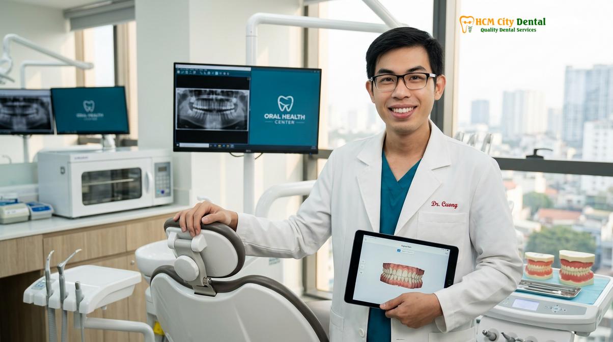
“Effective management of tooth sensitivity requires a transition from symptom suppression to structural preservation. By identifying the root cause early, we can implement conservative therapies that protect the vitality of the tooth for a lifetime.”
Frequently Asked Questions
Can tooth sensitivity go away on its own without treatment?
Mild tooth sensitivity may temporarily subside if the underlying trigger, such as an acidic diet or aggressive brushing, is removed. However, because lost enamel and receded gums do not regenerate naturally, the structural vulnerability remains. Without professional clinical intervention or the consistent use of desensitizing agents, the hypersensitivity is highly likely to return or progressively worsen over time as the dentinal tubules remain exposed to the oral environment.
What is the most effective toothpaste for sensitive teeth?
The most effective desensitizing toothpastes contain active ingredients like potassium nitrate, stannous fluoride, or calcium sodium phosphosilicate (NovaMin). Potassium nitrate works by depolarizing the nerve endings to block pain signals, while stannous fluoride and NovaMin physically occlude the exposed dentinal tubules. Consistent use twice daily is required to build and maintain the protective barrier; discontinuing use often leads to a relapse of symptoms.
Does professional teeth whitening cause permanent tooth sensitivity?
Professional teeth whitening can cause transient tooth sensitivity, but it is rarely permanent. The bleaching agents, typically hydrogen or carbamide peroxide, temporarily increase enamel permeability, allowing stimuli to reach the dentin. This sensation usually resolves within a few days to a couple of weeks post-treatment. Dentists often apply fluoride or potassium nitrate varnishes immediately after the whitening procedure to mitigate this temporary discomfort and accelerate remineralization.
How long does professional fluoride varnish take to work?
Professional fluoride varnish often provides immediate or same-day relief by rapidly forming a calcium fluoride barrier over exposed dentinal tubules. For optimal results, patients are typically advised to avoid eating hard foods, drinking hot liquids, or brushing for at least 4 to 6 hours after application, allowing the varnish to fully adhere and integrate with the tooth structure. The desensitizing effect can last for several months depending on the patient’s dietary and brushing habits.
Is root canal therapy the only cure for severe tooth sensitivity?
Root canal therapy is not the only treatment, but it is the definitive solution for severe, lingering sensitivity caused by irreversible pulpitis. If the dental pulp is permanently damaged or infected, topical agents and fillings will no longer be effective. By removing the inflamed nerve tissue, endodontic therapy completely eliminates the source of the pain while preserving the natural tooth structure, which is then typically protected with a custom dental crown.
References
- Journal of Clinical Periodontology. Dentin hypersensitivity: etiology, prevalence and treatment modalities. (2021).
- Journal of the American Dental Association. Efficacy of topical interventions for dentin hypersensitivity. (2022).
- International Endodontic Journal. Diagnostic accuracy of pulp testing in determining the status of the dental pulp. (2020).
- Operative Dentistry. Clinical evaluation of cervical composite restorations in non-carious cervical lesions. (2019).
- Journal of Periodontology. Periodontal plastic surgery for the treatment of gingival recession and dentin hypersensitivity. (2023).