Enamel shaving for veneers is a highly precise, minimally invasive clinical procedure that involves removing a microscopic layer of the tooth’s outermost surface. This essential preparatory step creates the necessary space for the ceramic restoration, helps ensure a seamless marginal fit, and maximizes the adhesive bond strength for long-lasting aesthetic results.
Clinical Summary:
Tooth preparation for porcelain veneers is a meticulously controlled process aimed at preserving the maximum amount of natural tooth structure. By utilizing modern depth-cutter burrs and advanced digital scanning technology, prosthodontists can achieve optimal enamel reduction. This conservative approach helps prevent bulky restorations while safeguarding the dentin-enamel junction (DEJ), supporting the goal that final veneers look natural, function properly, and integrate with the surrounding gingival tissues without causing unnecessary structural compromise.
Key Takeaways:
- Veneer preparation typically requires removing only 0.3mm to 0.7mm of the outer enamel layer.
- Preserving the dentin-enamel junction (DEJ) is critical for maximizing long-term bond strength.
- Modern clinical techniques utilize an enamel depth-cutter to prevent accidental over-preparation.
- Digital workflows with advanced intraoral scanners offer a highly accurate alternative to traditional impressions.
- Proper preparation helps prevent bulky margins, protects the biological width, and supports gingival health.
The Role of Enamel in Veneer Bonding Strength
Enamel provides the ideal structural substrate for dental adhesives, offering a significantly stronger and more predictable micromechanical bond than exposed dentin.
Understanding the anatomical and chemical properties of tooth enamel is fundamental to the success of any cosmetic dental restoration. Enamel is the hardest substance in the human body, composed of highly mineralized inorganic hydroxyapatite crystals. This rigid structure is what makes Porcelain Veneers incredibly durable when bonded correctly. During the preparation phase, the goal of enamel shaving for veneers is not merely to reduce the tooth’s size, but to create an optimized surface topography that facilitates superior adhesion[1].
When a prosthodontist prepares a tooth, they are essentially creating microscopic retentive features. The subsequent application of phosphoric acid during the adhesive bonding protocol etches the enamel, creating a porous, honeycomb-like surface. When the bonding resin is applied, it flows into these microporosities and polymerizes, forming resin tags. This micromechanical interlocking is the cornerstone of veneer retention. If the enamel is entirely removed and the underlying dentin is exposed, this predictable bonding mechanism is compromised because dentin contains significantly more water and organic material.
Furthermore, maintaining a robust layer of enamel helps ensure that the tooth remains rigid. Enamel acts as a protective shell that distributes occlusal forces evenly across the tooth structure. When a high-strength ceramic is bonded to this rigid enamel shell, the two materials function as a cohesive biomechanical unit, highlighting why conservative enamel preservation is a standard in contemporary cosmetic dentistry.
While rare under modern protocols, If nerve damage occurs, a root canal may be required to protect the tooth.
Traditional Prep vs. Minimal-Prep Thickness (0.3mm to 0.7mm)
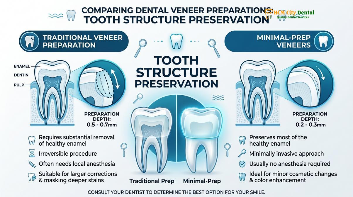
While traditional veneers may require up to 0.7mm of reduction to mask severe discoloration, minimal-prep options demand as little as 0.2mm to 0.3mm of enamel removal.
The extent of tooth preparation depth varies significantly based on the patient’s initial clinical presentation and their desired aesthetic outcome. Historically, older veneer techniques required more aggressive reduction to accommodate thicker ceramics. Today, advancements in material science have introduced ultra-thin, highly translucent ceramics that require minimal tooth reduction, fundamentally changing the approach to smile makeovers[2].
Traditional preparation typically involves removing 0.5mm to 0.7mm of enamel. This depth is often clinically necessary when dealing with complex cases, such as masking dark intrinsic stains, correcting moderate misalignments, or when transitioning from composite veneers to stronger porcelain alternatives. The ceramist requires this specific thickness to layer different opacities of porcelain, effectively blocking out the underlying dark tooth structure.
Conversely, minimally invasive preparation focuses on maximum enamel preservation. Techniques utilizing ultra-thin restorations require only 0.2mm to 0.3mm of reduction. This approach is ideal for patients with naturally light-colored teeth who wish to enhance the shape, close minor gaps, or restore slight incisal wear. Because almost the entirety of the preparation remains within the enamel layer, the bond strength remains exceptionally high.
| Preparation Type | Enamel Reduction Depth | Primary Clinical Indications | Anesthesia Requirement |
|---|---|---|---|
| Traditional Prep | 0.5mm – 0.7mm | Severe discoloration, moderate crowding, masking dark stains. | Usually required |
| Minimal-Prep | 0.2mm – 0.3mm | Mild discoloration, minor shape adjustments, slight wear. | Optional / Minimal |
| No-Prep (Prep-less) | 0.0mm | Microdontia, diastema closure, adding volume to the arch. | Not required |
It is crucial to understand that the choice between traditional and minimal-prep is a clinical decision dictated by the physical space required to achieve the desired aesthetic without creating a bulky contour that could trap plaque.
Micro-abrasion and Depth-Cutter Burrs: Modern Precision Tools
Advanced rotary instruments and digital intraoral scanners allow dentists to calibrate enamel reduction with microscopic accuracy, supporting highly conservative treatment.
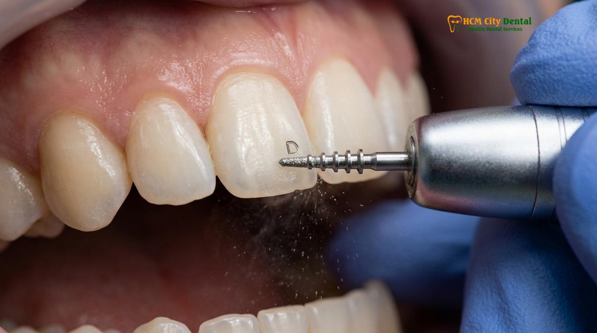
The precision of enamel shaving for veneers relies heavily on the specialized armamentarium utilized by the prosthodontist. The procedure begins with the use of an enamel depth-cutter burr. This specialized diamond rotary instrument features specific non-cutting guide pins that physically prevent the burr from penetrating deeper than a predetermined depth. By creating these initial depth grooves across the labial surface of the tooth, the dentist establishes a precise map for the subsequent reduction.
Once the depth grooves are established, fine-grit round-end diamond burrs are used to gently blend the grooves and create a smooth, uniform surface. The margins of the preparation are typically finished with a light chamfer. This specific margin design provides a distinct finishing line for the ceramist and helps ensure a seamless transition between the ceramic and the tooth.
“The integration of digital intraoral scanners and precision depth-cutting burs has transformed cosmetic dentistry, allowing practitioners to preserve healthy tooth structure while achieving excellent marginal adaptation.”
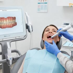
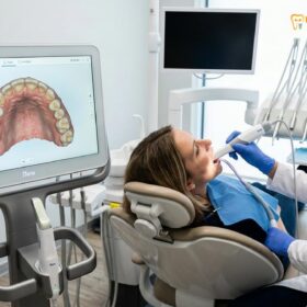
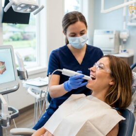
Following the physical preparation, the workflow transitions to the digital realm. Modern clinics utilize state-of-the-art optical technology to capture thousands of images per second, creating a highly accurate 3D digital replica of the prepared teeth[4]. This digital file is instantly transmitted to the dental laboratory, where CAD/CAM milling technology and expert ceramists design and fabricate the final restorations.
Why Over-Preparation Leads to Sensitivity and Bond Failure
Removing too much enamel exposes the underlying dentin, which reduces adhesive retention and increases the risk of post-operative thermal sensitivity.
The phrase “shaved down to pegs” is a common misconception associated with veneers, but it actually describes the aggressive preparation required for full-coverage dental crowns. When evaluating a dental crown vs veneer, it is important to note that veneers require far less reduction. When a tooth is over-prepared for a veneer, the primary risk is the exposure of the dentin layer. Dentin is composed of thousands of microscopic tubules that lead directly to the tooth’s central nerve. When these tubules are exposed, changes in temperature can cause fluid movement within the tubules, triggering dentinal hypersensitivity.
Beyond patient discomfort, over-preparation compromises the longevity of the restoration. Dental adhesives bond exceptionally well to the highly mineralized structure of enamel. Dentin, however, is highly organic and moist. While modern bonding agents can adhere to dentin, the bond strength is generally weaker and degrades faster over time compared to an enamel bond[3].
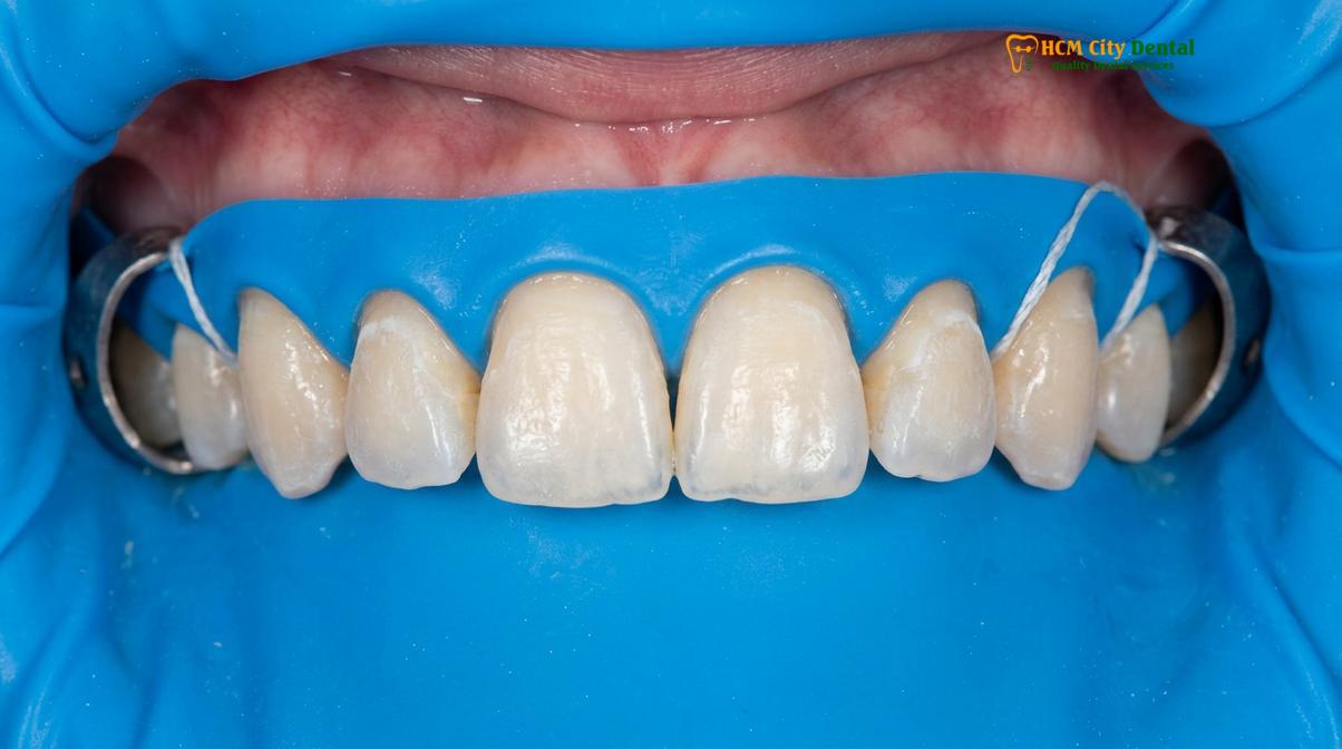
Furthermore, improper preparation can disrupt the gingival zenith calibration. If the margins of the veneer are placed too deep beneath the gumline in an attempt to hide a bulky restoration, it invades the biological width. The body’s immune system recognizes this as an intrusion, which can lead to chronic redness, bleeding, and eventual gum recession[5]. This underscores why planning your smile with a conservative approach is paramount.
Preserving the Dentin-Enamel Junction (DEJ)
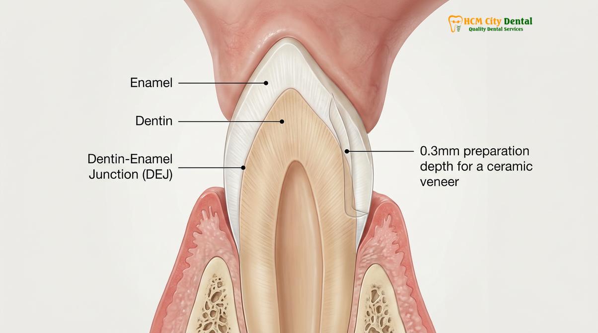
The DEJ is a critical anatomical boundary; keeping the preparation strictly within the enamel layer prevents structural weakening and supports long-term veneer stability.
The dentin-enamel junction (DEJ) is the complex, scalloped interface where the hard outer enamel meets the softer inner dentin. This junction acts as a biological shock absorber. When occlusal forces are applied to the tooth, the DEJ dissipates these stresses, helping to prevent cracks from propagating from the brittle enamel into the vulnerable dentin. Preserving this vital structure is a primary objective during minimally invasive preparation.
If the preparation depth breaches the DEJ, the tooth loses a portion of its natural biomechanical resilience. The tooth becomes more susceptible to flexure under load, which can transfer excessive stress to the rigid ceramic veneer. Therefore, a skilled prosthodontist will constantly evaluate the depth of the preparation, utilizing magnification to ensure the burr remains safely within the enamel layer.
Clinical Case Study: A 28-year-old patient visited HCMC Dental Clinic in Ho Chi Minh City seeking to correct mild crowding and uneven incisal edges. Utilizing Digital Smile Design (DSD), the clinical team planned a highly conservative treatment. By employing precision depth-cutters, the preparation was kept strictly to 0.3mm, fully preserving the DEJ. The patient experienced minimal post-operative sensitivity and achieved a natural-looking smile with ultra-thin restorations.
To protect the prepared teeth while the final ceramics are being fabricated, the application of provisional restorations is a standard clinical protocol. These temporary veneers seal the freshly cut enamel, help maintain the position of the gingival tissues, and provide the patient with a preview of their new smile.
Important Clinical Considerations and When to Consult a Prosthodontist
Seeking professional evaluation is crucial to determine if you are a suitable candidate for minimally invasive veneers or if alternative treatments are required.
Not every patient is an immediate candidate for minimal-prep veneers. Individuals with severe bruxism (teeth grinding), significant structural damage, or active periodontal disease require comprehensive evaluation before any cosmetic procedure is initiated. In some cases, exploring an alternative for veneers, such as orthodontic alignment prior to restorative work, can help minimize the amount of enamel shaving required.
According to the guidelines set forth by the Vietnam Odonto-Stomatology Association (VOSA) regarding aesthetic restorations, preserving natural tooth structure should always be the primary clinical objective[6]. Patients experiencing persistent pain, severe thermal sensitivity, or signs of gum inflammation after previous dental work should consult a qualified prosthodontist promptly to assess the integrity of their restorations and underlying tooth structure.
During a consultation, the dental team will utilize panoramic X-rays and 3D imaging to assess bone levels, root health, and enamel thickness. This diagnostic phase is essential for developing a customized treatment plan that aligns with both aesthetic desires and biological limitations.
Clinical Workflow and Material Selection at HCMC Dental Clinic
A successful smile makeover relies on certified premium materials, direct laboratory collaboration, and a transparent approach tailored for patient accessibility.
The execution of a flawless veneer case requires a seamless integration of clinical expertise and advanced dental technology. Dr. Nguyen Van Cuong, a leading specialist in aesthetic restorations, emphasizes that thorough clinical evaluation is paramount. Dr. Cuong and his team utilize advanced diagnostics to determine the exact amount of enamel reduction required, ensuring each patient receives a highly customized, biologically sound treatment plan. At HCMC Dental Clinic, the workflow is optimized to deliver excellent results, particularly catering to international patients and dental tourists who require efficiency without compromising on quality.
The clinic’s direct partnership with a leading laboratory in Ho Chi Minh City enables strict quality control and real-time collaboration between the prosthodontist and the master ceramist. This streamlined communication allows for an efficient turnaround. Within a short timeframe, a patient can undergo comprehensive digital diagnostics, precise tooth preparation, and the final bonding of custom-characterized veneers.
“By partnering directly with top-tier ceramists in Ho Chi Minh City, we maintain strict quality control over every restoration, ensuring our patients receive excellent aesthetics and precision fit.”
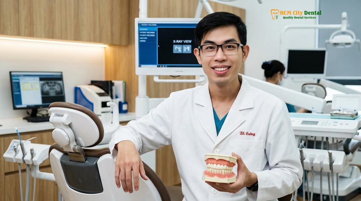
Material certification is strictly adhered to, utilizing authentic, globally recognized ceramics. Depending on the patient’s functional needs and aesthetic goals, the clinic utilizes high-strength Zirconia for areas requiring high flexural strength, and lithium disilicate for anterior teeth where outstanding lifelike aesthetics and light dynamics are paramount. Understanding how long porcelain veneers last is directly tied to this combination of conservative preparation and premium material selection.
References
- Journal of Esthetic and Restorative Dentistry. The influence of enamel preservation on veneer bond strength. (2021).
- International Journal of Prosthodontics. Clinical outcomes of minimally invasive porcelain veneers. (2020).
- The Journal of Prosthetic Dentistry. Biomechanical properties of the dentin-enamel junction. (2019).
- Clinical Oral Investigations. Digital intraoral scanning accuracy in fixed prosthodontics. (2022).
- Journal of Clinical Periodontology. Maintaining biological width in aesthetic dental restorations. (2018).
- Vietnam Odonto-Stomatology Association (VOSA). National guidelines on minimally invasive prosthodontics and aesthetic restorations. (2023).
Are you considering a smile makeover and want to learn more about minimally invasive veneer preparation? Contact HCMC Dental Clinic in Ho Chi Minh City today to schedule a comprehensive consultation. Discover how our conservative approach to porcelain veneers vs porcelain crowns can help you achieve a beautiful, lasting smile while protecting your natural teeth.