Zygomatic implants are specialized, extra-long titanium posts anchored directly into the dense cheekbone, providing a secure foundation for full-arch teeth replacement in patients with severe upper jaw bone loss. This advanced technique bypasses the need for extensive bone grafting, allowing for immediate functional restoration.
Clinical Summary:
Zygomatic implantology represents a paradigm shift for edentulous patients suffering from extreme maxillary atrophy. By utilizing the zygomatic arch for anchorage, clinicians can achieve high primary stability even when the alveolar ridge is severely resorbed. This approach significantly reduces overall treatment time by eliminating complex sinus augmentation procedures. With computer-guided surgical protocols, the predictability and safety of placing these specialized implants have dramatically improved. This graftless solution offers a reliable pathway to immediate occlusal loading, allowing patients to receive a fixed provisional prosthesis on the day of surgery and rapidly restoring their quality of life, masticatory function, and facial aesthetics.
Key Takeaways:
- Anchors securely into the dense, high-quality cortical bone of the zygomatic arch rather than the resorbed upper jaw.
- Eliminates the need for invasive sinus lifts and prolonged bone graft healing periods, streamlining the treatment timeline.
- Facilitates immediate loading protocols, allowing patients to receive a fixed provisional prosthesis on the same day as surgery.
- Requires advanced 3D CBCT imaging, virtual surgical planning, and specialized maxillofacial expertise for safe and precise placement.
- Demonstrates exceptionally high long-term survival rates that are comparable to, or exceed, conventional implant therapies in compromised bone.
The Challenge of Severe Upper Jaw Bone Resorption
Severe maxillary bone loss occurs rapidly after tooth extraction, often leaving insufficient bone volume for standard implants and necessitating advanced anchorage solutions to restore oral function.
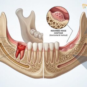
The human upper jaw, or maxilla, is highly susceptible to structural changes following the loss of natural teeth. When teeth are extracted or lost due to advanced periodontal disease, the alveolar bone—the specialized ridge of bone that surrounds and supports the tooth roots—no longer receives the mechanical stimulation required to maintain its density and volume. Without the biomechanical forces of chewing transmitted through the periodontal ligament, the body initiates a natural physiological process known as bone resorption. Over time, this leads to severe maxillary bone loss, drastically reducing both the height and width of the upper jaw.[1]
In the posterior regions of the upper jaw, this resorptive process is further complicated by the presence of the maxillary sinuses. These air-filled cavities, located just above the upper premolars and molars, tend to expand downward (a process called sinus pneumatization) as the alveolar bone shrinks upward. The convergence of these two phenomena often leaves a paper-thin layer of bone separating the oral cavity from the sinus floor. For patients who have worn traditional upper dentures for decades, the continuous pressure exerted by the acrylic baseplate on the gum tissue accelerates this bone degradation, leading to a completely flat or even concave maxillary ridge.
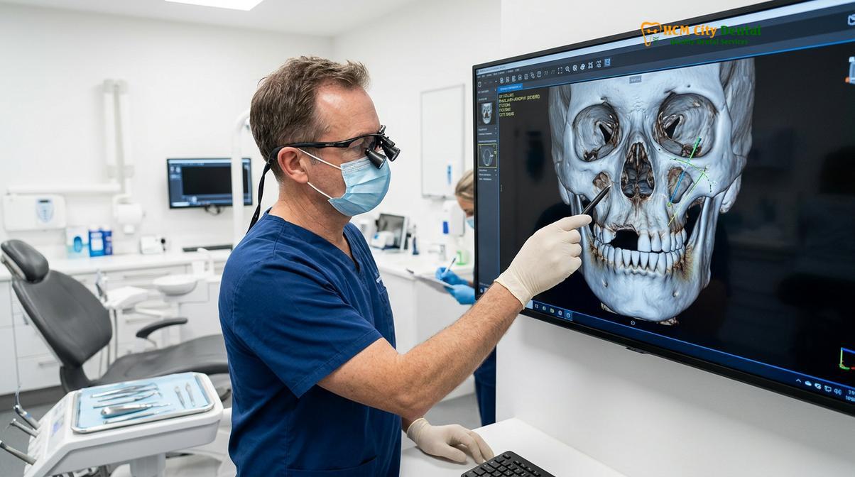
When severe maxillary bone loss reaches this advanced stage, placing standard cylindrical dental implants becomes biomechanically impossible without extensive reconstructive surgery. Traditional protocols dictate that the deficient bone must be rebuilt using autogenous bone blocks harvested from the hip or jaw, combined with bilateral sinus lift procedures. While effective in some cases, these massive grafting surgeries are highly invasive, carry significant risks of morbidity, and require prolonged healing periods—often leaving the patient without fixed teeth for up to a year or more.[2]
“The psychological and physical burden of severe maxillary atrophy cannot be overstated. Patients often present with compromised facial aesthetics, collapsed vertical dimension, and an inability to chew nutritious foods. Traditional grafting methods, while historically standard, subject these patients to extended periods of discomfort and uncertainty.”
Furthermore, the quality of the residual bone in a severely atrophic maxilla is typically poor. The maxilla is primarily composed of cancellous (spongy) bone, which is less dense than the cortical bone found in the lower jaw. When this already porous bone resorbs, achieving the primary stability necessary for successful osseointegration becomes a profound clinical challenge. This anatomical reality forces implantologists to look beyond the alveolar ridge for stable anatomical structures capable of supporting a full-arch prosthesis, leading to the development of extra-maxillary anchorage techniques. Understanding these limitations is crucial when evaluating the advantages and limitations of implant therapy for complex cases.
What are Zygomatic Implants? Anchoring into the Cheekbone
These are extra-long titanium implants, ranging from 30mm to 60mm, designed to bypass the deficient upper jaw and anchor securely into the dense zygomatic bone.
Zygomatic implants represent a sophisticated evolution in maxillofacial prosthetics. Unlike conventional dental implants, which are typically 8mm to 18mm in length and designed to sit entirely within the alveolar bone of the jaw, zygomatic fixtures are significantly longer. Ranging from 30mm to over 60mm, these specialized titanium posts are engineered to traverse the maxillary sinus cavity—or pass along its outer wall—to engage the body of the zygomatic bone (the cheekbone). This technique, known as zygoma bone anchorage, capitalizes on the unique anatomical properties of the facial skeleton to provide unparalleled support for dental prosthetics.[3]
The zygomatic bone is a robust, dense structure composed primarily of thick cortical bone. Unlike the maxilla, which resorbs rapidly following tooth loss, the cheekbone maintains its dense architecture throughout a patient’s life, regardless of dental status. This makes it an ideal anatomical buttress for anchoring load-bearing prosthetics. When a surgeon utilizes zygoma bone anchorage, the apical (tip) portion of the implant is firmly embedded into this dense cortical bone, while the coronal (head) portion emerges in the mouth at the ideal position to support a dental bridge.
The design of cheekbone dental implants is specifically tailored to their unique anatomical pathway. They often feature a smooth, unthreaded mid-section to prevent irritation of the sinus membrane (the Schneiderian membrane) if the implant passes through the sinus cavity, while the apical portion is aggressively threaded to maximize engagement with the zygomatic bone. The head of the implant is typically angled—often at 45 or 55 degrees—to compensate for the steep trajectory of the implant body, allowing the prosthetic connection to align perfectly with the occlusal plane of the mouth.
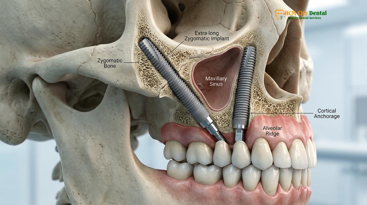
Depending on the severity of the bone loss, a patient may require different configurations of cheekbone dental implants. In cases where the anterior (front) maxilla still retains sufficient bone volume, a surgeon may place two conventional implants in the front and two zygomatic implants in the back (one on each side). This hybrid approach provides excellent cross-arch stabilization. However, in cases of extreme, generalized maxillary atrophy where no usable bone remains in the upper jaw, a “Quad Zygoma” approach may be utilized. This involves placing four zygomatic implants—two on each side—to entirely support the upper prosthesis without relying on the maxilla at all.[4]
| Feature | Conventional Implants | Zygomatic Implants |
|---|---|---|
| Length | 8mm – 18mm | 30mm – 60mm+ |
| Anchorage Site | Alveolar bone of the maxilla/mandible | Cortical bone of the zygomatic arch (cheekbone) |
| Bone Density Required | Moderate to high local bone volume | Bypasses local bone; relies on dense remote bone |
| Need for Sinus Lift | Often required in the posterior maxilla | Eliminates the need for sinus augmentation |
| Primary Stability | Variable, depends on local bone quality | Exceptionally high due to cortical bone engagement |
The biomechanical advantage of this approach is profound. Because the implants are anchored in dense cortical bone, they achieve exceptionally high insertion torque values (often exceeding 45 Ncm). This high primary stability is the critical prerequisite for immediate loading protocols, allowing patients to transition from edentulism to a fixed, functional smile in a fraction of the time required by traditional methods. This level of advanced restorative dentistry has revolutionized the way clinicians approach the most challenging anatomical presentations.
Eliminating the Need for Massive Sinus Lifts and Bone Grafts
By utilizing remote bone anchorage, this technique allows patients to bypass complex sinus augmentation, significantly reducing surgical morbidity and total treatment time.
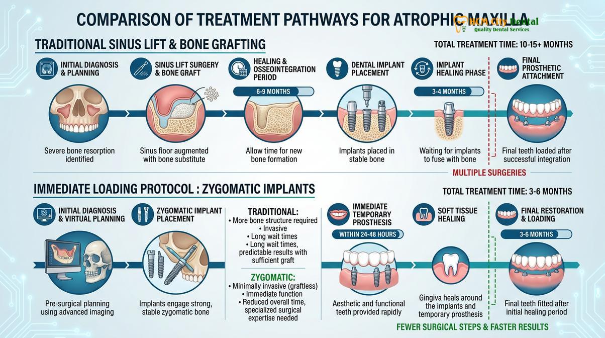
For decades, the standard of care for rehabilitating the severely atrophic posterior maxilla involved extensive bone grafting, specifically the lateral window sinus lift procedure. In this traditional approach, the surgeon creates a window in the lateral wall of the maxilla, delicately elevates the delicate sinus membrane, and packs the newly created space with particulate bone graft material (often a mix of autogenous bone, allografts, or xenografts). The patient must then wait six to nine months for this graft material to consolidate and form new, viable bone before conventional implants can even be placed. Following implant placement, another three to six months of healing is required before the final teeth can be attached.[5]
This prolonged, multi-stage process is not only exhausting for the patient but also carries inherent risks. Sinus membrane perforations, graft infections, and unpredictable bone regeneration can compromise the entire treatment plan. Furthermore, harvesting autogenous bone blocks from the patient’s hip (iliac crest) or lower jaw (mandibular ramus) introduces a second surgical site, significantly increasing post-operative pain, swelling, and the overall risk of morbidity.
The advent of zygomatic implantology offers a highly elegant alternative: the ability to completely avoid sinus lift surgery. By changing the paradigm from “building bone to fit the implant” to “finding existing bone to anchor the implant,” clinicians can dramatically streamline the patient journey. The extra-long fixtures simply bypass the pneumatized sinus cavities entirely, seeking out the pristine, unresorbed bone of the zygomatic arch. This graftless approach not only eliminates the need for donor site surgery but also removes the unpredictable variables associated with large-volume bone regeneration.
From a biological perspective, avoiding massive bone grafts preserves the delicate vascular supply of the maxilla. Extensive periosteal stripping required for large block grafts can compromise blood flow, leading to graft necrosis or delayed healing. By utilizing a minimally invasive or anatomy-guided approach for zygomatic placement, the surgeon preserves the local microcirculation, promoting faster soft tissue healing and reducing post-operative discomfort. This is particularly beneficial when restoring oral function in older adults, who may have reduced healing capacity or underlying systemic conditions that make extensive grafting contraindicated.
Moreover, the psychological benefit to the patient is immense. The prospect of undergoing multiple surgeries over a two-year period is a significant deterrent for many individuals seeking full-arch rehabilitation. The ability to avoid sinus lift surgery and achieve a definitive result in a single surgical intervention transforms a daunting medical ordeal into a highly manageable, life-changing procedure. The reduction in total treatment time also has favorable economic implications, making this advanced therapy a highly strategic investment in comprehensive oral rehabilitation.
The Surgical Procedure and Safety Standards
The placement of zygomatic fixtures requires meticulous 3D planning, computer-guided surgical templates, and execution by highly trained maxillofacial specialists to ensure anatomical safety.
The surgical placement of zygomatic implants is one of the most advanced and technically demanding procedures in the field of implant dentistry. Because the surgical field involves critical anatomical structures—including the maxillary sinus, the infraorbital nerve, and the orbital floor (the bone beneath the eye)—absolute precision is mandatory. This procedure is not performed by general dentists; it requires the expertise of a highly trained oral and maxillofacial surgeon or a specialized implantologist with extensive experience in extra-maxillary anchorage.
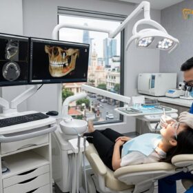
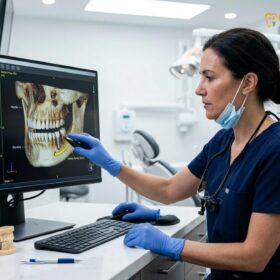
The foundation of a safe and successful procedure lies in meticulous pre-operative diagnostics. A high-resolution 3D Cone Beam Computed Tomography (CBCT) scan is absolutely essential. This imaging modality allows the surgical team to visualize the patient’s craniofacial anatomy in three dimensions, accurately measuring the volume, density, and trajectory of the zygomatic bone. Using advanced virtual planning software, the surgeon can simulate the exact placement of the implants, ensuring they engage maximum bone mass while maintaining a safe distance from critical nerves and the orbital cavity.[1]
Clinical Safety Warning: Zygomatic implant surgery carries specific risks, including acute or chronic sinusitis, altered nerve sensation (paresthesia) in the cheek or lip, and orbital complications if placed incorrectly. It is imperative that this procedure is performed only by credentialed maxillofacial specialists utilizing 3D computer-guided surgical protocols to mitigate these risks.
Modern surgical protocols have evolved significantly to enhance safety and predictability. Historically, the classic “intrasinus” approach required the implant to pass directly through the maxillary sinus cavity, which occasionally led to post-operative sinus complications. Today, leading experts, including Dr. Nguyen Van Cuong, frequently utilize the ZAGA (Zygoma Anatomy-Guided Approach). This modern technique adapts the implant trajectory to the patient’s specific anatomy. Depending on the concavity of the maxillary wall, the implant may be placed entirely outside the sinus (extramaxillary), partially within the sinus wall, or through the sinus, thereby minimizing membrane disruption and drastically reducing the incidence of post-operative sinusitis.[2]
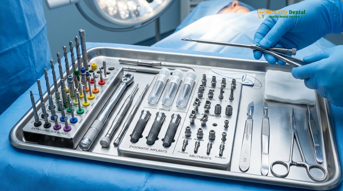
During the procedure, which is typically performed under intravenous (IV) sedation or general anesthesia for maximum patient comfort, the surgeon makes a precise incision along the crest of the maxillary ridge. The soft tissue is carefully elevated to expose the lateral wall of the maxilla and the base of the zygomatic arch. Using a sequence of specialized, extra-long drills, the osteotomy (bone preparation) is created along the pre-planned trajectory. The surgical site is continuously irrigated with sterile saline to prevent bone overheating, which is critical for maintaining the vitality of the bone cells required for osseointegration.
Once the osteotomy is complete, the titanium implant is slowly threaded into position. The surgeon carefully monitors the insertion torque, aiming for a high degree of primary stability. After the implants are securely anchored, multi-unit abutments are attached to the heads of the implants. These abutments correct the angulation of the implants, providing a parallel foundation upon which the prosthetic bridge can be securely screwed. The precision required during this phase highlights the importance of utilizing premium implant systems that offer specialized zygomatic components engineered for maximum biomechanical strength.
Post-operative care is rigorously managed. Patients are typically prescribed a course of broad-spectrum antibiotics, corticosteroids to minimize facial swelling, and appropriate analgesics. Dr. Cuong emphasizes that strict adherence to post-operative instructions, including a soft diet and specific oral hygiene protocols, is vital during the initial healing phase to prevent infection and ensure the long-term success of the implants.
Immediate Loading: Getting Teeth-in-a-Day with Zygomatic Posts
The exceptional primary stability achieved in the cheekbone allows for the immediate attachment of a fixed provisional bridge, restoring function and aesthetics in a single day.
One of the most profound benefits of utilizing the zygomatic arch for anchorage is the ability to execute immediate loading protocols. In traditional implantology, especially when bone grafting is involved, patients are often required to wear a removable temporary denture for months while the implants integrate with the bone. These temporary dentures can be uncomfortable, unstable, and psychologically distressing. However, because zygomatic implants engage the dense, highly mineralized cortical bone of the cheekbone, they routinely achieve insertion torque values that far exceed the threshold required for immediate loading.[3]
Immediate loading, often referred to as “Teeth-in-a-Day,” is a biomechanical concept where a rigid, full-arch provisional prosthesis is attached to the implants within 24 to 48 hours of surgery. This provisional bridge is typically milled from high-density PMMA (polymethyl methacrylate) and reinforced with a titanium cylinder framework. By splinting the implants together with a rigid bridge, the biomechanical forces of chewing are evenly distributed across the entire arch. This cross-arch stabilization prevents micro-movement of individual implants, which is the primary cause of early implant failure, and actually promotes favorable bone remodeling during the osseointegration phase.
Clinical Case Example: A 62-year-old patient presented to HCMC Dental Clinic in Ho Chi Minh City with a history of severe periodontal disease and decades of wearing an ill-fitting upper denture. CBCT imaging revealed extreme maxillary atrophy (Cawood and Howell Class VI), making conventional implants impossible without massive hip grafts. Dr. Nguyen Van Cuong designed a hybrid treatment plan utilizing two anterior conventional implants and two posterior zygomatic implants. The surgery was completed in a single morning, and by the afternoon, the patient walked out with a fixed, screw-retained provisional bridge, completely restoring her smile and ability to chew without the need for a single bone graft.
This immediate functional restoration is life-changing for patients. They enter the clinic edentulous or with failing dentition and leave with a fixed, non-removable set of teeth that look and feel natural. The provisional bridge is designed to be slightly out of heavy occlusion (bite contact) to protect the implants during the initial healing phase, but patients can immediately enjoy a soft diet and speak with confidence. This approach is a cornerstone of modern full-arch fixed prosthetics, providing a seamless transition to a restored quality of life.
After a healing period of approximately four to six months, during which the titanium fixtures fuse completely with the zygomatic bone at a cellular level, the patient returns to the clinic. The provisional bridge is removed, and final impressions (often digital intraoral scans) are taken to fabricate the definitive prosthesis. The final bridge is typically constructed from ultra-durable materials such as monolithic zirconia layered with porcelain, or a precision-milled titanium framework wrapped in high-impact acrylic. This final restoration is meticulously engineered to provide optimal facial support, perfect phonetics, and the strength to withstand the full forces of natural mastication, proving superior when comparing removable prosthetics to fixed solutions.
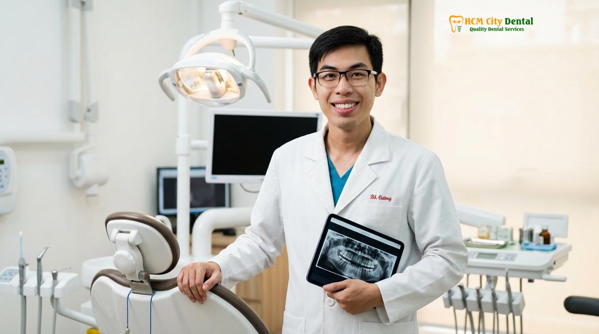
When to Consult a Specialist for Advanced Implantology
Patients who have been told they lack sufficient bone for traditional implants or who wish to avoid extensive bone grafting should seek a specialized clinical evaluation.
Determining candidacy for advanced extra-maxillary anchorage requires a comprehensive clinical and radiographic evaluation. You should consider consulting a maxillofacial specialist if you fall into any of the following categories:
- Severe Bone Atrophy: You have been wearing upper dentures for many years and have experienced significant facial collapse or “sunken” facial features due to bone loss.
- Failed Previous Procedures: You have undergone traditional sinus lift surgeries or bone grafts that failed to integrate or became infected.
- Desire to Avoid Grafting: You have been told you need massive bone grafts from your hip or jaw and wish to explore less invasive, graftless alternatives.
- Trauma or Pathology: You have lost significant maxillary bone due to facial trauma, tumor resection, or severe congenital conditions like cleft palate.
However, this procedure is not suitable for everyone. Absolute contraindications include active, untreated acute sinusitis, severe uncontrolled systemic diseases (such as uncontrolled diabetes or recent cardiovascular events), and a history of high-dose intravenous bisphosphonate therapy. A thorough medical history review and an endoscopic evaluation of the sinus health may be required prior to surgical clearance. It is highly recommended to schedule a personalized consultation at a specialized center, such as HCMC Dental Clinic, where a multidisciplinary team can assess your unique anatomical presentation and design a safe, predictable treatment pathway tailored to your specific needs.
Frequently Asked Questions
Clear, evidence-based answers to the most common patient inquiries regarding advanced cheekbone implant procedures and recovery expectations.
Who is a candidate for zygomatic implants?
Candidates are typically patients with severe upper jaw bone loss who have been told they lack sufficient bone for conventional implants. This includes individuals who have worn upper dentures for many years, those who have experienced failed sinus lifts, or patients who have lost bone due to trauma or pathology. A comprehensive 3D CBCT scan is required to evaluate the density and volume of the cheekbone to confirm candidacy. The specialist will also assess your overall health to ensure you can safely undergo the surgical procedure.
Are zygomatic implants safe?
Yes, when performed by highly trained maxillofacial specialists using 3D computer-guided planning, this procedure is considered highly safe and predictable. While it is a more complex surgery than standard implant placement, modern anatomical approaches have significantly minimized risks such as sinusitis or nerve irritation. Thorough pre-operative diagnostics and adherence to strict surgical protocols ensure a high long-term success rate. However, choosing an experienced surgical team is the most critical factor in ensuring patient safety.
What is the healing time for cheekbone implants?
Initial soft tissue healing typically takes about one to two weeks, while complete osseointegration into the cheekbone requires four to six months. However, because these implants achieve exceptional primary stability in dense bone, patients usually receive a fixed provisional bridge on the same day as surgery. This means you can eat soft foods and smile confidently while the permanent biological integration occurs. Full functional recovery with the final permanent bridge is achieved after the integration phase is complete.
Do zygomatic implants feel like natural teeth?
Once fully integrated and restored with a final fixed prosthesis, they function and feel remarkably similar to natural teeth. The rigid anchorage provided by the cheekbone allows for a strong bite force, enabling you to chew a normal diet without the slipping or discomfort associated with removable dentures. The prosthetic bridge is also custom-designed to support your facial soft tissues naturally, restoring your lip and cheek profile to a youthful contour.
How do I maintain and clean my zygomatic implant bridge?
Maintenance requires a meticulous daily oral hygiene routine, including the use of water flossers, super floss, and soft-bristled toothbrushes to clean beneath the prosthetic bridge. Because the implants are longer and positioned differently, preventing plaque accumulation around the abutments is critical. Regular professional cleanings and clinical examinations every six months are mandatory to ensure the long-term health of the supporting tissues and the mechanical integrity of the prosthetic screws.
References
- International Journal of Oral and Maxillofacial Surgery. Long-term survival rates of zygomatic implants in severely atrophic maxillae. (2021).
- Journal of Clinical Periodontology. Extramaxillary versus intrasinus approach for zygomatic implant placement: A systematic review. (2020).
- Clinical Implant Dentistry and Related Research. Immediate loading of zygomatic implants for full-arch rehabilitation. (2019).
- Journal of Prosthetic Dentistry. Biomechanical evaluation of quad zygoma configurations in edentulous patients. (2022).
- Journal of Oral Implantology. Management of sinus complications following zygomatic implant surgery. (2018).