The earliest signs of gum disease include red, swollen gums that bleed easily during brushing or flossing. Often painless in its initial stages, this bacterial infection requires prompt clinical intervention to prevent irreversible tissue damage, bone loss, and eventual tooth mobility.
Clinical Summary:
Gum disease, medically known as periodontal disease, initiates as a localized inflammation of the gingival tissues (gingivitis) caused by bacterial plaque accumulation. Because the early stages are largely asymptomatic regarding pain, patients frequently overlook critical warning signs such as bleeding on probing, persistent halitosis, and subtle changes in gum color and texture. If the bacterial biofilm is not disrupted through professional intervention and rigorous home care, the infection penetrates deeper, destroying the periodontal ligament and alveolar bone (periodontitis). Recognizing the clinical manifestations early allows dental professionals to implement non-surgical therapies, such as scaling and root planing, to halt disease progression and restore optimal oral health.
Key Takeaways:
- Bleeding during routine brushing or flossing is never normal and is the primary indicator of active gingival inflammation.
- Early-stage gum disease (gingivitis) is entirely reversible with professional cleaning and improved oral hygiene.
- Advanced gum disease (periodontitis) causes irreversible bone loss and is a leading cause of adult tooth loss.
- Systemic health conditions, including diabetes and cardiovascular issues, can exacerbate periodontal infections.
- Routine periodontal probing by a dental professional is essential for diagnosing the disease before visible symptoms appear.
The Silent Nature of Periodontal Infection
Periodontal infection often progresses without causing significant pain, making it a “silent” disease that patients frequently overlook until severe structural damage occurs.
One of the most challenging aspects of managing periodontal health is the insidious, silent nature of the disease. Unlike dental caries (cavities), which often present with acute sensitivity to temperature or sweets as the decay approaches the nerve, gum disease primarily affects the supporting structures of the teeth. The initial inflammatory response is confined to the soft tissues. Because the gingival tissues have a different distribution of pain receptors compared to the dental pulp, significant bacterial infiltration and tissue destruction can occur without triggering a strong nociceptive (pain) response.
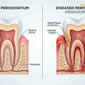
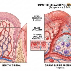
The pathogenesis of periodontal disease begins with the formation of a sticky, colorless biofilm known as dental plaque. This biofilm is a complex microbial community that adheres to the tooth surfaces, particularly along the gingival margin. When plaque is not adequately removed, the bacteria release toxins and metabolic byproducts that irritate the gingival epithelium. In response, the body’s immune system initiates an inflammatory cascade. White blood cells rush to the area to combat the bacterial invasion, leading to the classic signs of inflammation: increased blood flow (erythema or redness), fluid accumulation (edema or swelling), and a breakdown of the delicate microvasculature, which results in bleeding.[1]
Dr. Nguyen Van Cuong, a leading expert in periodontal care, frequently emphasizes to his patients that the absence of pain does not equate to the absence of disease. Many individuals harbor chronic, low-grade periodontal infections for years. During this time, the early symptoms of gum disease may intermittently flare up and subside, leading patients into a false sense of security. They might notice a bit of pink in the sink when brushing but dismiss it as brushing too hard. However, this chronic inflammation slowly degrades the collagen fibers of the periodontal ligament and stimulates osteoclasts—cells that break down bone tissue—leading to the gradual resorption of the alveolar bone.
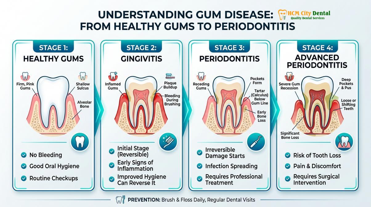
Understanding the distinction between gingivitis and periodontitis is crucial for grasping the silent threat of this condition. Gingivitis is the reversible stage, characterized solely by soft tissue inflammation without any loss of clinical attachment or bone. If intercepted at this stage, the tissues can heal completely. However, when the microbial dysbiosis shifts towards more virulent, anaerobic bacteria, the disease crosses a critical threshold into periodontitis. At this stage, the inflammatory front moves apically (down the root), creating periodontal pockets—spaces between the tooth and gum where even more bacteria can thrive in an oxygen-depleted environment. This downward spiral of infection and immune response is what ultimately leads to tooth mobility and loss, often catching the patient completely by surprise.
10 Early Signs of Gum Disease to Watch For
Recognizing the initial symptoms of gingivitis, such as bleeding, persistent bad breath, and localized swelling, is crucial for halting the progression of periodontal disease.
Because periodontal disease does not typically announce itself with severe pain, vigilance is required to detect its subtle onset. Patients must learn how to check for gum disease by observing changes in their oral cavity during their daily hygiene routines. Identifying these signs early can mean the difference between a simple professional cleaning and the need for complex surgical intervention. Below is a comprehensive clinical breakdown of the ten most common early signs of gum disease.
1. Bleeding Gums (Bleeding on Probing or Brushing)
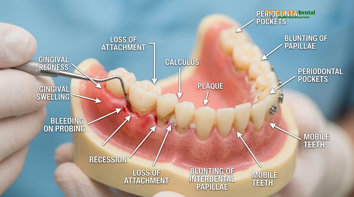
Bleeding is the hallmark sign of gingival inflammation. Healthy gums do not bleed when subjected to the normal mechanical forces of a soft-bristled toothbrush or dental floss. When plaque bacteria irritate the gingival sulcus (the shallow crevice between the tooth and gum), the tissue becomes ulcerated and the underlying capillaries become engorged and fragile. Consequently, even minor friction causes these micro-vessels to rupture. If you notice blood on your toothbrush, dental floss, or in your saliva after brushing, it is a definitive sign of an active inflammatory process that requires attention.
2. Erythema (Redness of the Gums)
Healthy gingival tissue is typically a pale, coral pink color, though natural pigmentation varies among individuals. When an infection takes hold, the body increases blood flow to the affected area to deliver immune cells. This vasodilation causes the gums to appear darker red, purplish, or intensely inflamed, particularly at the margins where the gum meets the tooth. This color change is often most noticeable in the interdental papillae—the small, triangular pieces of gum tissue between the teeth.
3. Edema (Swollen or Puffy Gums)
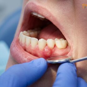
Alongside increased blood flow, the inflammatory response causes fluid to leak from the blood vessels into the surrounding connective tissues. This results in edema, making the gums look swollen, puffy, and enlarged. Swollen gums lose their tight, knife-edged adaptation to the tooth surface and may appear rolled or bulbous. This swelling can also create pseudo-pockets, making it easier for more plaque to accumulate beneath the gumline.
“The earliest clinical manifestation of periodontal disease is the loss of the tight, protective seal between the gingival margin and the tooth enamel, allowing pathogenic bacteria to colonize the subgingival space.”
4. Persistent Halitosis (Bad Breath)
While bad breath can occasionally result from dietary choices or gastrointestinal issues, chronic halitosis is frequently a byproduct of periodontal infection. The anaerobic bacteria responsible for gum disease thrive in the deep, oxygen-deprived pockets around the teeth. As these bacteria metabolize proteins from food debris and dead tissue cells, they release volatile sulfur compounds (VSCs), such as hydrogen sulfide and methyl mercaptan. These gases produce a distinct, foul odor that cannot be masked for long by mouthwash or mints.[2]
5. Gingival Recession (Gums Pulling Away)
Gum recession occurs when the margin of the gingival tissue pulls back, exposing more of the tooth crown and eventually the root surface. While recession can be caused by aggressive brushing or anatomical factors, it is a common consequence of periodontal disease. As the underlying bone is destroyed by the inflammatory process, the overlying gum tissue follows the receding bone level. This can make the teeth appear longer than normal and create dark triangular spaces (black triangles) between the teeth.
6. Increased Tooth Sensitivity
As the gums recede and expose the root surfaces, patients often experience increased sensitivity to hot, cold, sweet, or acidic stimuli. Unlike the crown of the tooth, which is protected by a dense layer of enamel, the root is covered by cementum, a much softer material. Beneath the cementum lies the dentin, which contains microscopic tubules leading directly to the tooth’s nerve center (the pulp). When these tubules are exposed to the oral environment due to gum recession, stimuli are transmitted directly to the nerve, causing sharp, transient pain.
7. Exudate (Pus Between Teeth and Gums)
The presence of pus, or purulent exudate, is a clear sign of a severe, active bacterial infection. Pus is a collection of dead white blood cells, tissue debris, and bacteria. In the context of gum disease, it may be visible as a yellowish or whitish fluid oozing from the gingival margin when pressure is applied to the gums. This indicates that the body is actively fighting a significant localized infection, and immediate clinical intervention is necessary to drain the area and eliminate the bacterial source.
8. Changes in the Bite (Occlusal Discrepancies)
As periodontal disease progresses and destroys the alveolar bone and periodontal ligaments that anchor the teeth, the teeth may begin to shift or migrate. Patients might notice that their teeth no longer fit together exactly as they used to when biting down. This change in occlusion can cause uneven wear on the enamel, muscle tension in the jaw, and further accelerate the breakdown of the weakened periodontal structures due to traumatic occlusal forces.
9. Loose or Mobile Teeth
Tooth mobility is a late-stage sign of early gum disease transitioning into advanced periodontitis. The teeth are held in their sockets by a complex network of periodontal ligament fibers attaching the root cementum to the alveolar bone. When this attachment apparatus is severely degraded by chronic inflammation, the teeth lose their structural support. Initially, the mobility may be slight (only detectable by a dentist), but as bone loss continues, the teeth may become noticeably loose to the patient, eventually requiring extraction if left untreated.
10. Itchy or Tender Gums
While severe pain is rare in early gum disease, patients often report a sensation of itchiness, dull aching, or tenderness in the gums. This discomfort is typically localized to areas of active inflammation and may be exacerbated during eating or brushing. The tenderness is a direct result of the pressure exerted by the swollen, fluid-filled tissues and the ongoing immune response within the gingiva.
How Gums Change: Color, Texture, and Position
As periodontal disease advances, healthy stippled, pink gums transform into smooth, dark red, and inflamed tissues that gradually pull away from the tooth crowns.
A visual inspection of the oral cavity can reveal significant information about a patient’s periodontal health. Dental professionals are trained to evaluate the gingiva based on specific clinical parameters: color, contour, consistency, and surface texture. Understanding these changes can help patients recognize gum infection signs early in the disease process.
Healthy gingiva is typically described as being coral pink, though melanin pigmentation can cause natural variations ranging from light brown to dark purple, depending on the individual’s ethnicity. The tissue should be firm, resilient, and tightly bound to the underlying bone. The gingival margin should form a scalloped, knife-edge contour around the neck of each tooth, and the interdental papillae should completely fill the spaces between the teeth. Furthermore, healthy attached gingiva often exhibits a stippled texture, resembling the surface of an orange peel. This stippling is a sign of strong, healthy collagen bundles anchoring the tissue.
When gingivitis sets in, these characteristics undergo a marked transformation. The color shifts from pale pink to erythematous (red) or cyanotic (bluish-red) due to vascular engorgement and sluggish blood flow. The contour changes as the tissue becomes edematous (swollen); the margins become rolled or bulbous, and the papillae may become blunted or cratered. The consistency of the tissue changes from firm to soft, spongy, and friable, meaning it tears and bleeds easily. Crucially, the healthy stippled texture is lost as the swelling stretches the tissue, leaving it looking smooth and shiny.
| Clinical Parameter | Healthy Gums | Diseased Gums (Gingivitis/Periodontitis) |
|---|---|---|
| Color | Pale coral pink (or naturally pigmented) | Dark red, purplish, or cyanotic |
| Contour | Knife-edged, scalloped, tight to the tooth | Rolled margins, bulbous, blunted papillae |
| Consistency | Firm, resilient, tightly bound | Soft, spongy, friable, easily compressible |
| Texture | Stippled (like an orange peel) | Smooth, shiny, stretched appearance |
| Bleeding | No bleeding upon gentle probing | Bleeds easily upon probing or brushing |
As the disease progresses to periodontitis, the position of the gums changes dramatically. The inflammatory destruction of the connective tissue attachment causes the gingival margin to migrate apically (downward toward the root apex), resulting in clinical recession. This not only compromises the aesthetic appearance of the smile but also exposes the vulnerable root surfaces to decay and sensitivity. Monitoring these visual and tactile changes is a fundamental aspect of General Dentistry and preventive care.
The Role of Plaque and Tartar Accumulation
The primary catalyst for gingival inflammation is the accumulation of bacterial plaque, which hardens into porous tartar that cannot be removed by standard brushing.
To truly understand the signs of gum disease, one must understand its primary etiology: the accumulation of dental plaque and calculus (tartar). The oral cavity is a complex ecosystem harboring hundreds of different bacterial species. In a healthy state, these microorganisms exist in a symbiotic balance. However, when oral hygiene is inadequate, this balance is disrupted, leading to a pathogenic shift in the microbial flora.
Dental plaque is not merely a loose collection of bacteria; it is a highly organized, complex biofilm. Within minutes of brushing, a thin layer of salivary proteins called the acquired pellicle coats the teeth. Pioneer bacteria, primarily Gram-positive aerobic cocci, quickly adhere to this pellicle. If left undisturbed, these bacteria multiply and excrete an extracellular matrix—a sticky, protective slime layer that shields them from the body’s immune system and antimicrobial agents. As the biofilm matures over several days, the environment at the base of the plaque layer becomes oxygen-depleted. This allows highly virulent, Gram-negative anaerobic bacteria (such as Porphyromonas gingivalis and Treponema denticola) to colonize and thrive.[3]
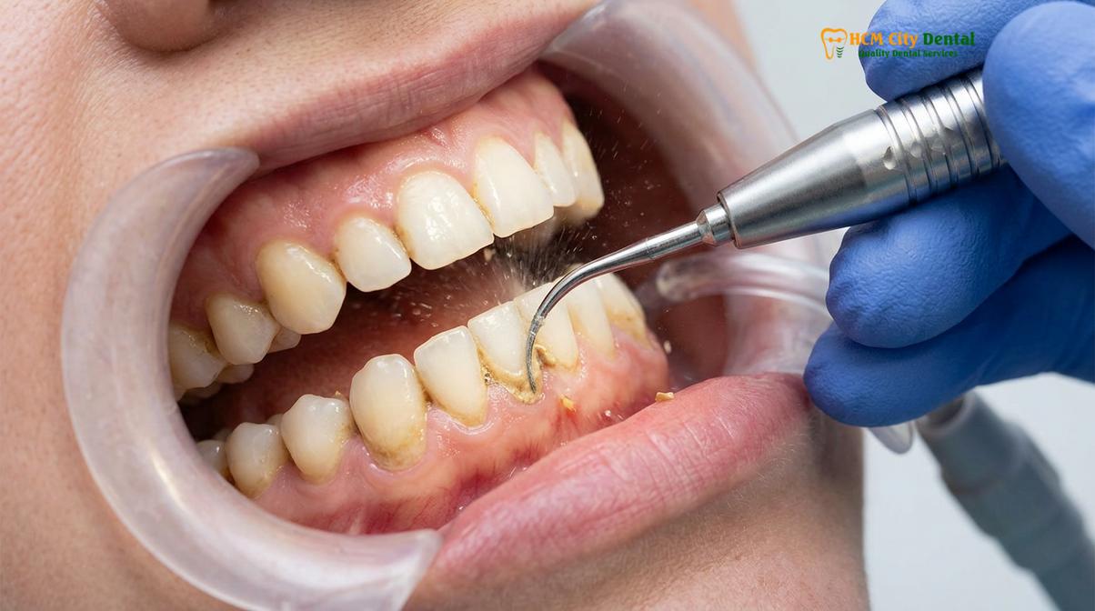
These anaerobic pathogens are the primary drivers of periodontal destruction. They produce potent endotoxins, enzymes (like collagenases and proteases), and metabolic byproducts that directly degrade the gingival tissues and trigger a hyperactive immune response. The body’s attempt to fight off the infection inadvertently causes collateral damage to its own tissues, leading to the breakdown of the periodontal ligament and alveolar bone.
If plaque is not removed mechanically through brushing and flossing within 24 to 72 hours, it begins to mineralize. Calcium and phosphate ions from the saliva precipitate into the biofilm matrix, hardening it into calculus, commonly known as tartar. Calculus can form above the gumline (supragingival) or below the gumline (subgingival). Subgingival calculus is particularly detrimental because it is dark, hard, and firmly attached to the root surface. Its rough, porous surface acts as a perfect retention site for even more pathogenic plaque, creating a vicious cycle of infection and inflammation. Once calculus has formed, it cannot be removed by home care methods; it requires professional scaling by a dental hygienist or dentist using specialized ultrasonic and hand instruments.
How Dentists Diagnose Early-Stage Gum Disease
Dental professionals utilize periodontal probing to measure pocket depths and radiographic imaging to assess underlying bone levels, ensuring an accurate diagnosis of gingival health.
While patients can observe the superficial signs of gum disease at home, a definitive diagnosis requires a comprehensive clinical evaluation by a dental professional. Because the most severe damage occurs beneath the gumline, out of sight, dentists rely on specific diagnostic tools and metrics to assess the true extent of periodontal tissue destruction.
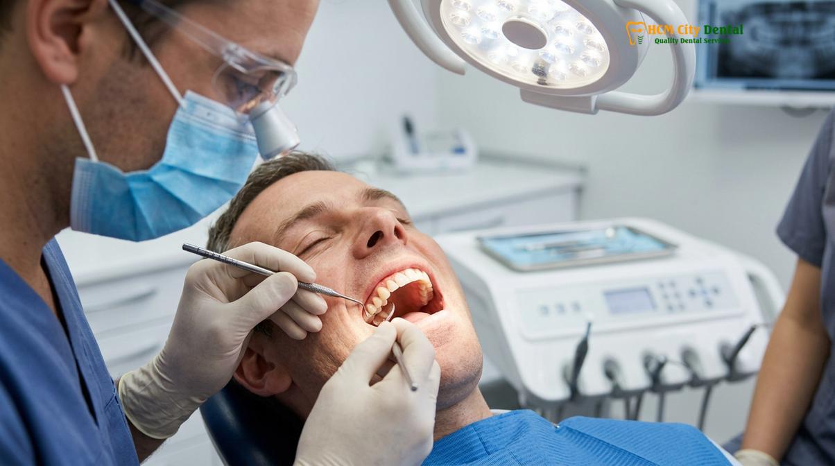
The cornerstone of periodontal diagnosis is the periodontal charting process. During this examination, the dentist or hygienist uses a specialized instrument called a periodontal probe. This probe is essentially a tiny measuring stick with millimeter markings. It is gently inserted into the gingival sulcus—the space between the tooth and the gum—to measure its depth. In a healthy state, this sulcus is typically 1 to 3 millimeters deep. When inflammation causes the tissue to detach from the tooth, this space deepens, forming a periodontal pocket. Pocket depths of 4 millimeters indicate early periodontitis, while depths of 5 millimeters or greater signify moderate to advanced disease.[4]
Clinical Case Review: Early Detection at HCMC Dental Clinic
A 35-year-old patient visited HCMC Dental Clinic in Ho Chi Minh City for a routine check-up, reporting no pain but noticing occasional bleeding when flossing. During the clinical examination, Dr. Nguyen Van Cuong performed a comprehensive periodontal probing. While the patient’s gums appeared relatively normal visually, the probing revealed generalized 4mm pockets and localized bleeding on probing (BOP). Digital radiographs confirmed the very beginning stages of crestal bone loss. Thanks to this early diagnostic intervention, Dr. Cuong was able to implement a conservative scaling and root planing protocol, successfully reversing the active infection and preventing irreversible structural damage.
In addition to measuring pocket depths, the clinician will assess several other critical parameters:
- Bleeding on Probing (BOP): As the probe is walked around the tooth, the clinician notes any areas that bleed. BOP is a highly reliable indicator of active, ongoing inflammation in the base of the pocket.
- Clinical Attachment Level (CAL): This measurement calculates the total amount of tissue detachment from the root surface, factoring in both the pocket depth and any visible gum recession. CAL is the most accurate metric for tracking disease progression over time.
- Furcation Involvement: For multi-rooted teeth (like molars), the dentist will check if the bone loss has extended into the area where the roots divide (the furcation). Infection in these areas is particularly challenging to treat.
- Tooth Mobility: The dentist will apply gentle pressure to the teeth to check for looseness, grading the mobility on a clinical scale.
Radiographic evaluation (X-rays) is the second essential component of periodontal diagnosis. While probing measures the soft tissue, X-rays reveal the hard tissue—the alveolar bone supporting the teeth. Digital bitewing and periapical radiographs allow the dentist to visualize the crestal bone levels and identify any patterns of horizontal or vertical bone loss. The combination of clinical probing and radiographic imaging provides a complete, three-dimensional understanding of the patient’s periodontal status, allowing for the formulation of a targeted, effective treatment plan.
Immediate Steps to Take if You Notice Symptoms
Upon detecting gingival abnormalities, patients must immediately optimize their daily oral hygiene routine and schedule a professional periodontal evaluation to remove calcified deposits.
If you identify any gingivitis warning signs, such as bleeding, redness, or persistent bad breath, taking immediate action is critical to prevent the condition from escalating into irreversible periodontitis. The management of early gum disease requires a dual approach: rigorous, optimized home care and prompt professional clinical intervention.
The first step is to critically evaluate and upgrade your daily oral hygiene routine. Many patients brush twice a day but use incorrect techniques that fail to disrupt the biofilm at the critical junction where the gum meets the tooth. Dental professionals recommend the Modified Bass brushing technique. This involves placing a soft-bristled toothbrush at a 45-degree angle to the gumline and using small, gentle, circular or vibratory motions. This technique allows the bristles to slightly penetrate the gingival sulcus, sweeping away the plaque without traumatizing the delicate tissue.
Furthermore, brushing alone only cleans about 60% of the tooth surfaces. The interproximal areas (between the teeth) are prime real estate for plaque accumulation and are often where periodontal disease initiates. Daily interdental cleaning is non-negotiable. Depending on the size of the spaces between your teeth, this may involve traditional dental floss, water flossers, or interdental brushes. Interdental brushes are particularly effective for patients who already have some degree of gum recession or larger embrasure spaces.
“Effective periodontal management is a partnership; the most advanced clinical therapies will fail if the patient does not maintain meticulous daily biofilm control at home.”
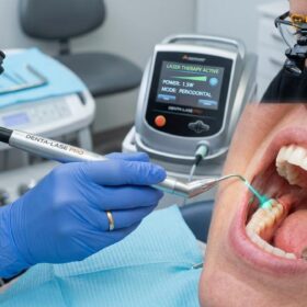
While optimizing home care is essential, it is not sufficient to treat established gum disease. Once plaque has mineralized into calculus, no amount of brushing or flossing will remove it. Therefore, the most crucial immediate step is to schedule an appointment for a professional evaluation and Gum Treatment. A dental hygienist or dentist will perform a thorough prophylaxis (cleaning) or, if necessary, a deeper cleaning known as scaling and root planing (SRP). SRP involves using specialized instruments to carefully remove plaque and calculus from the crown and root surfaces, deep within the periodontal pockets, and smoothing the root surfaces to discourage further bacterial attachment.
In some cases, the dentist may also recommend the short-term use of an antimicrobial mouthrinse containing chlorhexidine gluconate to help reduce the bacterial load and soothe inflamed tissues. However, mouthwashes are adjuncts, not replacements, for mechanical plaque removal.
When to See a Doctor for Periodontal Evaluation
Understanding when to transition from home monitoring to seeking professional medical advice is vital for preserving your dentition. While mild, occasional bleeding might prompt an improvement in flossing habits, certain clinical presentations require urgent evaluation by a dental professional to prevent systemic complications and tooth loss.[5]
Important Clinical Considerations:
You should seek immediate professional dental care if you experience any of the following severe symptoms:
- Spontaneous bleeding from the gums that occurs without brushing or provocation.
- The presence of purulent exudate (pus) oozing from the gumline.
- A sudden, noticeable loosening or shifting of one or more teeth.
- Severe, localized pain, swelling, or the formation of a gum boil (abscess).
- Fever, facial swelling, or difficulty swallowing associated with oral pain.
Even in the absence of severe symptoms, regular dental check-ups are the most effective strategy for preventing periodontal disease. It is highly recommended to undergo a professional cleaning and examination at least twice a year. Patients with a history of periodontal disease, diabetes, cardiovascular conditions, or those who smoke may require more frequent maintenance visits (every 3 to 4 months) to keep the bacterial load under control and monitor the stability of the periodontal tissues.
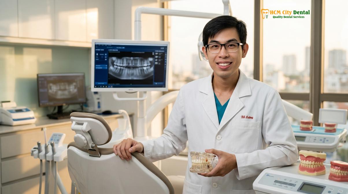
If you are experiencing any of the early symptoms of gum disease, do not wait for the condition to become painful. Early diagnosis and intervention are the keys to a successful, minimally invasive outcome. Based on clinical diagnostics, the team at HCMC Dental Clinic can provide a personalized treatment plan to restore your gingival health and protect your smile for the long term. Contact our clinic today to schedule a comprehensive periodontal evaluation.
Frequently Asked Questions
What does early gum disease look like?
Early gum disease typically presents as red, swollen, and puffy gums that may bleed easily when you brush or floss. At this initial stage, known as gingivitis, the gum tissue loses its firm, pale pink appearance and healthy stippled texture. Clinically, dentists look for localized inflammation along the gingival margin and a lack of tight adaptation of the tissue to the tooth surface. Because it is often painless, visual inspection is critical.
Can you have gum disease without knowing it?
Yes, you can have gum disease for a long time without experiencing any noticeable pain or obvious symptoms. Periodontal infection is notoriously silent in its early and moderate stages. The bacterial inflammatory process slowly destroys the supporting bone and connective tissues beneath the gum line without triggering significant nerve pain. Many patients only realize there is a problem when teeth become loose or an acute abscess forms.
Are sore gums a sign of gum disease?
Sore, tender, or itchy gums are common indicators of an active periodontal infection or gingival inflammation. While occasional soreness might result from aggressive brushing or a minor abrasion, persistent tenderness usually signifies that bacterial plaque has triggered an immune response. The localized swelling puts pressure on the gingival tissues, leading to a dull ache or sensitivity, especially when eating or applying pressure during oral hygiene routines.
How fast does gum disease progress?
The progression of gum disease varies significantly depending on individual oral hygiene, genetics, systemic health, and lifestyle factors like smoking. Gingivitis can develop within a few days of plaque accumulation. If left untreated, it can advance into mild periodontitis over several months or years. However, certain aggressive forms of periodontitis can cause rapid attachment loss and bone destruction in a matter of weeks, highlighting the need for regular monitoring.
What should I do if I have early signs of gum disease?
If you notice early signs of gum disease, immediately improve your oral hygiene by brushing twice daily and flossing thoroughly, then schedule a professional dental evaluation. A dentist needs to perform a comprehensive periodontal examination to measure pocket depths and remove hardened tartar (calculus) that you cannot remove at home. Early intervention can completely reverse gingivitis before it causes permanent bone loss.
References
- Journal of Periodontology. Pathogenesis of periodontal disease and the inflammatory immune response. (2021).
- American Academy of Periodontology. Comprehensive periodontal therapy: a statement on early diagnosis and clinical signs. (2020).
- Clinical Oral Investigations. The role of dental plaque biofilm and calculus in gingival inflammation. (2019).
- International Journal of Dentistry. Diagnostic accuracy of periodontal probing and radiographic assessment. (2022).
- Journal of Clinical Periodontology. Systemic implications of untreated periodontal infections. (2018).