Is there a clinical link between periodontitis and Alzheimer’s disease? Yes, emerging medical evidence suggests that chronic gum inflammation, specifically driven by the pathogen Porphyromonas gingivalis, may accelerate neurodegeneration and cognitive decline by introducing neurotoxic bacteria and inflammatory cytokines into the brain.
Clinical Summary:
The intersection of oral health and neurology has revealed a profound connection between severe periodontitis and Alzheimer’s disease. Chronic periodontal infections act as a continuous reservoir of Gram-negative anaerobic bacteria, most notably Porphyromonas gingivalis. These pathogens, along with their toxic byproducts known as gingipains, can enter the systemic circulation through ulcerated gingival tissues. Once in the bloodstream, they possess the capability to compromise the blood-brain barrier. Inside the central nervous system, these bacterial toxins trigger a hyperactive immune response, leading to microglial activation, chronic neuroinflammation, and the accelerated accumulation of beta-amyloid plaques and tau tangles—the hallmark pathological features of Alzheimer’s disease. While periodontal therapy is not a cure for dementia, proactive management of gum disease through professional scaling, root planing, and advanced laser therapies is increasingly recognized as a vital component of comprehensive systemic health maintenance, potentially mitigating one of the modifiable risk factors for cognitive decline.
Key Takeaways:
- Severe periodontitis creates a systemic inflammatory burden that extends far beyond the oral cavity.
- Porphyromonas gingivalis, a primary periodontal pathogen, has been identified in the brains of Alzheimer’s patients.
- Bacterial toxins called gingipains directly damage neurons and promote the formation of beta-amyloid plaques.
- Chronic oral inflammation hyperactivates microglial cells in the brain, shifting them from a protective to a neurotoxic state.
- Rigorous periodontal treatment may help reduce systemic bacterial loads, serving as a potential preventive measure against accelerated cognitive decline.
The Brain-Mouth Axis: Exploring the Science
The brain-mouth axis refers to the systemic pathway where oral pathogens and inflammatory mediators travel from infected periodontal tissues to the central nervous system, potentially triggering neurodegenerative processes.
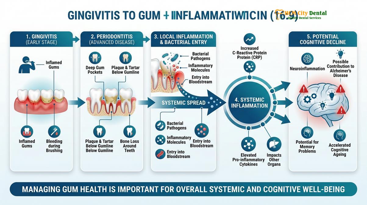
For decades, the oral cavity was viewed in clinical isolation from the rest of the human body. However, modern medical science has firmly established the concept of the brain-mouth axis, a complex bidirectional relationship where oral health profoundly influences neurological well-being. At the center of this paradigm shift is the growing understanding of the gum disease dementia link. Periodontitis is not merely a localized infection of the gums and alveolar bone; it is a chronic, low-grade systemic inflammatory disease. When the periodontium becomes infected, the highly vascularized nature of the gingival tissues provides a direct gateway for oral pathogens to enter the systemic circulation.
The anatomical pathways facilitating this microbial migration are multifaceted. The most prominent route is hematogenous spread. In patients with severe periodontitis, the epithelial lining of the periodontal pockets becomes ulcerated and highly permeable. Simple daily activities, such as mastication (chewing) or aggressive tooth brushing, can force millions of bacteria into the bloodstream, creating a state of transient bacteremia. While a healthy immune system rapidly clears these microbes, the sheer volume and continuous nature of the bacterial influx in untreated periodontitis overwhelm systemic defenses. Over time, these circulating pathogens reach the cerebral vasculature. [1]
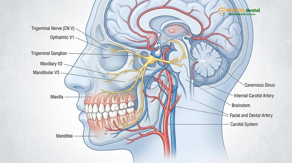
Beyond the bloodstream, researchers are investigating neural and lymphatic pathways. The oral cavity is densely innervated by the trigeminal nerve, one of the largest cranial nerves connecting directly to the brainstem. Certain neurotropic viruses and bacteria are known to utilize peripheral nerves as conduits to bypass systemic immune surveillance, migrating retrogradely along the nerve fibers directly into the central nervous system. Furthermore, the lymphatic drainage of the head and neck provides another potential avenue for inflammatory cytokines and bacterial antigens to reach the cerebral environment. Understanding these pathways is crucial for dental professionals providing comprehensive gum treatment, as the goal extends beyond saving teeth to preserving overall systemic integrity.
The systemic burden of periodontitis is characterized by the continuous release of pro-inflammatory mediators, including C-reactive protein (CRP), interleukins, and tumor necrosis factor-alpha (TNF-α). These molecules circulate systemically, promoting endothelial dysfunction and altering the permeability of various physiological barriers, including the highly selective blood-brain barrier. This systemic inflammatory priming sets the stage for neurodegenerative vulnerability, making the management of periodontal health a critical factor in holistic patient care.
How P. gingivalis Penetrates the Blood-Brain Barrier
Porphyromonas gingivalis breaches the blood-brain barrier through transient bacteremia, utilizing specialized virulence factors to degrade endothelial tight junctions and invade cerebral tissues.
The blood-brain barrier (BBB) is a highly specialized, semi-permeable border of endothelial cells that prevents solutes in the circulating blood from non-selectively crossing into the extracellular fluid of the central nervous system. It is the brain’s primary defense mechanism against systemic infections. However, the pathogen Porphyromonas gingivalis (P. gingivalis), a keystone bacterium in the development of severe periodontitis, has evolved sophisticated mechanisms to circumvent and dismantle this vital barrier.
The journey of porphyromonas gingivalis brain invasion begins in the deep, anaerobic periodontal pockets. Here, the bacteria multiply and express a multitude of virulence factors. When transient bacteremia occurs, P. gingivalis enters the bloodstream. Unlike many benign oral bacteria that are quickly neutralized by serum complement proteins, P. gingivalis possesses a unique capsule and secretes enzymes that degrade these immune proteins, allowing it to survive and circulate freely. [2]
Upon reaching the cerebral microvasculature, P. gingivalis must cross the endothelial lining. The endothelial cells of the BBB are bound together by complex protein structures known as tight junctions (primarily composed of claudins, occludins, and junctional adhesion molecules). P. gingivalis secretes potent proteolytic enzymes that specifically target and cleave these tight junction proteins. By degrading the structural integrity of the BBB, the bacteria create microscopic paracellular gaps, allowing them to slip between the endothelial cells and enter the brain parenchyma.
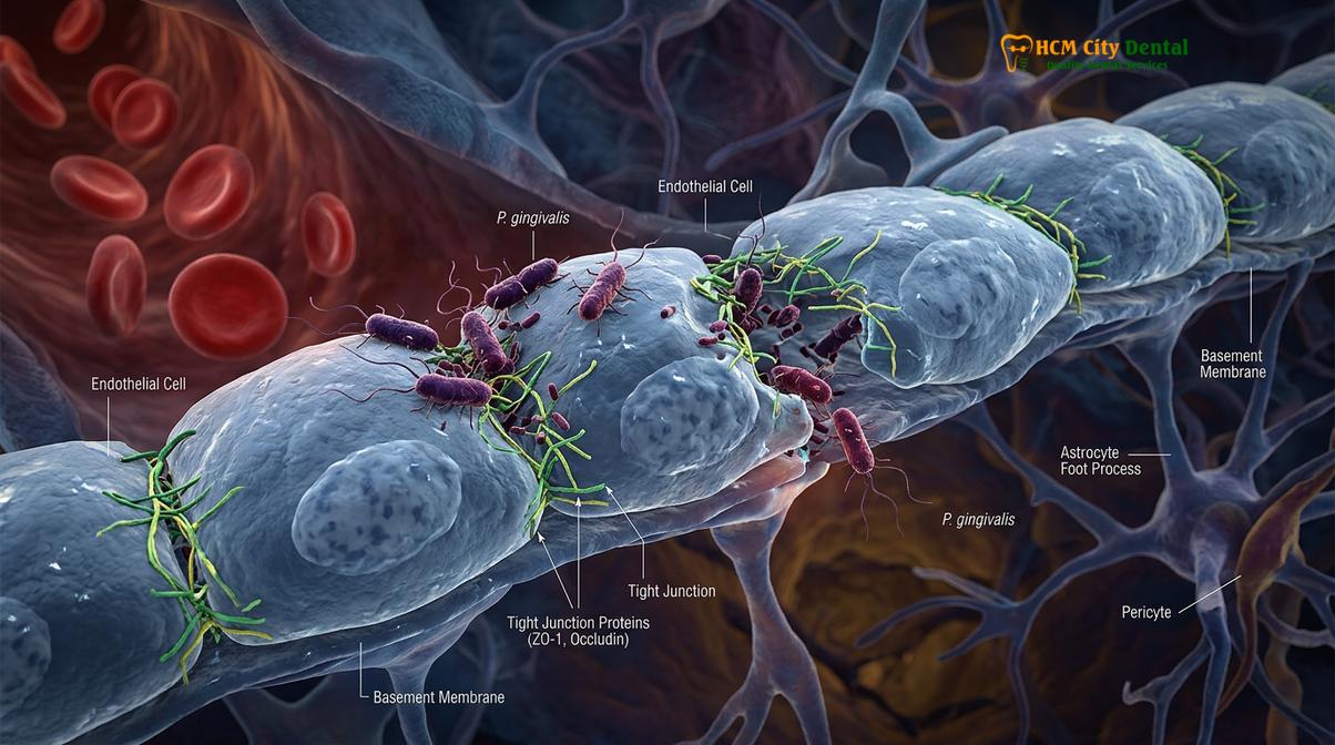
Furthermore, P. gingivalis is an intracellular pathogen. It can actively invade the endothelial cells themselves through a process called receptor-mediated endocytosis. Once inside the host cell, the bacterium can evade immune detection, replicate, and eventually exit the cell on the abluminal side, directly into the brain tissue. This transcellular migration is facilitated by the bacterium’s fimbriae—hair-like appendages that bind to specific receptors on the surface of the endothelial cells. The ability of this oral pathogen to so effectively breach the brain’s ultimate defense highlights the severe systemic implications of untreated periodontal disease.
P. gingivalis Gingipains and Beta-Amyloid Plaque Accumulation
Gingipains, toxic proteases secreted by P. gingivalis, directly damage neurons and provoke the brain to produce beta-amyloid proteins as an antimicrobial defense, inadvertently forming Alzheimer’s plaques.
Once P. gingivalis successfully infiltrates the brain tissue, the true neurodegenerative damage begins, largely driven by its primary virulence factors: gingipains. Gingipains are cysteine proteases—enzymes that break down proteins—secreted in massive quantities by the bacteria to acquire nutrients (specifically iron and amino acids) from the host and to paralyze local immune responses. In the context of Alzheimer’s disease, the discovery of gingipains in the brains of deceased patients has been a monumental breakthrough in understanding the pathogenesis of cognitive decline.
There are two main types of gingipains produced by P. gingivalis: Arginine-specific gingipains (RgpA and RgpB) and Lysine-specific gingipains (Kgp). These enzymes are highly destructive to human tissue. In the brain, gingipains have been shown to directly cleave and fragment tau proteins. Normal tau proteins are essential for stabilizing the internal microtubule structure of neurons. When gingipains degrade tau, the proteins collapse and tangle together, forming neurofibrillary tangles—one of the two primary pathological hallmarks of Alzheimer’s disease. This structural collapse leads to the death of the neuron and the disruption of synaptic communication. [3]
| Gingipain Type | Primary Function in Periodontitis | Neurotoxic Effect in the Brain |
|---|---|---|
| RgpA (Arginine-specific) | Degrades host tissue for nutrients; evades immune detection. | Cleaves tau proteins, contributing to neurofibrillary tangle formation. |
| RgpB (Arginine-specific) | Disrupts inflammatory signaling and degrades complement proteins. | Induces severe neuroinflammation and microglial hyperactivation. |
| Kgp (Lysine-specific) | Scavenges iron from red blood cells (causing bleeding gums). | Directly damages neuronal synapses and promotes beta-amyloid aggregation. |
The relationship between gingipains and beta-amyloid plaques—the other major hallmark of Alzheimer’s—is equally complex and fascinating. Historically, beta-amyloid was viewed strictly as a toxic metabolic byproduct that accumulated in the aging brain. However, recent neurobiological research suggests the “Antimicrobial Protection Hypothesis.” According to this model, beta-amyloid is actually an antimicrobial peptide produced by the brain’s immune system in response to invading pathogens like P. gingivalis.
“The accumulation of beta-amyloid plaques may initially serve as a protective mechanism, a biological net cast by the brain to trap and neutralize invading oral pathogens. However, chronic exposure to these bacteria leads to an overproduction of amyloid, which eventually becomes neurotoxic and drives the progression of dementia.”
When P. gingivalis enters the brain, the local immune system responds by rapidly producing beta-amyloid to encapsulate and sequester the bacteria. While this mechanism may temporarily halt the infection, the continuous influx of bacteria from chronic, untreated periodontitis causes an unrelenting overproduction of beta-amyloid. These amyloid clusters eventually aggregate into dense, insoluble plaques that suffocate surrounding neurons, disrupt cell-to-cell signaling, and trigger a cascade of neuroinflammation. Thus, the brain’s attempt to defend itself against an oral infection inadvertently accelerates its own degeneration.
Chronic Inflammation, Microglial Activation, and Dementia Risk
Systemic inflammatory cytokines from severe periodontal disease cross into the brain, hyperactivating microglial cells which then destroy healthy neurons instead of protecting them.
The physical presence of bacteria in the brain is only one facet of the cognitive decline oral health connection. The other critical component is the profound impact of chronic systemic inflammation. Periodontitis is characterized by a persistent, low-grade inflammatory state. The ulcerated periodontal pockets constantly release pro-inflammatory cytokines—such as Interleukin-1 beta (IL-1β), Interleukin-6 (IL-6), and Tumor Necrosis Factor-alpha (TNF-α)—into the bloodstream. These circulating inflammatory markers can cross the blood-brain barrier, even in the absence of intact bacteria, and profoundly alter the neuro-immune landscape.
The primary immune cells of the central nervous system are microglia. In a healthy brain, microglia exist in a resting, neuroprotective state (often referred to as the M2 phenotype). They act as the brain’s custodians, quietly patrolling the neural tissue, clearing away metabolic debris, pruning unused synapses, and supporting neuronal health. However, when exposed to systemic inflammatory signals originating from severe gum disease, these microglia undergo a radical transformation.
The influx of peripheral cytokines, combined with the presence of bacterial lipopolysaccharides (LPS) from Gram-negative oral pathogens, triggers microglial priming and activation. The microglia shift into an aggressive, pro-inflammatory state (the M1 phenotype). Instead of clearing debris, hyperactivated microglia begin to secrete their own massive quantities of neurotoxic cytokines and reactive oxygen species. This localized cytokine storm within the brain creates a highly hostile environment for neurons. [4]
Clinical Warning: Ignoring chronic bleeding gums allows systemic inflammation to persist unchecked. This continuous inflammatory burden not only destroys the jawbone but may also prime the brain’s immune system for accelerated neurodegeneration. Early intervention is critical to halt this systemic cascade.
This state of chronic neuroinflammation is a primary driver of dementia risk. The hyperactivated microglia lose their ability to effectively clear beta-amyloid plaques, allowing the plaques to accumulate faster. Furthermore, the inflammatory mediators directly damage the synaptic connections between neurons, particularly in the hippocampus and cerebral cortex—the regions of the brain responsible for memory formation and cognitive processing. Over years and decades, this sustained inflammatory assault, fueled by untreated periodontal disease, significantly lowers the threshold for cognitive decline and accelerates the onset of Alzheimer’s symptoms.
Review of Clinical Studies Linking Periodontitis to Cognitive Decline
Extensive longitudinal studies demonstrate that patients with chronic, untreated periodontitis experience a significantly faster rate of cognitive decline compared to those with healthy periodontium.
The theoretical mechanisms connecting oral pathogens to neurodegeneration are strongly supported by a growing body of epidemiological and clinical research. Retrospective cohort studies and longitudinal analyses tracking thousands of patients over decades have consistently revealed a statistically significant correlation between the severity of periodontal disease and the incidence of cognitive impairment.
One of the most compelling pieces of evidence comes from post-mortem analyses of human brain tissue. Researchers have successfully identified the DNA of Porphyromonas gingivalis, as well as its toxic gingipain enzymes, in the brains of individuals who died from Alzheimer’s disease. Crucially, the concentration of these gingipains correlated directly with the severity of tau pathology and cognitive decline observed in the patients prior to death. Furthermore, these bacterial markers were found in the brains of individuals who exhibited early Alzheimer’s pathology but had not yet been diagnosed with dementia, suggesting that the bacterial infection precedes and potentially initiates the neurodegenerative process, rather than merely being a consequence of it.
Clinical Case Observation
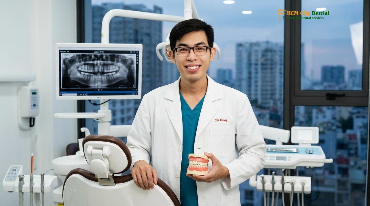
At HCMC Dental Clinic in Ho Chi Minh City, Dr. Nguyen Van Cuong frequently evaluates senior patients presenting with advanced periodontitis and early signs of memory impairment. In one notable case, a 68-year-old patient with severe, untreated gum disease and a recent diagnosis of mild cognitive impairment underwent comprehensive periodontal rehabilitation. While dental treatment cannot reverse neurological damage, stabilizing the oral infection significantly reduced the patient’s systemic inflammatory markers (CRP levels), demonstrating the critical role of oral health in managing systemic inflammatory burdens associated with cognitive decline.
Longitudinal studies tracking cognitive function in older adults have shown that individuals with severe periodontitis decline at a rate significantly faster than those with healthy gums. The risk is particularly pronounced in patients who have experienced extensive tooth loss. Tooth loss is generally the end-stage result of decades of chronic periodontal inflammation. Studies indicate that individuals with fewer than 20 remaining natural teeth face a substantially higher risk of developing dementia. This is attributed not only to the historical burden of systemic inflammation but also to the loss of masticatory function. Chewing stimulates cerebral blood flow; as teeth are lost and chewing efficiency decreases, the reduced blood flow to the brain may further compound cognitive vulnerabilities.
These clinical findings underscore the importance of early diagnosis and intervention. By the time cognitive symptoms manifest, significant neurological damage has already occurred. Therefore, identifying and treating chronic periodontitis in middle age may represent a crucial window of opportunity for mitigating long-term dementia risk.
Could Rigorous Gum Care Help Delay Alzheimer’s Symptoms?
While not a cure, aggressive management of periodontal disease through professional scaling, root planing, and laser therapy may reduce the systemic bacterial load, potentially slowing neurodegenerative progression.
Given the compelling evidence linking oral pathogens to neuroinflammation, the clinical focus naturally shifts to prevention and management. Protecting brain through gum care is emerging as a proactive strategy in holistic health maintenance. While periodontal therapy cannot cure Alzheimer’s disease or reverse existing brain damage, rigorous gum care is essential for eliminating the chronic source of systemic inflammation and halting the continuous influx of neurotoxic bacteria into the bloodstream.
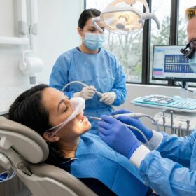
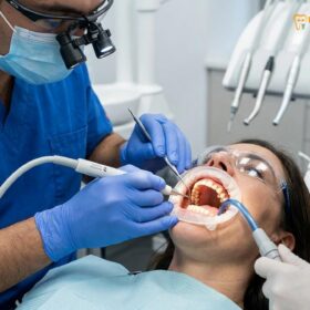
The foundation of periodontal management is non-surgical therapy, primarily scaling and root planing (SRP). This meticulous procedure involves the mechanical removal of calcified plaque (tartar) and bacterial biofilms from deep within the periodontal pockets, followed by the smoothing of the tooth roots to prevent further bacterial adherence. By physically debriding the infection site, SRP significantly reduces the population of Gram-negative anaerobes, including P. gingivalis, thereby lowering the systemic bacterial load. [5]
For more advanced cases where deep pockets persist, modern dentistry offers sophisticated interventions. Laser gum treatment utilizes targeted light energy to selectively vaporize diseased, infected tissue while leaving healthy tissue intact. The laser energy also provides a profound bactericidal effect, penetrating the soft tissue to eradicate pathogens that mechanical instruments cannot reach. This minimally invasive approach promotes faster healing and significantly reduces the inflammatory burden.
“Eradicating the periodontal infection is not just about saving the dentition; it is about closing the gateway that allows chronic inflammation and neurotoxic pathogens to assault the systemic circulation and, ultimately, the brain.”
In cases of severe architectural damage to the periodontium, surgical interventions such as gum flap surgery may be necessary to access deep root surfaces, reshape damaged bone, and eliminate the anatomical pockets where bacteria thrive. The ultimate clinical objective across all these modalities is to achieve a stable, maintainable periodontium free of active inflammation. By achieving this, patients can drastically reduce their systemic levels of CRP and pro-inflammatory cytokines, potentially creating a more favorable physiological environment that resists, rather than promotes, neurodegeneration.
Important Clinical Considerations (When to See a Doctor)
Periodontal disease is often described as a “silent” condition because it can progress significantly before causing noticeable pain. However, recognizing the early clinical signs is vital for preventing both oral tissue destruction and the associated systemic complications. Patients should seek immediate evaluation from a qualified dental professional if they experience any of the following symptoms.
Persistent bleeding during brushing or flossing is the most common early indicator of active gingival inflammation. Healthy gums do not bleed. If the inflammation progresses, patients may notice chronic halitosis (bad breath) that does not improve with oral hygiene, a condition deeply tied to the volatile sulfur compounds produced by anaerobic bacteria. For a deeper understanding of this symptom, patients can review the clinical causes of bad breath related to gum disease.
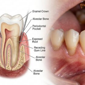
More advanced signs requiring urgent clinical attention include swollen, purulent gums around a tooth, gingival recession (teeth appearing longer), and increased tooth mobility. These symptoms indicate that the underlying alveolar bone is actively being destroyed. Dr. Nguyen Van Cuong emphasizes that personalized clinical diagnostics, including comprehensive periodontal probing and full-mouth radiographic analysis, are essential to determine the exact severity of the disease and to formulate an appropriate, evidence-based treatment plan. Regular maintenance visits, typically every three to four months for patients with a history of periodontitis, are crucial to monitor stability and prevent disease relapse.
Frequently Asked Questions
Can gum disease cause dementia or Alzheimer’s?
Gum disease does not directly cause Alzheimer’s, but severe periodontitis is considered a significant risk factor that may accelerate cognitive decline. Chronic oral inflammation allows neurotoxic bacteria to enter the bloodstream, potentially triggering or worsening the neurodegenerative processes associated with dementia over time. The continuous systemic inflammatory burden lowers the body’s overall resilience, making the brain more susceptible to the accumulation of amyloid plaques and tau tangles.
What is Porphyromonas gingivalis?
Porphyromonas gingivalis is a highly destructive, Gram-negative anaerobic bacterium that serves as a primary pathogen in severe periodontal disease. It produces toxic enzymes called gingipains, which degrade gum tissue, suppress local immune responses, and have been discovered in the brains of patients suffering from Alzheimer’s disease. This bacterium is particularly adept at evading host defenses and breaching the blood-brain barrier, making it a focal point in neuro-dental research.
How does mouth bacteria travel to the brain?
Oral bacteria primarily travel to the brain through the bloodstream following transient bacteremia caused by chewing or brushing infected gums. Additionally, pathogens may migrate along the cranial nerves, such as the trigeminal nerve, or through the lymphatic system, eventually breaching the blood-brain barrier to infect cerebral tissues. Once inside the brain, these bacteria can initiate a localized immune response that contributes to chronic neuroinflammation.
Does deep cleaning protect against cognitive decline?
Professional deep cleaning, or scaling and root planing, helps reduce the systemic bacterial load and lowers chronic inflammation, which may theoretically help protect against cognitive decline. By eliminating the source of periodontal infection, patients can minimize the volume of neurotoxic pathogens entering their systemic circulation. While it is not a guaranteed preventive measure for dementia, maintaining a healthy periodontium is a critical component of reducing overall systemic inflammatory risk factors.
Is there a link between tooth loss and memory loss?
Yes, extensive clinical studies indicate a strong correlation between significant tooth loss and an increased risk of memory loss or dementia. Tooth loss is often the end-stage result of chronic periodontitis, reflecting decades of systemic inflammation, and it also reduces masticatory stimulation, which is vital for maintaining cerebral blood flow. Preserving natural teeth through proactive periodontal care is therefore considered beneficial for both oral function and long-term cognitive health.
References
- Journal of Alzheimer’s Disease. Porphyromonas gingivalis in Alzheimer’s disease brains: Evidence for disease causation and treatment with small-molecule inhibitors. (2019).
- Journal of Periodontology. The bidirectional relationship between periodontitis and neurodegenerative diseases. (2021).
- Clinical Oral Investigations. Systemic inflammatory markers in severe periodontitis and their impact on cognitive function. (2020).
- International Journal of Oral Science. Mechanisms of blood-brain barrier penetration by periodontal pathogens. (2022).
- American Dental Association (ADA). Clinical practice guidelines for the non-surgical treatment of chronic periodontitis. (2018).