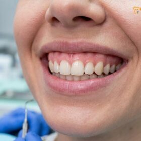
Laser gingivoplasty in Ho Chi Minh City utilizes advanced diode technology to reshape uneven gum tissue, enhancing smile symmetry without scalpels or sutures. This minimally invasive procedure instantly coagulates blood vessels, ensuring a rapid, comfortable recovery while preserving the critical biological width of your dental anatomy.
Clinical Summary:
Laser gingivoplasty is a highly precise periodontal procedure designed to recontour the gingival margins for optimal aesthetic and functional outcomes. By employing 810nm diode lasers, clinicians can sculpt scalloped gum contours with minimal thermal damage, immediate hemostasis, and virtually no postoperative swelling. The treatment strictly adheres to biological width parameters, ensuring long-term periodontal health. For complex cases involving vertical maxillary excess, multidisciplinary approaches including orthognathic surgery or neuromodulator injections are integrated to achieve comprehensive smile harmony.
Key Takeaways:
- Utilizes 810nm AMD Picasso diode lasers for bloodless, suture-free tissue sculpting.
- Preserves the minimum 2mm supracrestal tissue attachment to prevent bone resorption.
- Offers rapid 48-hour recovery with significantly reduced postoperative pain and swelling.
- Integrates Digital Smile Design (DSD) to accurately calibrate gingival zenith points.
- Collaborates with maxillofacial specialists for severe skeletal gummy smile cases.
What is Gingivoplasty and How Does It Differ From Gingivectomy?
Gingivoplasty focuses on reshaping and thinning healthy gum tissue for aesthetic contouring, whereas a gingivectomy primarily involves the complete removal of diseased or excess gingival pockets.
In the realm of aesthetic periodontics, understanding the distinction between Gum Contouring & Gummy Smile procedures is vital for proper treatment planning. While patients often use the terms interchangeably, the clinical objectives of gingivoplasty vs gingivectomy are distinctly different, even though both can be performed using advanced laser technology. Gingivoplasty is fundamentally a resective and recontouring procedure. Its primary goal is to thin the attached gingiva, create physiological sluiceways, and establish an aesthetically pleasing, knife-edged gingival margin. This is often required when patients present with thick, fibrotic gum tissue or minor asymmetries that disrupt the visual harmony of the smile. [1]
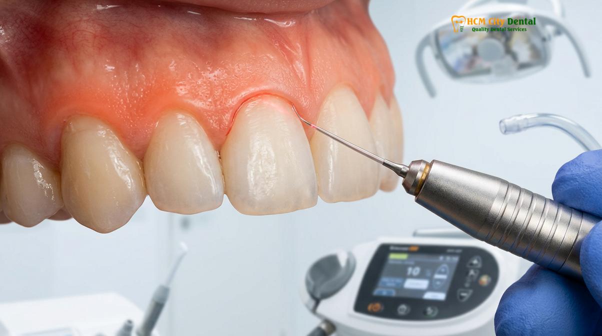
Conversely, a gingivectomy is an excisional procedure designed to eliminate suprabony periodontal pockets or remove significant amounts of hyperplastic tissue (often induced by medications or chronic inflammation). In a gingivectomy, the clinician excises the soft tissue wall of the pocket to reduce its depth, facilitating better oral hygiene and halting the progression of periodontal disease. When addressing a gummy smile caused by altered passive eruption, a clinician will often perform a gingivectomy to expose the anatomical crown, followed immediately by a gingivoplasty to refine and bevel the newly established margins. [2]
The advent of soft tissue lasers has revolutionized both procedures. Traditional scalpel techniques required secondary intention healing, often leaving the patient with exposed connective tissue, bleeding, and the need for periodontal dressing (Coe-Pak). Today, the use of targeted light energy allows for simultaneous cutting and coagulation, drastically altering the postoperative experience. To fully grasp the Laser vs Scalpel Gingivectomy: Clinical Comparison & Recovery, one must look at the histological response of the tissue, which shows significantly less inflammatory infiltrate when a laser is utilized.
| Clinical Parameter | Gingivoplasty | Gingivectomy |
|---|---|---|
| Primary Objective | Aesthetic reshaping, thinning, and contouring of healthy tissue. | Excision of diseased pocket walls or hyperplastic overgrowth. |
| Tissue State | Generally healthy, fibrotic, or asymmetrical. | Often inflamed, hyperplastic, or forming deep pockets. |
| Laser Application | Surface ablation and beveling to create knife-edged margins. | Deep excision to the base of the pocket to expose tooth structure. |
| Postoperative Healing | Rapid epithelialization (48-72 hours) with minimal discomfort. | Slightly longer healing depending on the volume of tissue removed. |
Fine-Tuning Asymmetric Gum Zenith Margins
Achieving a harmonious smile requires precise calibration of the gingival zenith points, ensuring the highest point of the gum scallop aligns slightly distal to the tooth’s long axis.
The aesthetic success of any anterior dental restoration or periodontal surgery hinges heavily on the architecture of the soft tissue frame. The gingival zenith—the most apical point of the free gingival margin—plays a critical role in this visual balance. In a naturally aesthetic adult dentition, the zenith of the maxillary central incisors and canines is typically located 1mm distal to the vertical midline of the tooth, while the zenith of the lateral incisors is usually centered. When these points are blunted, uneven, or misaligned, the smile appears asymmetrical, regardless of how perfectly aligned the teeth themselves may be. Correcting this requires a minor gum asymmetry fix, meticulously planned through Digital Smile Design (DSD).
However, altering the gingival zenith is not merely a matter of cutting away tissue until it looks right. The clinician must strictly adhere to the anatomical boundaries of the periodontium. Dr. Nguyen Van Cuong, a leading expert at HCMC Dental Clinic, emphasizes that zenith calibration must always respect the biological width sounding to prevent chronic inflammation. The biological width, now more accurately termed the supracrestal tissue attachment, consists of the junctional epithelium (approximately 0.97mm) and the supracrestal connective tissue attachment (approximately 1.07mm). Together, they form a mandatory 2mm barrier above the alveolar bone crest. [3]
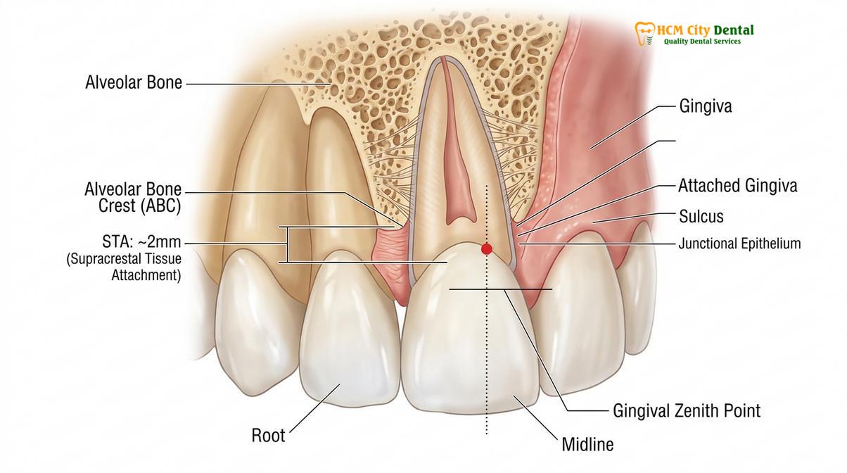
“The preservation of at least 2mm of supracrestal tissue attachment is non-negotiable in aesthetic crown lengthening; violating this space inevitably leads to unpredictable bone resorption and chronic gingival inflammation.”
If a patient requires a zenith adjustment that would encroach upon this 2mm safety zone, soft tissue laser ablation alone is contraindicated. In such cases, the procedure must be upgraded to an aesthetic crown lengthening, which involves raising a mucoperiosteal flap and performing an osteotomy (removal of supporting bone) or osteoplasty (reshaping of non-supporting bone) to re-establish the correct distance between the bone crest and the new gingival margin. Failure to perform this osseous resective work will result in the gum tissue rebounding to its original position or remaining in a state of perpetual, bleeding inflammation. Proper diagnostic sounding with a periodontal probe under local anesthesia is the only way to map this hidden topography before the laser is ever activated.
The Role of Soft Tissue Lasers in Precision Sculpting
The 810nm AMD Picasso diode laser provides a sterile, minimally invasive beam that instantly cauterizes tissue, eliminating the need for scalpels and sutures while accelerating epithelialization.
The transition from traditional steel scalpels to photonic energy represents a paradigm shift in periodontal plastic surgery. At the forefront of this evolution are the AMD Picasso Lasers, specifically the 810nm diode soft tissue laser. This specific wavelength is highly absorbed by tissue chromophores—namely melanin and hemoglobin—making it exceptionally efficient at cutting highly vascularized oral mucosa. When the laser energy interacts with the gingiva, it causes rapid photothermal vaporization of the intracellular water, resulting in a clean, precise incision.
The most profound advantage of laser gums sculpting is its ability to achieve immediate hemostasis. As the laser cuts, the peripheral thermal energy instantly coagulates small blood vessels and capillaries. This creates a virtually bloodless surgical field, allowing the clinician to evaluate the aesthetic contours in real-time without the visual obstruction of hemorrhage. Furthermore, the laser energy seals lymphatic vessels and nerve endings, which directly translates to a massive reduction in postoperative pain and swelling. Patients who undergo this procedure often require nothing more than over-the-counter analgesics for the first 24 hours. For a deeper understanding of postoperative care, patients can review the Gum Contouring Swelling: Clinical Recovery & Laser Healing Guide.
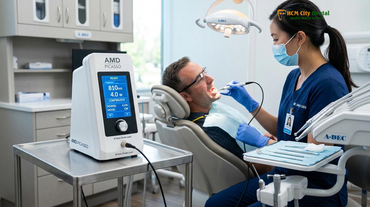
In addition to its cutting and coagulating properties, the diode laser emits a sterile beam. The high-energy light destroys bacteria at the surgical site, significantly minimizing the risk of postoperative infection. This bactericidal effect is particularly beneficial when treating patients with a history of mild gingivitis or those undergoing concurrent restorative work. The resulting wound edge is characterized by a thin layer of denatured protein that acts as a natural biological dressing, protecting the underlying connective tissue and promoting rapid epithelialization. Within 48 hours, a new layer of epithelial cells begins to migrate across the treated area, leading to a swift and uneventful recovery. This efficiency is a cornerstone of modern Waterlase Laser Gums: Clinical Efficacy in Painless Gum Contouring protocols.
What to Expect During a Quick In-Office Reshaping Session
A standard laser contouring session involves digital smile mapping, profound local anesthesia, and rapid laser ablation, typically completed within a single visit with immediate aesthetic results.
For patients seeking to enhance their smile, the workflow for a laser gingivoplasty is highly streamlined, designed to maximize precision while minimizing chair time. The journey begins with a comprehensive clinical evaluation. Prior to any surgical intervention, a thorough dental scaling and prophylaxis must be performed to ensure the gingival tissues are free of plaque, calculus, and active inflammation. Operating on inflamed tissue compromises the predictability of the final margins.
Once the oral environment is optimized, the clinician utilizes Digital Smile Design (DSD) software to map the ideal scalloped gum contours over high-resolution photographs of the patient’s face and teeth. This digital blueprint is then transferred to the mouth using a surgical stent or precise marking pens. Profound local anesthesia (typically Lidocaine 2% with epinephrine 1:100,000) is administered via a virtually painless computerized injection system. Once the tissue is completely numb, the clinician employs the AMD Picasso diode laser to meticulously trace the predetermined zenith points, vaporizing the excess tissue in sweeping, brush-like strokes.
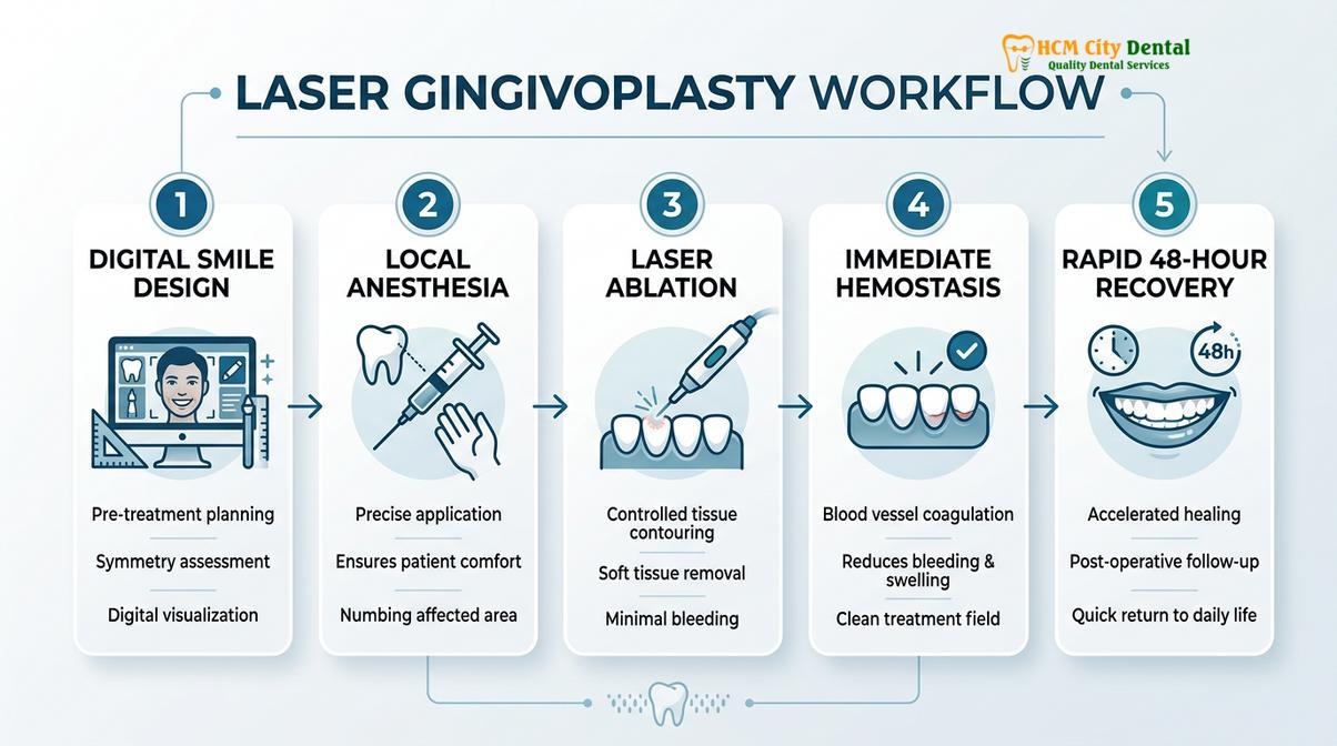
The entire laser ablation process for the anterior sextant (the front six teeth) often takes less than 30 minutes. Because there are no sutures to place and no bleeding to control, the patient can immediately view their new smile in the mirror. The clinic provides detailed postoperative instructions, which can be further explored in the Gum Contouring Recovery: Clinical Healing Timeline & Care Guide.
Understanding the financial investment is crucial for patients, especially those combining their treatment with dental tourism in Vietnam. According to the latest clinic fee schedule, the pricing structure at HCMC Dental Clinic is highly transparent and competitive:
- Laser Gingivectomy (per tooth): 1,500,000 VND (~$60 USD) (Walk-in: 2,500,000 VND).
- Full-Arch Laser Gum Contouring (up to 10 teeth): 12,000,000 VND (~$480 USD) (Walk-in: 20,000,000 VND).
- Functional/Esthetic Crown Lengthening (per tooth, including bone contouring): 3,000,000 VND (~$120 USD) (Walk-in: 5,000,000 VND).
- Surgical Lip Repositioning / Lip Lowering: 15,000,000 VND (~$600 USD) (Walk-in: 25,000,000 VND).
- Botox Gummy Smile Correction (both sides): 4,500,000 VND (~$180 USD) (Walk-in: 7,500,000 VND).
Note: International and local patients booking in advance via WhatsApp are eligible for the -40% discounted rates listed above.
Clinical Case Study: Full-Arch Aesthetic Contouring
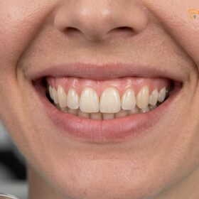
A 28-year-old female patient visited HCMC Dental Clinic in Ho Chi Minh City presenting with altered passive eruption, resulting in short, square-looking teeth and a prominent gummy smile. Under the expert care of Dr. Nguyen Van Cuong, clinical sounding confirmed adequate bone levels, allowing for a purely soft-tissue approach. A full-arch laser gum contouring was performed using the 810nm diode laser. The procedure was completed in 45 minutes with zero bleeding. The patient reported a pain score of 1/10 the following day and achieved a beautifully proportioned, harmonious smile that dramatically enhanced her facial aesthetics.
Advanced Cases: Addressing Skeletal and Muscular Factors
When excessive gingival display stems from vertical maxillary excess or a hyperactive levator muscle, soft tissue lasers alone are insufficient, requiring orthognathic or neuromodulator interventions.
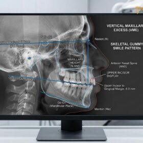
While laser gingivoplasty is highly effective for soft tissue discrepancies, a comprehensive diagnosis must rule out underlying skeletal or muscular etiologies. A gummy smile is not always a gum problem. In cases of Vertical Maxillary Excess (VME), the entire upper jaw bone has grown too far downward. No amount of laser sculpting can correct this severe skeletal discrepancy without exposing the root surfaces of the teeth. For these complex cases, HCMC Dental Clinic relies on its Surgical Jaw Partners. The clinic works in direct collaboration with elite maxillofacial specialists at the National Hospital of Odonto-Stomatology in Ho Chi Minh City (Bệnh viện Răng Hàm Mặt Trung Ương TP.HCM), the highest clinical authority in the region. [4]
Treatment for severe VME involves a Le Fort I osteotomy, a major orthognathic surgery where the maxilla is sectioned, impacted upward, and stabilized with titanium plates. This physically raises the entire dental arch, permanently eliminating the gummy smile at its skeletal root. While invasive, it provides life-changing functional and aesthetic results for patients with severe facial disproportions.
Clinical Warning: Misdiagnosis of Gummy Smiles
Attempting to treat a skeletal gummy smile (VME) with aggressive laser gingivectomy will result in severe root exposure, extreme dentin hypersensitivity, and compromised structural integrity of the teeth. Accurate cephalometric and periodontal diagnosis is mandatory before initiating any irreversible tissue removal.
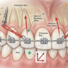
Another common non-gingival cause of excessive gum display is a hyperactive levator muscle (specifically the levator labii superioris alaeque nasi). In these patients, the gum and bone anatomy are perfectly normal, but the upper lip pulls up too high when smiling. This dynamic issue can be managed non-surgically with Botox Gummy Smile Correction. By injecting a precise dose of botulinum toxin into the hyperactive muscles, the lip elevation is temporarily weakened, allowing the lip to rest lower and cover the gums during a full smile. Alternatively, a Surgical Lip Repositioning procedure can be performed to permanently restrict the upward movement of the lip by removing a strip of mucosa from the maxillary vestibule and suturing the lip to a lower position. [5]
When to See a Doctor for Gingival Evaluation
A professional periodontal evaluation is necessary if you experience uneven gum lines, excessive gingival display, or suspect underlying periodontal disease that requires clinical intervention.
Not every patient is an immediate candidate for aesthetic laser contouring. It is crucial to seek a professional clinical evaluation if you notice persistent redness, bleeding during brushing, or if your teeth appear unusually short (a condition known as delayed passive eruption). A qualified periodontist or cosmetic dentist must assess your periodontal biotype—whether your gums are thick and flat or thin and scalloped—as this dictates the surgical approach and healing predictability.
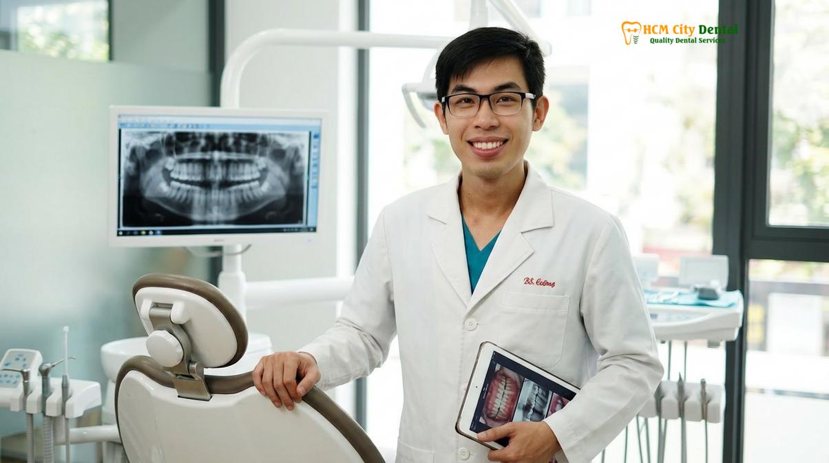
“A comprehensive periodontal evaluation is mandatory before any aesthetic gingival surgery to rule out active periodontitis and assess the underlying osseous crest topography.”
Contraindications for immediate laser gingivoplasty include active periodontal disease, inadequate zones of attached keratinized gingiva, and poor oral hygiene. If a patient undergoes laser contouring while suffering from active periodontitis, the risk of postoperative infection and unpredictable tissue sloughing increases exponentially. Patients experiencing any signs of postoperative complications from previous surgeries should immediately consult the Gingivectomy Infection Signs: Clinical Warnings & Healing Guide. Always prioritize a thorough diagnostic consultation, including periodontal probing and radiographic imaging, to ensure that aesthetic enhancements are built upon a foundation of absolute oral health. For those managing post-procedural discomfort, the Gum Contouring Pain: Clinical Recovery Guide & Management provides essential clinical advice.
Frequently Asked Questions
Find evidence-based answers to the most common patient inquiries regarding laser gingivoplasty, recovery timelines, and procedural expectations.
How long does a gingivoplasty session take?
A standard laser gingivoplasty session typically takes 30 to 60 minutes, depending on the number of teeth being contoured. Because the diode laser cuts and coagulates simultaneously, the procedure is highly efficient. Patients undergo digital smile mapping and local anesthesia before the rapid laser ablation begins, allowing for a swift, single-visit treatment that fits easily into a busy schedule.
Is anesthetic required for a laser gingivoplasty?
Yes, local anesthesia is required to ensure a completely comfortable and painless experience during the laser ablation process. While soft tissue lasers are far gentler than traditional scalpels, numbing the gingival margins prevents any thermal discomfort. The anesthesia wears off within a few hours, after which patients rarely report significant pain, usually managing any mild tenderness with standard over-the-counter ibuprofen.
How does it heal compared to traditional tools?
Laser gingivoplasty heals significantly faster than traditional scalpel surgery, typically within 48 to 72 hours, with virtually no bleeding or need for sutures. The laser’s photothermal effect instantly seals blood vessels and nerve endings, drastically reducing postoperative swelling and pain while creating a sterile wound environment that accelerates epithelialization and minimizes the risk of infection.
Can I combine this procedure with dental tourism in Vietnam?
Absolutely. Because laser gum contouring requires minimal downtime and offers a rapid 48-hour recovery, it is an ideal procedure for international patients engaging in dental tourism. Patients can have their smile aesthetically enhanced in a single visit and comfortably enjoy their travel itinerary in Ho Chi Minh City shortly after, taking advantage of world-class technology at highly competitive pricing.
Will the gum tissue grow back after the procedure?
When performed correctly by respecting the biological width and underlying bone crest, the contoured gum tissue will not grow back. However, if the supracrestal tissue attachment is violated without performing necessary bone recontouring (osteoplasty), the body may respond with chronic inflammation or tissue rebound. Proper clinical sounding prevents this, ensuring permanent, stable aesthetic results.
References
- Journal of Clinical Periodontology. Esthetic crown lengthening outcomes and biological width. (2021).
- International Journal of Periodontics & Restorative Dentistry. Diode laser vs scalpel gingivectomy: A comparative clinical trial. (2020).
- Journal of Aesthetic and Restorative Dentistry. Management of gingival zenith points in digital smile design. (2019).
- Journal of Oral and Maxillofacial Surgery. Le Fort I osteotomy stability in vertical maxillary excess. (2022).
- Clinical Oral Investigations. Surgical lip repositioning longevity and patient satisfaction. (2018).