Correcting a gummy smile through TADs intrusion involves using temporary anchorage devices (mini-screws) to apply upward orthopedic force, intruding the maxillary anterior teeth into the alveolar bone. This minimally invasive biomechanical approach effectively reduces excessive gingival display without requiring complex orthognathic surgery.
Clinical Summary:
TADs (Temporary Anchorage Devices) have revolutionized the management of vertical maxillary excess and deep overbites. By providing absolute skeletal anchorage, orthodontists can achieve true vertical orthodontic intrusion of the maxillary segment. This technique mitigates the need for highly invasive Le Fort I osteotomies in mild to moderate cases. When combined with AMD Picasso Lasers for soft tissue zenith calibration, clinicians can predictably restore the biological width and optimize the supracrestal tissue attachment. Dr. Nguyen Van Cuong emphasizes that precise force vectors and digital smile design (DSD) are critical for achieving stable, esthetic outcomes in gummy smile correction.
Key Takeaways:
- TADs provide absolute skeletal anchorage, preventing unwanted extrusion of posterior teeth during anterior intrusion.
- Vertical orthodontic intrusion can reduce gingival display by 2 to 5 millimeters depending on alveolar bone density.
- Biomechanical force vectors must pass near the center of resistance of the maxillary anterior segment for true translation.
- Soft tissue recontouring with an 810nm diode laser often complements TADs to finalize gingival zenith points.
- Severe skeletal discrepancies may still require collaboration with maxillofacial surgical jaw partners for optimal results.
The Biomechanics of Orthodontic Intrusion
Orthodontic intrusion using TADs relies on precise force vectors directed through the center of resistance of the maxillary anterior teeth to achieve true vertical displacement.
The correction of excessive gingival display, commonly known as a gummy smile, has historically presented a significant biomechanical challenge in orthodontics. Traditional mechanics utilizing continuous archwires or utility arches often result in reciprocal extrusion of the posterior teeth. When the posterior teeth extrude, the mandible rotates downward and backward, increasing the lower anterior facial height and potentially worsening the aesthetic profile. The introduction of TADs dental biomechanics has fundamentally altered this paradigm by decoupling the anterior intrusion forces from the posterior reactive forces[1].
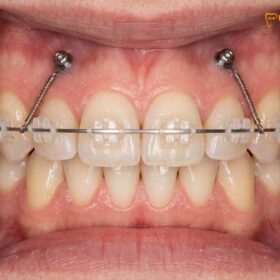
To achieve true vertical orthodontic intrusion, the applied force must pass directly through or very close to the center of resistance of the target teeth. For the six maxillary anterior teeth (canine to canine), the center of resistance is typically located apically and distally to the roots of the central incisors. When temporary anchorage devices are placed high in the alveolar mucosa—often between the lateral incisors and canines, or between the premolars—they serve as fixed points from which elastomeric chains or nickel-titanium coil springs can deliver a continuous, light force.
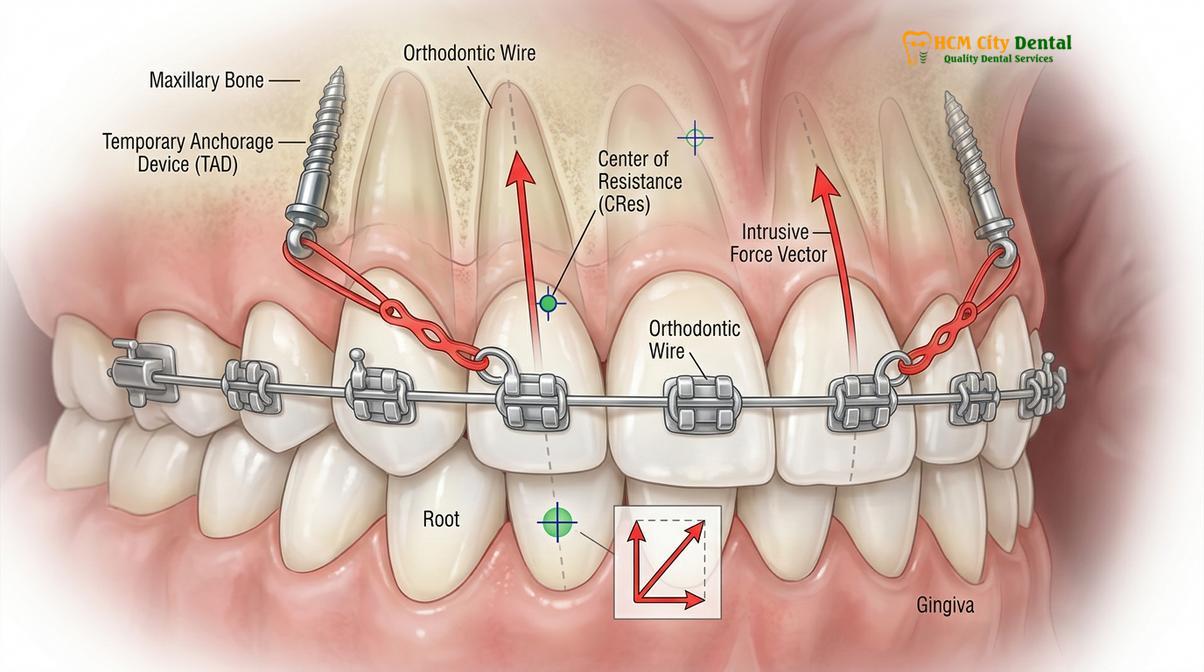
Clinical protocols generally dictate that the optimal force magnitude for intruding anterior teeth ranges from 40 to 60 grams per tooth. Therefore, intruding the entire anterior segment requires a carefully calibrated force of approximately 200 to 250 grams distributed bilaterally. Applying excessive force does not accelerate tooth movement; rather, it increases the risk of root resorption, periodontal ligament hyalinization, and loss of vitality. The alveolar bone undergoes a complex process of osteoclastic resorption at the apex and osteoblastic deposition at the crest, allowing the entire dentoalveolar complex to move superiorly[2].
“The predictability of anterior intrusion relies entirely on controlling the moment-to-force ratio. By utilizing skeletal anchorage, we eliminate the reciprocal posterior extrusion that historically compromised our vertical control in gummy smile patients.”
Furthermore, the use of segmented archwires in conjunction with TADs allows for simultaneous intrusion and retraction. By altering the angle of the force vector relative to the archwire, clinicians can resolve the force into vertical (intrusive) and horizontal (retractive) components. This is particularly beneficial for patients presenting with both a gummy smile and bimaxillary protrusion, allowing for comprehensive three-dimensional control of the anterior segment.
How TADs Provide Absolute Anchorage in the Alveolar Bone
By integrating directly into the cortical bone, temporary anchorage devices bypass the periodontal ligament, offering an immovable anchor point for complex orthodontic movements.
Anchorage in orthodontics is defined as the resistance to unwanted tooth movement. According to Newton’s third law of motion, every applied force generates an equal and opposite reactive force. In traditional orthodontics, teeth are pitted against other teeth, meaning that moving the front teeth backward or upward inevitably pulls the back teeth forward or downward. Mini implant anchorage solves this dilemma by utilizing the rigid cortical bone of the maxilla or mandible to absorb the reactive forces, providing what is clinically termed “absolute anchorage.”
TADs are typically manufactured from medical-grade titanium alloy (Ti-6Al-4V), which offers excellent biocompatibility and mechanical strength. Unlike traditional dental implants designed for prosthetic replacement, TADs are not intended to undergo complete osseointegration. Instead, they rely on mechanical retention—primary stability—achieved through the interlocking of the screw threads with the dense cortical bone. This design allows them to withstand immediate orthodontic loading while ensuring they can be easily unscrewed and removed once the treatment objectives are met[3].
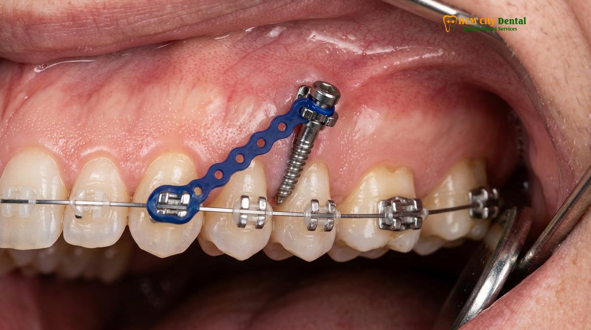
The anatomical placement of gummy smile mini screws requires meticulous planning, often aided by Cone Beam Computed Tomography (CBCT). The most common insertion sites for maxillary intrusion include the interradicular spaces between the second premolars and first molars, the anterior region between the lateral incisors and canines, and extra-alveolar sites such as the infrazygomatic crest. The choice of site depends on the required force vector, the volume of available bone, and the proximity to adjacent tooth roots and the maxillary sinus.
| Feature | Traditional Anchorage (Teeth/Headgear) | TADs (Skeletal Anchorage) |
|---|---|---|
| Anchorage Source | Periodontal ligament of anchor teeth | Cortical alveolar bone |
| Reactive Movement | High risk of posterior extrusion/mesialization | Zero reactive tooth movement (Absolute) |
| Patient Compliance | High (requires wearing elastics or headgear) | None required after placement |
| Intrusion Capacity | Limited (1-2mm) with side effects | Significant (2-5mm) true vertical intrusion |
| Invasiveness | Non-invasive | Minimally invasive (micro-screw insertion) |
During the insertion procedure, the orthodontist will apply a topical anesthetic followed by a minimal amount of local infiltration. The TAD is then driven through the attached gingiva and into the bone using a specialized manual driver or a low-speed handpiece. The entire process takes only a few minutes per screw and is generally well-tolerated by patients, with post-operative discomfort being minimal and easily managed with over-the-counter analgesics.
Minimizing Upper Jaw Display Without Surgical Osteotomy
TAD-supported intrusion offers a conservative alternative to orthognathic surgery for patients with mild to moderate vertical maxillary excess, significantly reducing gingival display.
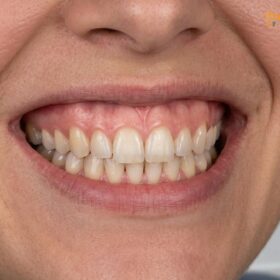
The etiology of a gummy smile is multifactorial and must be accurately diagnosed before initiating treatment. Causes include altered passive eruption (where the gums fail to recede normally during childhood), a hyperactive levator muscle of the upper lip, a short upper lip, and Vertical Maxillary Excess (VME). Historically, patients diagnosed with VME had only one definitive treatment option: a Le Fort I osteotomy with superior impaction. This major orthognathic surgery involves fracturing the upper jaw, removing a wedge of bone, and repositioning the maxilla upward using titanium plates and screws.
While Le Fort I osteotomy remains the gold standard for severe skeletal discrepancies, the advent of TADs has created a powerful middle ground. For patients exhibiting 3 to 6 millimeters of excessive gingival display, vertical orthodontic intrusion can often achieve highly satisfactory aesthetic results without the risks, costs, and extended recovery times associated with jaw surgery[4]. By intruding the maxillary dentoalveolar complex, the upper lip drapes more naturally over the teeth, reducing the amount of gum tissue exposed during a full smile.
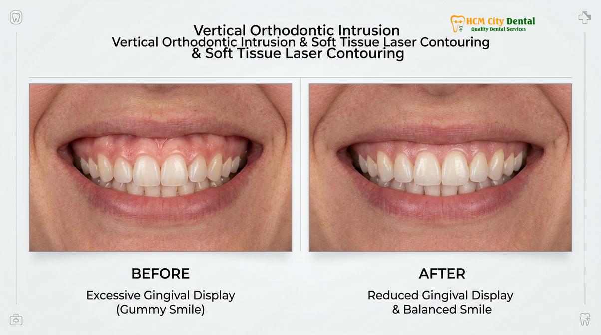
However, it is crucial to recognize the limitations of orthodontic intrusion. Dr. Nguyen Van Cuong notes that while TADs can successfully intrude the teeth and alveolar bone, they do not alter the basal skeletal structures of the face. Therefore, patients with severe VME (exceeding 7-8mm of gingival display), significant facial asymmetry, or severe obstructive sleep apnea may still require surgical intervention. For these complex cases, the clinic works in close collaboration with highly esteemed Surgical Jaw Partners at the National Hospital of Odonto-Stomatology in Ho Chi Minh City (Bệnh viện Răng Hàm Mặt Trung Ương TP.HCM), ensuring patients receive the highest standard of maxillofacial surgical care.
Clinical Case Review: Non-Surgical Gummy Smile Correction
A 26-year-old patient visited HCMC Dental Clinic in Ho Chi Minh City presenting with a 5mm gingival display upon smiling and a Class II deep bite. Instead of opting for orthognathic surgery, the patient underwent a comprehensive treatment plan utilizing fixed orthodontic appliances and four maxillary TADs. Over a period of 14 months, a continuous intrusive force of 200g was applied. The treatment successfully intruded the maxillary anterior segment by 4mm, completely resolving the deep bite and reducing the gummy smile to a highly aesthetic 1mm display, demonstrating the profound efficacy of skeletal anchorage.
The decision between TADs and surgery also involves evaluating the patient’s periodontal biotype and root morphology. Intruding teeth into dense cortical bone requires healthy periodontal tissues. Prior to any intrusion mechanics, patients must undergo thorough dental scaling and periodontal evaluation to ensure the absence of active inflammation, which could exacerbate bone loss during active tooth movement.
Integrating Laser Gum Contouring for Optimal Esthetics
Following orthodontic intrusion, diode laser gingivectomy refines the soft tissue architecture, establishing ideal zenith points and respecting the biological width.
While TADs successfully reposition the underlying bone and teeth, the soft tissue does not always follow the bone in a 1:1 ratio. After the completion of vertical orthodontic intrusion, patients may still present with uneven gingival margins, blunted papillae, or residual altered passive eruption. To achieve the ultimate aesthetic result, orthodontic treatment is frequently followed by soft tissue refinement using advanced laser technology.
At our facility, clinicians utilize AMD Picasso Lasers (Diode soft tissue laser, 810nm) to perform highly precise gingivectomies and gingivoplasties. The 810nm wavelength is highly absorbed by melanin and hemoglobin, making it exceptionally efficient for cutting oral soft tissues while simultaneously providing instant coagulation and cauterization. This results in bloodless, sutureless cuts, significantly reduced postoperative pain and swelling, and a rapid 48-hour recovery period. Furthermore, the sterile nature of the laser beam minimizes the risk of postoperative infection.
“The synergy between skeletal intrusion and laser soft tissue management is the cornerstone of modern smile design. The laser allows us to sculpt the gingival zenith points with micro-millimeter precision, framing the newly positioned teeth perfectly.”
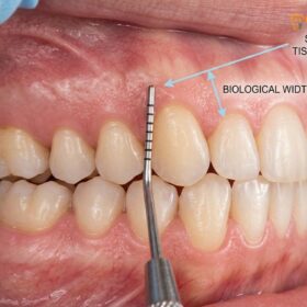
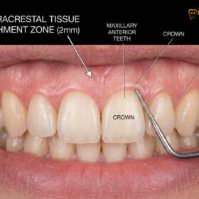
During this phase, the clinician must strictly respect the biological width—the minimum 2mm of supracrestal tissue attachment (comprising the junctional epithelium and connective tissue) required to prevent chronic inflammation and unpredictable bone resorption. If the required soft tissue removal encroaches upon this biological width, a simple laser gingivectomy is insufficient. In such cases, functional or esthetic crown lengthening involving osteoplasty (bone recontouring) is necessary to re-establish the proper distance between the alveolar crest and the gingival margin.
For patients considering these procedures, especially those engaging in dental tourism, understanding the financial investment is important. According to the latest clinic fee schedule, the pricing structure is highly transparent:
- Laser Gingivectomy (per tooth): 1,500,000 VND (~$60 USD) (Walk-in: 2,500,000 VND).
- Full-Arch Laser Gum Contouring (up to 10 teeth): 12,000,000 VND (~$480 USD) (Walk-in: 20,000,000 VND).
- Functional/Esthetic Crown Lengthening (per tooth, including bone contouring): 3,000,000 VND (~$120 USD) (Walk-in: 5,000,000 VND).
- Surgical Lip Repositioning / Lip Lowering: 15,000,000 VND (~$600 USD) (Walk-in: 25,000,000 VND).
- Botox Gummy Smile Correction (both sides): 4,500,000 VND (~$180 USD) (Walk-in: 7,500,000 VND).
Patients booking online or via messaging platforms can often take advantage of a -40% WhatsApp booking discount. For a comprehensive overview of these aesthetic procedures, patients are encouraged to explore the Gum Contouring & Gummy Smile services available.
Hygiene, Maintenance, and Risks of TADs During Braces Treatment
Meticulous peri-implant hygiene is critical to prevent localized inflammation, which is the primary cause of premature TAD failure during orthodontic treatment.
The success of temporary anchorage devices is heavily dependent on the health of the surrounding soft tissues. Because TADs protrude through the oral mucosa, they create a pathway where dental plaque and bacteria can accumulate. If plaque is allowed to build up around the head of the mini-screw, it can lead to peri-implant mucositis—a localized inflammation of the gums. If left untreated, this inflammation can progress down the threads of the screw, compromising the mechanical retention in the cortical bone and leading to premature loosening and failure of the device[5].
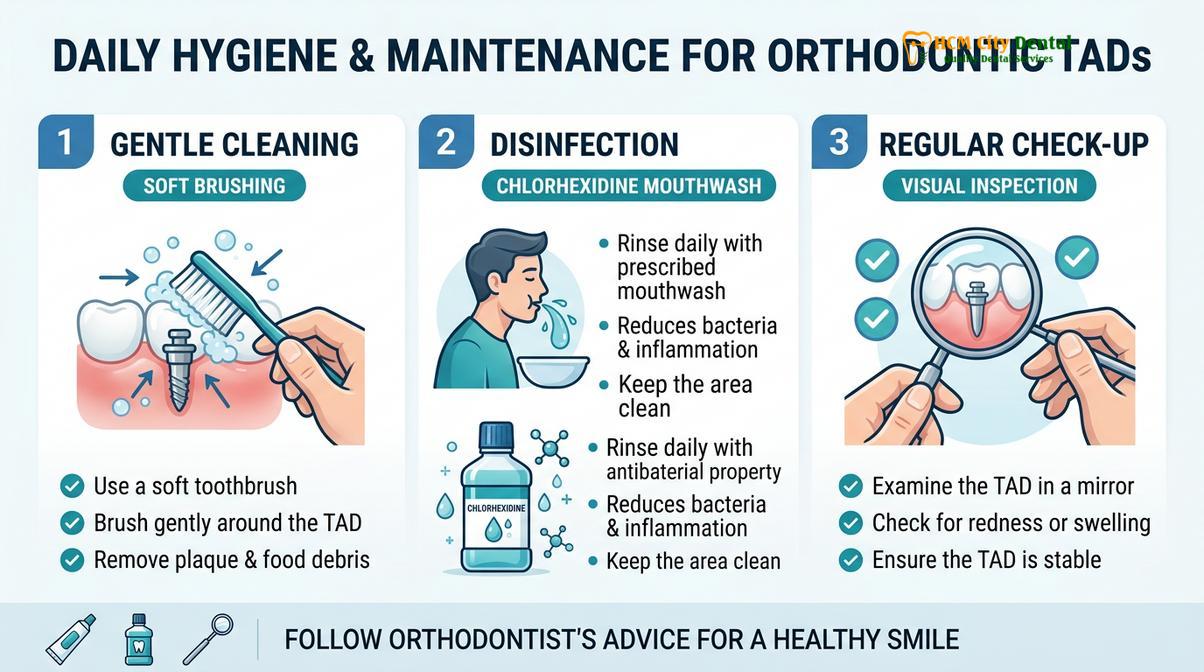
Patients must adopt a rigorous daily hygiene protocol. This includes using a soft-bristled toothbrush or an interdental brush to gently clean around the TAD head, ensuring that all food debris and plaque are removed. Vigorously scrubbing the area should be avoided, as excessive mechanical trauma can irritate the mucosa and destabilize the screw. In addition to mechanical cleaning, the use of a chlorhexidine gluconate mouthwash (typically 0.12%) applied locally with a cotton swab can significantly reduce bacterial load and promote tissue health.
Important Clinical Warning: Signs of TAD Failure
Patients should monitor their TADs regularly. If you experience persistent pain, noticeable mobility of the mini-screw, swelling, or purulent discharge (pus) around the insertion site, contact your orthodontist immediately. While slight tenderness for the first 24-48 hours after placement is normal, ongoing discomfort indicates potential peri-implant inflammation or root proximity issues that require prompt clinical evaluation.
Beyond hygiene-related failures, other clinical risks include root proximity. If a TAD is placed too close to the root of an adjacent tooth, it can cause localized root damage or impede the desired orthodontic movement. This is why Dr. Nguyen Van Cuong and the orthodontic team at HCMC Dental Clinic rely heavily on 3D CBCT imaging to map out the safest interradicular corridors prior to insertion. Should a TAD become loose during treatment, it is typically removed, the site is allowed to heal for a few weeks, and a new device is placed in an adjacent location to continue the biomechanical force application.
When to See a Doctor for Gummy Smile Correction
Determining the appropriate treatment for a gummy smile requires a comprehensive diagnostic approach. You should seek a professional consultation if your excessive gingival display causes you aesthetic concern, affects your self-confidence, or is accompanied by functional issues such as a deep overbite, lip incompetence (inability to close your lips comfortably at rest), or excessive tooth wear.
Because the etiology can range from a hyperactive levator muscle to altered passive eruption or skeletal vertical maxillary excess, a one-size-fits-all approach is ineffective. A qualified dental professional will utilize digital smile design (DSD), cephalometric analysis, and periodontal sounding to determine the exact cause of your condition. Based on these clinical diagnostics, they will recommend a personalized treatment plan—whether that involves Botox, laser gingivectomy, TAD-supported orthodontic intrusion, or a referral to maxillofacial surgical partners. Always consult with a certified specialist to understand the candidacy, scope, and potential contraindications of each procedure.
Frequently Asked Questions
Are TADs safe?
Yes, TADs are highly safe when placed by a qualified orthodontist using proper anatomical guidelines. They are made of biocompatible medical-grade titanium alloy, minimizing the risk of allergic reactions, and are placed in safe interradicular zones to avoid root damage. The procedure is minimally invasive, and complications are rare when patients adhere to prescribed oral hygiene protocols.
How long do TADs stay in my gums?
TADs typically remain in your gums for 6 to 12 months, depending on the complexity of the vertical orthodontic intrusion required. Once the maxillary anterior teeth have been sufficiently intruded and the gummy smile is corrected, the devices are promptly removed. They are only meant to provide temporary anchorage during the active phase of your orthodontic treatment.
Is local anesthesia required to remove TADs?
No, local anesthesia is rarely required to remove TADs. Because they do not osseointegrate like traditional dental implants, the removal process is incredibly fast and usually only requires a topical numbing gel to ensure complete patient comfort. The small puncture wound heals rapidly, often closing completely within 24 to 48 hours.
Can TADs completely replace jaw surgery for a gummy smile?
TADs can replace jaw surgery for mild to moderate cases of vertical maxillary excess (typically 3-6mm of display). However, severe skeletal discrepancies may still require a Le Fort I osteotomy performed by maxillofacial surgical specialists. A comprehensive cephalometric and clinical evaluation is necessary to determine if skeletal anchorage alone is sufficient to meet your aesthetic and functional goals.
Does laser gum contouring hurt after TAD removal?
Laser gum contouring is virtually painless due to the use of advanced diode lasers that seal nerve endings as they cut. Patients typically only require a mild local anesthetic during the procedure and experience a rapid, comfortable 48-hour recovery. The laser’s ability to instantly coagulate blood vessels means there is minimal postoperative swelling and no need for traditional sutures.
References
- American Journal of Orthodontics and Dentofacial Orthopedics. Biomechanics of true maxillary incisor intrusion using TADs. (2021).
- Journal of Clinical Periodontology. Supracrestal tissue attachment and biological width in aesthetic dentistry. (2020).
- International Journal of Periodontics & Restorative Dentistry. Diode laser vs scalpel for gingivectomy: Healing outcomes. (2019).
- Journal of Oral and Maxillofacial Surgery. Stability of Le Fort I osteotomy in vertical maxillary excess. (2022).
- Clinical Oral Investigations. Long-term stability of skeletal anchorage devices in orthodontics. (2018).