A laser gingivectomy in Saigon is a minimally invasive periodontal procedure utilizing advanced diode lasers to precisely reshape excess gum tissue. This bloodless, sutureless technique instantly coagulates the tissue, offering a rapid 48-hour recovery while effectively correcting gummy smiles and restoring optimal dentogingival aesthetics.
Clinical Summary:
Laser gingivectomy represents the gold standard in modern periodontal aesthetics, replacing traditional scalpel techniques with highly focused light energy. Utilizing systems like the AMD Picasso Laser (810nm), clinicians can vaporize excess gingival tissue while simultaneously sealing blood vessels and nerve endings. This dual-action process ensures a completely bloodless surgical field, eliminates the need for sutures, and drastically reduces postoperative edema. The procedure is highly effective for treating altered passive eruption, gingival hyperplasia, and asymmetrical gum lines. Success relies heavily on precise diagnostic sounding to respect the supracrestal tissue attachment (biological width) and accurate calibration of gingival zenith points. When combined with digital smile design, laser gum contouring delivers predictable, permanent, and highly aesthetic outcomes with minimal patient downtime.
Key Takeaways:
- Utilizes 810nm diode lasers for precise, bloodless, and sutureless tissue ablation.
- Offers a rapid 48-hour initial recovery phase with minimal postoperative discomfort.
- Requires strict adherence to biological width principles to prevent tissue rebound or bone loss.
- Highly effective for correcting asymmetrical gum lines and mild to moderate gummy smiles.
- Severe skeletal cases may require collaborative orthognathic surgery (Le Fort I osteotomy).
What is a Laser Gingivectomy and How Does It Work?
A laser gingivectomy utilizes focused light energy to vaporize excess gingival tissue, reshaping the gum line without traditional scalpels. This process relies on precise biological width calibration to ensure long-term periodontal health and aesthetic harmony.
The architecture of a beautiful smile is dictated not only by the shape and color of the teeth but also by the framing provided by the gingival tissues. A laser gingivectomy is a highly specialized periodontal procedure designed to excise and reshape excess or asymmetrical gum tissue. Unlike traditional methods that rely on steel blades, this modern approach harnesses the power of concentrated light energy to perform precise soft tissue ablation. The primary goal is to expose the natural anatomical crown of the tooth, which may be hidden due to conditions such as altered passive eruption, medication-induced gingival hyperplasia, or genetic predispositions that result in a “gummy smile.”
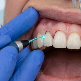
At the core of this procedure is the advanced physics of soft tissue dental lasers. The most frequently utilized device in premium clinical settings is the AMD Picasso Laser, a diode laser operating at an 810nm wavelength. This specific wavelength is highly absorbed by melanin and hemoglobin, making it exceptionally efficient for cutting highly vascularized oral tissues. As the laser energy interacts with the gingiva, it causes rapid intracellular vaporization, effectively “erasing” the excess tissue layer by layer. Simultaneously, the thermal energy coagulates the surrounding proteins, sealing capillaries and lymphatic vessels instantly. This creates a sterile, bloodless surgical field that provides the clinician with unparalleled visibility and control.[1]
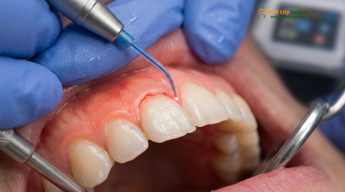
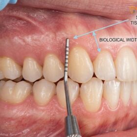
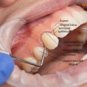
However, the success of a laser gingivectomy is not solely dependent on the technology; it requires a profound understanding of periodontal microanatomy. The most critical concept in gum contouring is the preservation of the supracrestal tissue attachment, historically referred to as the biological width. This dimension, typically measuring around 2.04 millimeters, comprises the junctional epithelium and the connective tissue attachment that sit above the alveolar bone crest. If a laser gingivectomy removes tissue too close to the bone, violating this attachment zone, the body will respond with chronic inflammation, unpredictable tissue rebound, or pathological bone resorption. Therefore, meticulous bone sounding is mandatory prior to any tissue removal to ensure that the final gingival margin remains at a safe physiological distance from the osseous crest.[2]
Furthermore, aesthetic success hinges on the precise calibration of the gingival zenith points. The zenith is the most apical (highest) point of the free gingival margin’s scallop. In a naturally aesthetic smile, the zenith of the maxillary central incisors and canines is displaced slightly distal to the long axis of the tooth, while the lateral incisors have a more centrally located zenith. During a laser gingivectomy, the clinician uses the laser tip like a micro-paintbrush to sculpt these zenith points, ensuring bilateral symmetry and a harmonious flow that complements the patient’s facial features and lip dynamics.
Diode Soft Tissue Lasers vs. Traditional Surgical Scalpels
Diode soft tissue lasers offer superior clinical advantages over traditional scalpels by providing instant cauterization, eliminating the need for sutures, and significantly reducing postoperative edema and infection risks.
The transition from traditional scalpel-based periodontics to laser-assisted surgery represents one of the most significant advancements in modern dentistry. For decades, the standard protocol for removing excess gum tissue involved the use of Kirkland or Orban periodontal knives, or standard surgical scalpels. While effective, these traditional methods inherently cause significant tissue trauma. Severing blood vessels with a cold steel blade results in immediate and often profuse bleeding, which obscures the surgical field and makes micro-aesthetic adjustments difficult. Furthermore, traditional gingivectomies leave raw, open connective tissue wounds that require the placement of periodontal dressings (gum packs) and sutures to manage bleeding and protect the site during the lengthy healing process.
In stark contrast, the diode laser fundamentally alters the surgical experience for both the clinician and the patient. Because the 810nm wavelength of the AMD Picasso Laser specifically targets the chromophores in the blood and tissue, it cuts and coagulates simultaneously. The thermal relaxation time of the tissue is carefully managed by the laser’s pulsed settings, ensuring that the zone of thermal necrosis (collateral heat damage) is kept to an absolute minimum—often less than 0.1 millimeters. This precision allows for what is commonly referred to as a painless gingivectomy, as the laser energy effectively seals the severed nerve endings, preventing the transmission of pain signals during the postoperative phase.
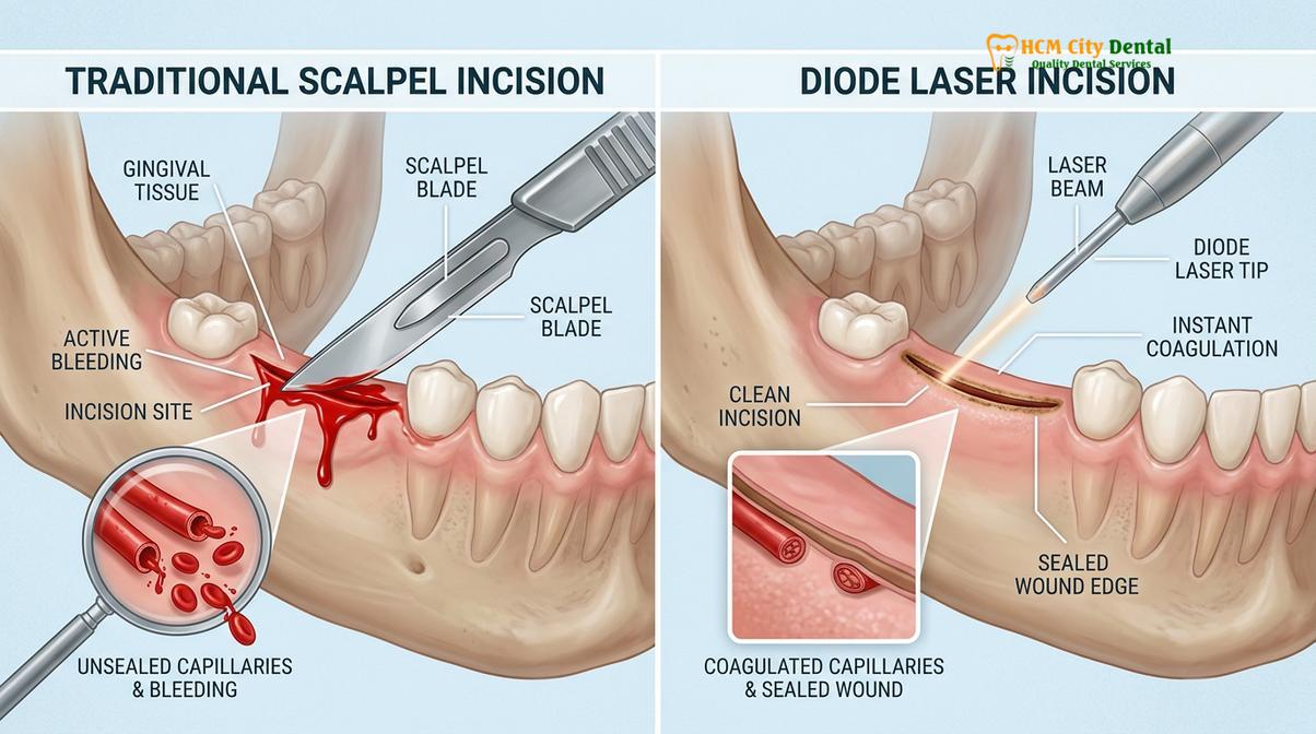
To clearly illustrate the clinical differences, the following table outlines the comparative metrics between diode laser ablation and traditional scalpel surgery:
| Clinical Parameter | Diode Soft Tissue Laser (810nm) | Traditional Surgical Scalpel |
|---|---|---|
| Hemostasis (Bleeding) | Instant coagulation; completely bloodless field. | Significant bleeding requiring manual control. |
| Sutures & Dressings | Sutureless; no periodontal dressing required. | Sutures and protective gum packs often necessary. |
| Infection Risk | Extremely low; the laser beam sterilizes the pocket. | Moderate; open wound susceptible to oral bacteria. |
| Postoperative Pain | Minimal to none; nerve endings are thermally sealed. | Moderate discomfort requiring analgesics. |
| Precision & Control | Micro-millimeter sculpting capabilities. | Limited by blade size and obscured visibility. |
Another profound advantage of the laser is its bactericidal property. The high-energy light beam effectively sterilizes the surgical site as it cuts, destroying periodontal pathogens residing in the gingival sulcus. This significantly reduces the risk of postoperative infections and promotes a cleaner, faster healing cascade. For patients seeking aesthetic gum trimming, the psychological benefit of avoiding scalpels, stitches, and bleeding cannot be overstated. The laser transforms a traditionally daunting surgical procedure into a streamlined, minimally invasive cosmetic enhancement.
“The true artistry in periodontal plastic surgery lies in the micro-millimeter adjustments. The diode laser provides us with a bloodless canvas, allowing for the precise calibration of gingival zenith points that a traditional scalpel simply cannot achieve without obscuring the field.”
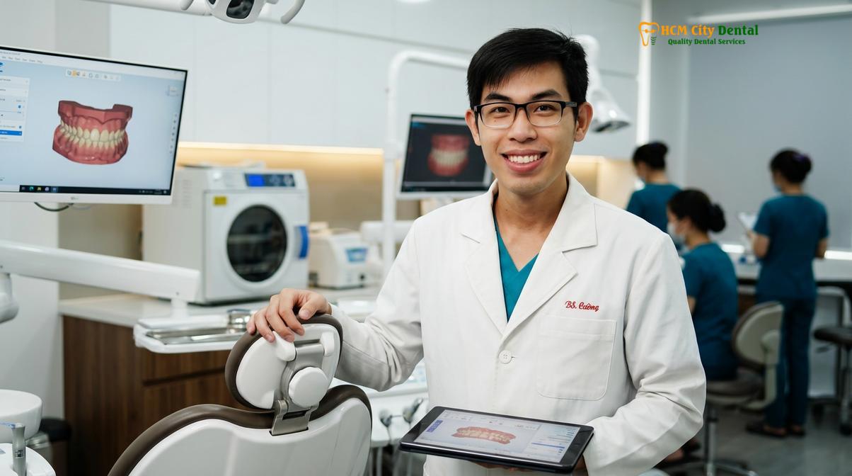
— Dr. Nguyen Van Cuong, Clinical Director
Step-by-Step Clinical Procedure at HCMC Dental Clinic
The clinical workflow for laser gum contouring involves comprehensive digital smile design, precise biological width sounding, and targeted laser ablation to achieve symmetrical, aesthetic gingival margins.
Achieving a flawless, natural-looking result requires a meticulous, multi-step clinical protocol. At HCMC Dental Clinic, the workflow for a laser gingivectomy is heavily rooted in comprehensive diagnostics and digital planning. The process begins with a detailed clinical examination and Digital Smile Design (DSD). High-resolution intraoral and extraoral photographs are taken to analyze the patient’s facial midline, interpupillary line, and the dynamic movement of the upper lip during a full smile. This digital analysis allows the clinician to virtually design the ideal gingival architecture before any physical treatment begins, ensuring that the proposed changes will harmonize with the patient’s unique facial proportions.
Once the aesthetic goals are established, the critical phase of periodontal diagnostics commences. The clinician performs bone sounding under local anesthesia. Using a specialized periodontal probe, the dentist measures the exact distance from the free gingival margin to the underlying alveolar bone crest. This step is non-negotiable, as it determines whether the patient is a candidate for a simple soft-tissue laser gingivectomy or if they require a more complex procedure. If there is more than 3 millimeters of tissue above the bone, a simple laser excision is safe. However, if the bone is too close to the cementoenamel junction (CEJ), removing gum tissue alone would violate the supracrestal tissue attachment. In such cases, a Functional or Esthetic Crown Lengthening procedure is required, which involves reflecting a flap and performing osteotomy (bone removal) and osteoplasty (bone reshaping) to re-establish the biological width at a more apical level.[3]
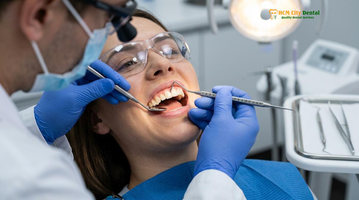
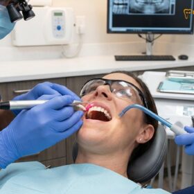
Prior to the surgical phase, thorough dental scaling and root planing are performed to remove any calculus or plaque deposits. The surgical environment must be pristine; operating on inflamed or infected gums compromises the healing process and the final aesthetic result. Once the tissues are healthy and profound local anesthesia is achieved, the laser ablation begins. The dentist utilizes the AMD Picasso Laser, typically set to a continuous or micro-pulsed mode depending on the tissue biotype. The laser tip is guided along the pre-marked zenith points, gently vaporizing the excess tissue. The clinician continuously wipes away the ablated tissue with a damp sterile gauze, revealing the newly sculpted, bloodless gingival margin.
In cases where a gummy smile is caused by severe skeletal discrepancies—specifically Vertical Maxillary Excess (VME), where the entire upper jaw has grown too far downward—soft tissue lasers and minor bone contouring are insufficient. For these complex orthognathic cases, HCMC Dental Clinic works in collaboration with elite maxillofacial surgical partners at the National Hospital of Odonto-Stomatology in Ho Chi Minh City. These severe cases require a Le Fort I osteotomy, a major hospital-based surgery where the maxilla is sectioned, impacted upward, and stabilized with titanium plates to correct the skeletal deformity at its source.[4]
Expected Healing Timelines and Sutureless Results
Patients undergoing laser gingivectomy typically experience a rapid 48-hour initial recovery phase, characterized by minimal discomfort and the complete absence of sutures, allowing for a swift return to normal activities.
The postoperative experience following a laser gingivectomy is remarkably mild, largely due to the atraumatic nature of the diode laser. Because the laser seals blood vessels and nerve endings simultaneously, the acute inflammatory response is significantly blunted. Within the first 24 to 48 hours, the treated gingival margins will form a protective fibrin clot. This thin, whitish-yellow layer is a normal and essential part of the histological healing cascade; it acts as a biological bandage over the vaporized tissue. Patients are strictly instructed not to brush or pick at this fibrin layer, as disturbing it can delay epithelialization and cause unnecessary sensitivity.
During this initial 48-hour window, patients may experience mild tenderness, similar to the sensation of a minor pizza burn on the roof of the mouth. This is easily managed with standard over-the-counter anti-inflammatory medications such as Ibuprofen. Because the procedure is entirely sutureless, there is no pulling or tension on the tissues, which further enhances patient comfort. Swelling is typically imperceptible to the naked eye. Patients are advised to maintain a soft diet, avoiding highly acidic, spicy, or sharp foods (like chips or crusty bread) that could mechanically traumatize the healing margins.
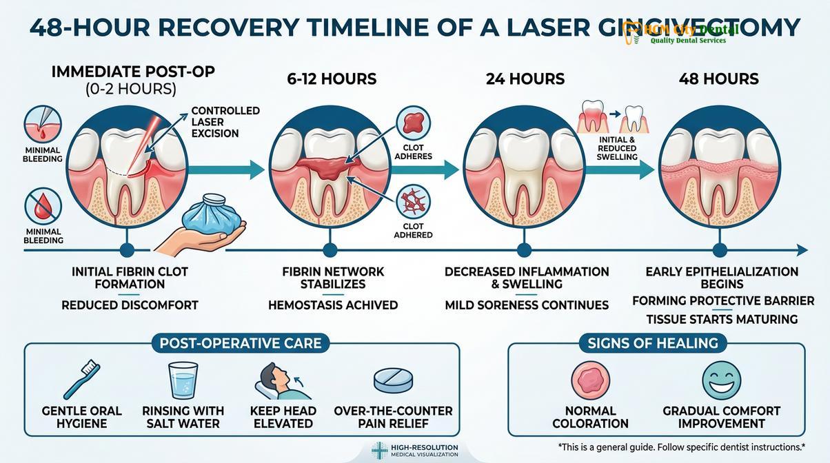
By the end of the first week, the fibrin clot sloughs off naturally, revealing newly formed, immature epithelial tissue that appears slightly redder than the surrounding gums. Over the subsequent three to four weeks, the connective tissue matures, and the gingiva regains its normal stippled texture and pale pink color. The final, stable position of the gingival margin is usually achieved by the sixth week post-operation. At this stage, the tissues have fully adapted to the new anatomical contours, and any planned restorative work, such as porcelain veneers or crowns, can be safely initiated without the risk of subsequent tissue shrinkage.
Clinical Case Study: Aesthetic Gum Trimming
A 28-year-old patient from Australia visited HCMC Dental Clinic as part of a dental tourism itinerary, presenting with a chief complaint of “short, square teeth” and an asymmetrical gum line. Clinical sounding confirmed adequate biological width, diagnosing the condition as delayed passive eruption. A comprehensive laser gums shaping HCMC procedure was performed on teeth #14 through #24 using the AMD Picasso Laser. The procedure was completed in under 45 minutes with zero bleeding. The patient reported no need for pain medication post-operatively and was able to enjoy the culinary offerings of Ho Chi Minh City the very next day. At the two-week follow-up, the gingival margins exhibited perfect symmetry and complete epithelial healing, dramatically enhancing the patient’s smile proportions.
Comprehensive Pricing Structure for Gum Contouring in Saigon
Ho Chi Minh City offers world-class periodontal treatments at highly competitive rates, providing transparent pricing for laser gingivectomy, crown lengthening, and comprehensive gummy smile correction procedures.
Vietnam has rapidly emerged as a premier destination for high-end dental tourism, offering advanced clinical technologies and elite specialist care at a fraction of the cost found in Western countries. For international patients and local expatriates alike, understanding the financial investment required for periodontal plastic surgery is a crucial step in the treatment planning process. The pricing structure is determined by the complexity of the diagnosis, the number of teeth involved, and whether the procedure requires soft tissue ablation alone or concurrent osseous (bone) recontouring.
According to the latest clinic fee schedule, the investment for a standard Laser Gingivectomy is 1,500,000 VND (approximately $60 USD) per tooth. For patients requiring comprehensive aesthetic correction across the entire visible smile zone, a Full-Arch Laser Gum Contouring package (covering up to 10 anterior teeth) is available at 12,000,000 VND (approximately $480 USD). It is important to note that these preferred rates are applicable for scheduled appointments; standard walk-in rates are higher (2,500,000 VND per tooth and 20,000,000 VND for the full arch, respectively). Patients are highly encouraged to utilize the -40% WhatsApp booking discount to secure these optimal rates prior to their arrival in Saigon.
When clinical sounding reveals that the alveolar bone is too close to the cementoenamel junction, a simple laser excision is contraindicated. In these instances, Functional or Esthetic Crown Lengthening is required. This more complex surgical procedure, which includes flap reflection and precise bone contouring (osteoplasty/osteotomy) to safely reposition the biological width, is priced at 3,000,000 VND (approximately $120 USD) per tooth (Walk-in: 5,000,000 VND). This ensures that the newly established gum line remains stable and free from chronic inflammation.
For patients whose gummy smile is primarily caused by muscular hypermobility rather than excess gum tissue, alternative therapies are available. A Botox Gummy Smile Correction, which temporarily relaxes the hyperactive levator muscle to prevent the upper lip from rising too high, is priced at 4,500,000 VND (approximately $180 USD) for both sides (Walk-in: 7,500,000 VND). For a more permanent muscular solution, Surgical Lip Repositioning (Lip Lowering) is offered at 15,000,000 VND (approximately $600 USD) (Walk-in: 25,000,000 VND). This procedure restricts the upward pull of the lip muscles, effectively masking the gingival display without altering the teeth or gums themselves.
When to See a Doctor (Important Clinical Considerations)
A professional clinical evaluation is essential to determine the exact etiology of a gummy smile, ensuring that the chosen treatment safely addresses soft tissue, muscular, or skeletal factors without compromising oral health.
While laser gum contouring is a highly safe and predictable procedure, it is not a universal cure for all types of excessive gingival display. The etiology of a gummy smile is multifactorial, and an accurate differential diagnosis is the cornerstone of successful treatment. Patients should seek a comprehensive evaluation from a qualified periodontist or cosmetic dentist to determine whether their condition is dentogingival (excess gums), muscular (hypermobile lip), or skeletal (vertical maxillary excess).
Clinical Contraindications & Warnings:
A laser gingivectomy should NOT be performed under the following conditions:
- Inadequate Keratinized Tissue: If removing the excess gum tissue would eliminate the protective band of attached, keratinized gingiva, leaving only fragile alveolar mucosa.
- Active Periodontitis: Untreated gum disease, characterized by deep periodontal pockets, bleeding, and bone loss, must be fully resolved before any cosmetic contouring is attempted.
- Biological Width Violation: If the distance from the proposed new gum line to the underlying bone is less than 3mm, a simple laser cut will cause chronic inflammation. Osseous surgery (crown lengthening) is mandatory in these cases.
If the excessive gingival display is caused by a hyperactive levator muscle—specifically the levator labii superioris alaeque nasi—the upper lip will retract excessively upon smiling, exposing normal-sized teeth and healthy gums. In these cases, a laser gingivectomy would inappropriately shorten the gums and expose the tooth roots. Instead, the clinical approach must target the musculature. Neuromodulator injections (Botox) offer a temporary, non-invasive solution by relaxing the elevator muscles. For a permanent anatomical correction, surgical lip repositioning is indicated. This procedure involves removing a strip of mucosa from the maxillary vestibule and suturing the lip to a lower position, physically restricting its upward mobility.[5]
Conversely, if the patient exhibits a significantly long lower third of the face and displays excessive gum tissue even when the lips are at rest, the underlying issue is likely skeletal Vertical Maxillary Excess (VME). Soft tissue lasers and lip repositioning cannot correct severe skeletal discrepancies. These complex cases require a multidisciplinary approach, combining orthodontics with orthognathic surgery to physically reposition the maxilla.
“The most common error in cosmetic periodontics is treating a muscular or skeletal gummy smile with a soft-tissue laser. Accurate differential diagnosis—distinguishing between delayed passive eruption, a hyperactive levator muscle, and vertical maxillary excess—is the absolute prerequisite for clinical success and patient safety.”
— Dr. Nguyen Van Cuong, Clinical Director
If you are self-conscious about a gummy smile or uneven gum margins, a personalized diagnostic consultation is the first step toward a predictable solution. By leveraging advanced digital smile design and precise periodontal sounding, the specialists at HCMC Dental Clinic can develop a tailored treatment plan that addresses the specific anatomical root cause of your condition. We invite you to explore our comprehensive gum contouring and gummy smile correction services to learn more about how modern laser technology can transform your smile architecture.
Frequently Asked Questions
Will my gums bleed during a laser gingivectomy?
No, your gums will experience virtually zero bleeding during a laser gingivectomy due to the instant cauterization effect of the diode laser. The 810nm wavelength specifically targets hemoglobin and melanin, sealing capillaries immediately as the tissue is vaporized. This bloodless surgical field not only enhances the dentist’s visibility for precise contouring but also eliminates the need for postoperative sutures, significantly reducing the risk of infection and accelerating the overall healing process.
How long does the laser cut recovery take?
The initial recovery phase for a laser gingivectomy takes approximately 48 hours, during which the superficial tissue forms a protective seal. Because the laser minimizes trauma and seals nerve endings, postoperative discomfort is remarkably low compared to traditional scalpel surgery. Patients can typically resume normal daily activities and consume soft foods within the first two days, while complete histological maturation of the gingival margins occurs over the following four to six weeks.
Does gum tissue grow back after a gingivectomy?
Gum tissue generally does not grow back if the procedure correctly respects the biological width and underlying bone architecture. If the excess tissue was caused by delayed passive eruption or medication-induced hyperplasia, removing it provides a permanent aesthetic result. However, if the supracrestal tissue attachment is violated without necessary bone recontouring (osteoplasty), the tissue may rebound as the body attempts to re-establish its natural biological dimensions.
Is a laser gingivectomy painful?
A laser gingivectomy is highly tolerable and considered virtually painless, as it is performed under profound local anesthesia. The laser itself seals nerve endings during the ablation process, which drastically reduces postoperative pain and swelling. Most patients report only mild tenderness or a slight tingling sensation in the treated area for a few days following the procedure, which is easily managed with over-the-counter anti-inflammatory medications.
Can I eat normally after laser gums shaping HCMC?
You should stick to a soft diet for the first 48 to 72 hours following your laser gums shaping procedure to protect the healing tissue. Avoid hot, spicy, acidic, or crunchy foods that could irritate the surgical site or disrupt the initial fibrin clot. After the first few days, as the gingival margins stabilize and sensitivity subsides, you can gradually reintroduce your normal diet based on your comfort level and clinical healing progress.
References
- Journal of Clinical Periodontology. Esthetic crown lengthening outcomes and biological width. (2021).
- International Journal of Periodontics & Restorative Dentistry. Diode laser vs scalpel gingivectomy: A clinical trial. (2020).
- Journal of Aesthetic and Restorative Dentistry. Supracrestal tissue attachment and gingival zenith calibration. (2019).
- Journal of Oral and Maxillofacial Surgery. Le Fort I osteotomy stability in vertical maxillary excess. (2022).
- Clinical Oral Investigations. Surgical lip repositioning longevity and hyperactive levator muscle management. (2018).