Orthognathic surgery for a gummy smile is a specialized maxillofacial procedure designed to correct severe vertical maxillary excess. By repositioning the upper jaw bone upwards, this intervention permanently resolves excessive gingival display that cannot be treated through soft tissue laser contouring or orthodontics alone.
Clinical Summary:
The management of a severe skeletal gummy smile requires a highly coordinated multidisciplinary approach. When excessive gingival display exceeds 4-5 millimeters and is driven by vertical maxillary excess (VME), traditional periodontal therapies are insufficient. The definitive treatment is a Le Fort I osteotomy, a surgical impaction of the upper jaw that restores facial proportions and functional occlusion. Following the skeletal correction, precise soft tissue refinement using advanced diode lasers ensures optimal gingival architecture. This comprehensive pathway, combining orthodontics, maxillofacial surgery, and advanced periodontics, delivers highly predictable, permanent aesthetic and functional rehabilitation for complex dentofacial deformities.
Key Takeaways:
- Severe skeletal gummy smiles are primarily caused by vertical maxillary excess (VME) and require orthognathic intervention.
- The Le Fort I osteotomy permanently repositions the maxilla upwards, reducing excessive gum exposure.
- Post-surgical soft tissue refinement utilizes 810nm diode lasers for bloodless, sutureless aesthetic contouring.
- Treatment requires collaborative care between orthodontists, maxillofacial surgeons, and periodontal specialists.
- Recovery involves a brief hospital stay followed by a structured 6-week dietary and healing protocol.
When is Gummy Smile Caused by Skeletal Vertical Maxillary Excess?
Vertical maxillary excess occurs when the upper jaw bone overdevelops downward, making orthognathic surgery the only definitive treatment for the resulting severe gingival display.
A gummy smile, clinically referred to as excessive gingival display, is a multifactorial aesthetic concern. To determine the appropriate treatment modality, clinicians must first establish an accurate etiology. While many cases of excessive gum tissue are strictly soft-tissue related, the most profound and complex presentations are rooted in skeletal discrepancies. Vertical Maxillary Excess (VME) is a developmental condition where the maxilla (upper jaw) grows excessively in the inferior direction. This downward overgrowth pushes the dentoalveolar complex lower in the face, resulting in a disproportionate amount of gum tissue being visible not only during a full smile but often while the lips are at rest.
Diagnosing VME requires comprehensive clinical and radiographic evaluation. Clinicians utilize cephalometric analysis to measure the precise relationship between the cranial base, the maxilla, and the mandible. In a normal anatomical presentation, the upper lip should rest near the gingival margin of the central incisors, with perhaps 1 to 2 millimeters of gingival display during a dynamic smile. In patients with a severe skeletal gummy smile, it is not uncommon to observe 5 to 10 millimeters of gingival exposure. Furthermore, VME is frequently accompanied by lip incompetence, a condition where the patient must strain their perioral muscles to close their lips completely over their teeth.
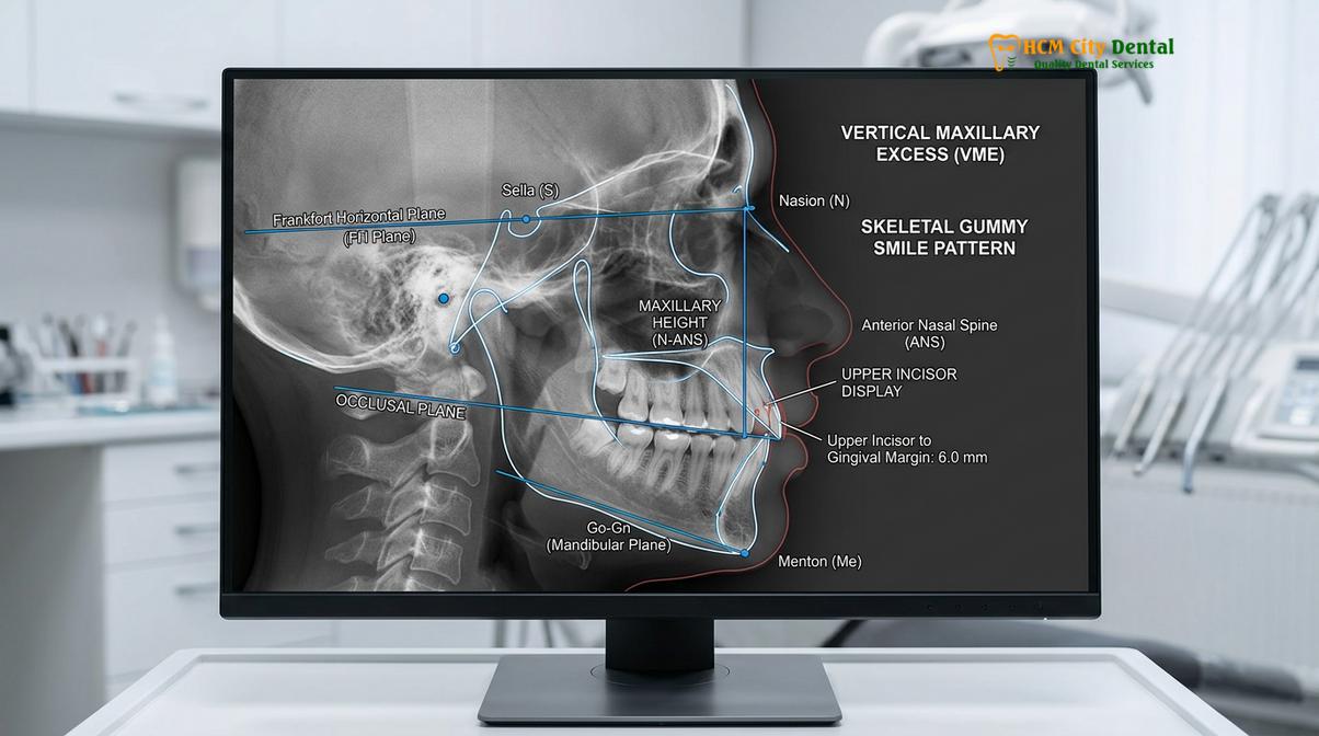
It is crucial to differentiate VME from other causes of a gummy smile to avoid inappropriate treatment planning. For instance, delayed passive eruption occurs when the gum tissue fails to recede normally during tooth development, leaving the anatomical crowns partially covered by gingiva. This condition mimics a skeletal issue but is effectively treated with a simple crown lengthening procedure. Similarly, a hyperactive levator muscle—the muscle responsible for elevating the upper lip—can draw the lip up too high during smiling. This muscular etiology is often managed conservatively with targeted Botox injections or a surgical lip repositioning procedure. However, when cephalometric data confirms that the entire skeletal base is positioned too low, these soft tissue interventions will yield unsatisfactory, compromised results. In such scenarios, jaw repositioning surgery becomes the mandatory clinical pathway.[1]
Routine dental scaling and standard periodontal maintenance, while essential for overall oral health, have no impact on the structural reality of VME. The underlying bone must be physically altered to achieve harmony. Understanding the distinction between soft tissue hyperplasia, muscular hyperactivity, and true skeletal overgrowth is the cornerstone of modern dentofacial orthopedics and aesthetic rehabilitation.
Understanding Le Fort I Osteotomy (Jaw Repositioning Surgery)
The Le Fort I osteotomy involves surgically separating and elevating the maxilla to reduce vertical height, effectively eliminating a severe skeletal gummy smile.
The Le Fort I osteotomy is the gold standard surgical intervention for correcting vertical maxillary excess. This sophisticated procedure, performed under general anesthesia, allows the maxillofacial surgeon to detach the entire upper jaw from the cranial base, reposition it into an anatomically ideal location, and secure it permanently. The primary objective in treating a severe skeletal gummy smile is maxillary impaction—moving the jaw upward to reduce the vertical dimension of the lower third of the face.
The surgical workflow begins with an incision made entirely intraorally, high in the buccal vestibule (the space between the upper lip and the gums). This approach ensures that there are no visible facial scars. Once the mucosal tissues are elevated, the surgeon exposes the anterior and lateral walls of the maxilla. Using specialized piezoelectric surgical instruments or micro-saws, a horizontal osteotomy (bone cut) is made above the apices of the tooth roots, extending from the piriform rim of the nose to the pterygomaxillary junction at the back of the jaw.[2]
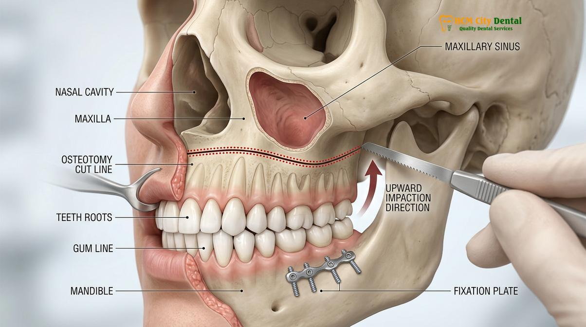
Following the osteotomy, the maxilla is carefully down-fractured, separating it from the nasal septum and the lateral nasal walls. At this critical juncture, the surgeon performs an osteoplasty, meticulously removing a pre-calculated wedge of bone from the superior aspect of the maxilla. The thickness of this bone wedge corresponds exactly to the amount of vertical reduction required to eliminate the gummy smile. Once the excess bone is removed, the maxilla is impacted (moved upward) into its new, superior position.
“The Le Fort I maxillary impaction remains the most highly predictable and stable orthognathic procedure for the correction of vertical maxillary excess, providing profound improvements in both facial aesthetics and functional occlusion.”
To ensure absolute stability, the newly positioned jaw is secured using Rigid Internal Fixation (RIF). Small, biocompatible titanium plates and screws are placed across the osteotomy lines. These plates hold the bone segments rigidly in place, allowing for primary bone healing without the need to wire the patient’s jaws shut (maxillomandibular fixation), which was common in historical techniques. The intraoral incisions are then closed with resorbable sutures. The result is a dramatic, immediate reduction in gingival display and a restoration of facial harmony.
Collaborative Care: Orthodontist and Maxillofacial Surgeon Workflows
Successful jaw repositioning surgery requires precise coordination between orthodontic decompensation, maxillofacial surgical impaction, and post-operative periodontal refinement.
The correction of a severe skeletal gummy smile is never an isolated surgical event; it is a carefully orchestrated journey requiring seamless collaboration between multiple dental specialties. The workflow is generally divided into three distinct phases: pre-surgical orthodontics, the surgical intervention, and post-surgical orthodontic and periodontal refinement. This multidisciplinary approach ensures that the final result is not only aesthetically pleasing but functionally stable.
The process begins with the orthodontist. In patients with skeletal discrepancies, the teeth often naturally tilt or angle themselves to compensate for the misaligned jaws—a phenomenon known as dental compensation. The goal of pre-surgical orthodontics is to reverse this process (decompensation). The orthodontist uses braces or clear aligners to move the teeth into their correct positions relative to their respective jawbones. Paradoxically, this phase may temporarily worsen the patient’s bite and facial appearance, as the true extent of the skeletal deformity is unmasked. This phase typically lasts between 12 to 18 months.[3]
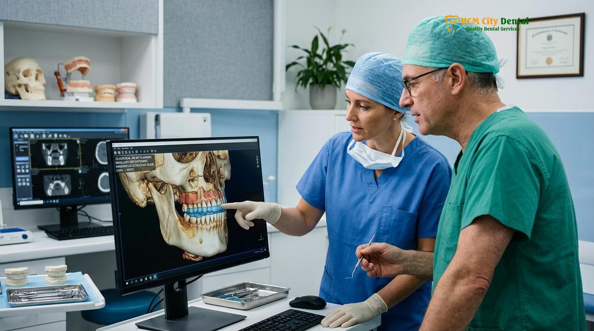
Once the teeth are properly aligned within the arches, the patient is ready for the surgical phase. For complex skeletal cases requiring Le Fort I osteotomy, clinical facilities often collaborate with highly specialized surgical centers. For instance, patients undergoing this level of treatment may be referred to Surgical Jaw Partners, such as the maxillofacial specialists at the National Hospital of Odonto-Stomatology in Ho Chi Minh City (Bệnh viện Răng Hàm Mặt Trung Ương TP.HCM). This institution represents the highest clinical authority for complex maxillofacial reconstructions in the region, ensuring that the jaw repositioning surgery is executed with maximum precision and safety.
Clinical Case Overview: Multidisciplinary Gummy Smile Correction
A 26-year-old patient presented with an 8mm gingival display and lip incompetence. Cephalometric analysis confirmed severe VME. Following 14 months of pre-surgical orthodontic decompensation, a Le Fort I osteotomy was performed at the National Hospital of Odonto-Stomatology HCMC, impacting the maxilla by 6mm. Six months post-surgery, the patient returned to HCMC Dental Clinic for final soft tissue detailing. Dr. Nguyen Van Cuong utilized a diode laser to refine the gingival zenith points, resulting in a perfectly balanced, aesthetic smile line with zero functional deficits.
Following the surgery and a brief healing period, the patient enters the post-surgical orthodontic phase. This final phase, lasting approximately 6 to 9 months, focuses on “settling” the bite, making micro-adjustments to the occlusion to ensure the upper and lower teeth interlock perfectly. It is during this final stage that the aesthetic details of the smile are finalized, often requiring the expertise of a periodontist or cosmetic dentist to perfect the soft tissue architecture.
Soft Tissue Refinement: Laser Contouring and Lip Repositioning
Following skeletal correction, diode laser technology is utilized to calibrate gingival zenith points and finalize the aesthetic smile design with minimal invasiveness.
Even after a successful Le Fort I osteotomy HCMC, the soft tissues of the gums may require fine-tuning to achieve a flawless aesthetic result. The surgery addresses the macro-aesthetics (the position of the jaw), but the micro-aesthetics (the shape and contour of the individual gum margins) must be meticulously calibrated. This is where advanced periodontal techniques and Digital Smile Design (DSD) come into play.
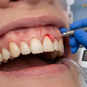
At the forefront of soft tissue refinement is the use of AMD Picasso Lasers. This specific diode soft tissue laser operates at an 810nm wavelength, which is highly absorbed by melanin and hemoglobin. From a clinical perspective, this means the laser can vaporize excess gum tissue while simultaneously sealing blood vessels and nerve endings. Dr. Nguyen Van Cuong frequently emphasizes the clinical advantages of this technology: it provides bloodless, sutureless cuts, instant coagulation, and a sterile beam that significantly minimizes infection risk. Patients benefit from reduced postoperative pain, minimal swelling, and a rapid 48-hour recovery period compared to traditional scalpel surgery.
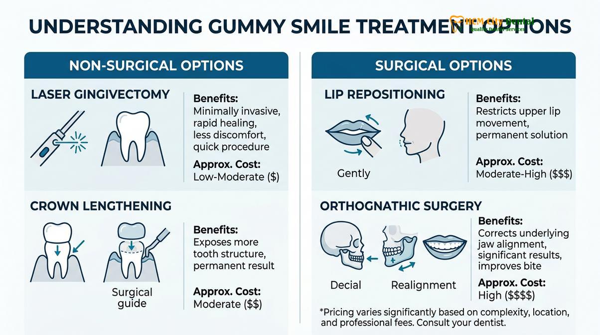
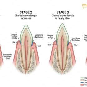
During the contouring process, the clinician must strictly respect the biological width—a critical anatomical dimension requiring a minimum of 2mm of supracrestal tissue attachment above the alveolar bone. Violating this space can lead to chronic inflammation or unpredictable bone resorption. Using the laser, the clinician carefully sculpts the gingival zenith points (the highest point of the gum scallop, which should be displaced slightly distal to the long axis of the tooth) to create perfect symmetry across the anterior sextant.[4]
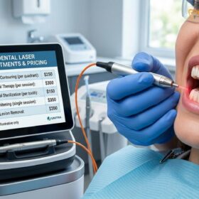
For patients seeking comprehensive comprehensive gum contouring procedures, understanding the financial investment is important. Below is the current pricing structure for soft tissue and adjunctive gummy smile treatments, noting that international patients often utilize a -40% WhatsApp booking discount prior to arrival:
| Clinical Procedure | Standard Walk-in Fee (VND) | Discounted Fee (VND) | Approx. USD |
|---|---|---|---|
| Laser Gingivectomy (per tooth) | 2,500,000 | 1,500,000 | ~$60 |
| Full-Arch Laser Gum Contouring (up to 10 teeth) | 20,000,000 | 12,000,000 | ~$480 |
| Functional/Esthetic Crown Lengthening (per tooth) | 5,000,000 | 3,000,000 | ~$120 |
| Surgical Lip Repositioning / Lip Lowering | 25,000,000 | 15,000,000 | ~$600 |
| Botox Gummy Smile Correction (both sides) | 7,500,000 | 4,500,000 | ~$180 |
In cases where a mild gummy smile persists due to muscular factors rather than skeletal, adjunctive treatments like Botox or Surgical Lip Repositioning may be recommended. Lip repositioning involves removing a strip of mucosa from the upper vestibule and suturing the lip in a lower position, restricting its upward pull during a smile. This provides a less invasive alternative for specific anatomical presentations.
Recovery Timelines and Hospitalization Guidelines in Vietnam
Patients undergoing maxillary impaction in Ho Chi Minh City can expect a structured recovery protocol, typically requiring a brief hospital stay followed by outpatient monitoring.
Undergoing orthognathic surgery is a significant medical event that requires a dedicated commitment to the recovery process. For patients engaging in dental tourism, understanding the timeline and logistical requirements is essential for a safe and successful outcome. When a Le Fort I osteotomy is performed, the initial phase of recovery is managed within a hospital setting to ensure optimal patient safety and comfort.
Immediately following surgery, patients typically remain hospitalized for 1 to 3 days. During this time, intravenous medications are administered to manage pain, prevent infection, and control swelling. Swelling is a natural and expected inflammatory response to bone surgery; it generally peaks around 48 to 72 hours post-operatively before gradually subsiding. Ice packs and specialized compressive dressings are utilized to mitigate edema. The surgical team closely monitors the patient’s airway, vital signs, and initial healing response.
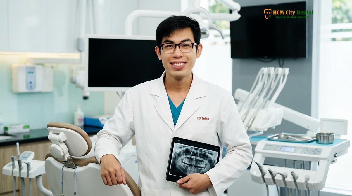
Upon discharge, the patient transitions to outpatient care. For international patients, it is highly recommended to remain in Ho Chi Minh City for at least 10 to 14 days post-surgery. This allows the surgical team and coordinating specialists at HCMC Dental Clinic to conduct essential follow-up examinations, monitor the intraoral incisions, and ensure the rigid internal fixation is stable. During the first two weeks, patients are restricted to a strict liquid or pureed diet to prevent any mechanical stress on the healing maxilla.[5]
“The success of orthognathic rehabilitation relies as much on strict adherence to post-operative dietary and functional protocols as it does on surgical precision. Patient compliance during the initial 6-week bone healing phase is non-negotiable.”
Between weeks 2 and 6, the diet is gradually advanced to soft foods (e.g., scrambled eggs, soft pasta). Hard, crunchy, or chewy foods must be strictly avoided until the surgeon confirms adequate bone consolidation, usually around the 6-to-8-week mark. While the majority of visible swelling resolves within the first month, residual micro-swelling can take up to six months to completely dissipate, at which point the final aesthetic results of the jaw repositioning surgery become fully apparent.
When to See a Doctor for a Severe Skeletal Gummy Smile
Determining whether a gummy smile requires surgical intervention or can be managed with conservative soft tissue therapies requires professional diagnostic imaging. Patients should seek a formal consultation with a maxillofacial specialist or an expert prosthodontist if they experience specific functional or aesthetic indicators that suggest underlying skeletal discrepancies.
Important Clinical Considerations:
You should schedule a comprehensive dentofacial evaluation if you experience any of the following signs associated with vertical maxillary excess:
- Displaying more than 4 millimeters of gum tissue when smiling naturally.
- Inability to close your lips comfortably at rest without straining the chin muscles (lip incompetence).
- Chronic mouth breathing or associated symptoms of obstructive sleep apnea.
- A facial profile that appears excessively long in the lower third.
- Difficulty biting or chewing due to an open bite or severe malocclusion accompanying the gummy smile.
Early diagnosis allows for the formulation of a comprehensive, phased treatment plan. A clinical examination, including 3D Cone Beam Computed Tomography (CBCT) and cephalometric tracing, will definitively map the supracrestal tissue attachment and skeletal architecture, ensuring that any proposed intervention—whether a Le Fort I osteotomy or a laser gingivectomy—is biologically sound and highly predictable.
Frequently Asked Questions
Is jaw surgery the only option for severe gummy smiles?
Orthognathic surgery is typically the only definitive treatment for severe skeletal gummy smiles caused by vertical maxillary excess. However, if the excessive gingival display is purely soft-tissue related, less invasive options like laser gingivectomy or lip repositioning may be sufficient. A thorough cephalometric analysis is required to determine the true etiology and prevent inappropriate treatment planning.
What is the recovery period for Le Fort I surgery?
The initial recovery period for a Le Fort I osteotomy spans two to three weeks, during which most visible swelling subsides. Complete bone healing and stabilization of the jaw structure generally require three to six months of careful monitoring. Patients must adhere to a strict soft-food diet for the first six weeks to protect the surgical site.
Does dental insurance cover orthognathic surgery?
Dental and medical insurance may cover orthognathic surgery if the procedure is deemed medically necessary to correct severe functional impairments, such as obstructive sleep apnea or severe malocclusion. Purely aesthetic gummy smile corrections are typically excluded from standard coverage. Patients should consult their specific insurance providers with detailed diagnostic records for pre-authorization.
How does a diode laser improve post-surgical gum aesthetics?
A diode laser precisely sculpts the gingival margins to create symmetrical zenith points without bleeding or the need for sutures. This technology ensures rapid healing and allows the clinician to finalize the aesthetic outcome following skeletal jaw repositioning. By utilizing an 810nm wavelength, the laser provides instant coagulation and minimizes postoperative discomfort.
Can Botox be used instead of jaw surgery for a gummy smile?
Botox can temporarily mask a gummy smile if the primary cause is a hyperactive levator muscle of the upper lip. It cannot, however, correct underlying skeletal discrepancies or vertical maxillary excess, which require surgical intervention for permanent resolution. Botox treatments typically last 3 to 4 months and require continuous maintenance.
References
- Journal of Oral and Maxillofacial Surgery. Long-term stability of Le Fort I osteotomy for vertical maxillary excess. (2022).
- International Journal of Periodontics & Restorative Dentistry. Diode laser applications in aesthetic gingival contouring. (2021).
- American Journal of Orthodontics and Dentofacial Orthopedics. Multidisciplinary management of the severe skeletal gummy smile. (2020).
- Journal of Clinical Periodontology. Biological width and supracrestal tissue attachment in restorative dentistry. (2019).
- Clinical Oral Investigations. Outcomes of surgical lip repositioning versus orthognathic interventions. (2018).