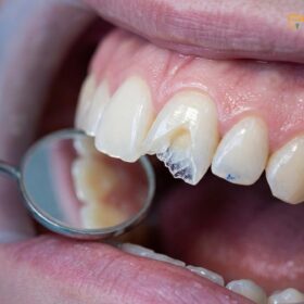
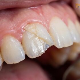
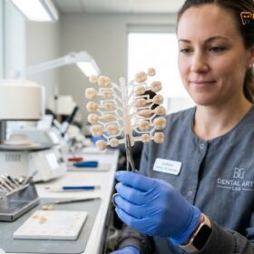
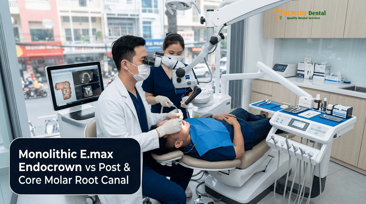
The restoration of endodontically treated teeth (ETT) represents one of the most complex biomechanical challenges in modern prosthodontics. Following root canal therapy, a molar suffers a profound loss of structural integrity, primarily due to the excavation of the pulp chamber, the removal of the pulpal roof, and the loss of vital dentinal fluid. Historically, the default restorative protocol involved drilling deep into the radicular canals to place a cast metal or glass fiber post, building up a composite core, and cementing a full-coverage crown. However, this invasive approach often necessitates the removal of sound radicular dentin, significantly increasing the risk of catastrophic root fractures and strip perforations. In 2026, the paradigm has shifted toward biomimetic, tissue-preserving dentistry. The debate between an endocrown monolithic IPS E.max zirconia vs post and core molar root canal restoration is now heavily weighted in favor of the adhesive endocrown—a single-piece, monoblock ceramic restoration that anchors directly into the pulp chamber without invading the root canals.
\n\n
At HCMC Dental Clinic in District 1, Ho Chi Minh City, our clinical protocols are guided by evidence-based biomimetic principles. Under the leadership of Dr. Nguyen Van Cuong, DDS, we utilize advanced CAD/CAM technology, 3D CBCT diagnostics, and strict adhesive dentistry protocols to restore severely compromised molars. By leveraging the macromechanical retention of the pulp chamber and the micromechanical adhesion of etched lithium disilicate (IPS E.max), the endocrown offers a highly predictable, minimally invasive alternative to traditional post-and-core crowns. This comprehensive clinical guide explores the biomechanics, material science, strict indications, and 2026 fee schedules for restoring root-canal-treated molars in Vietnam.
\n\n
Clinical Summary:
\n
The endocrown is a monolithic, adhesively bonded ceramic restoration designed specifically for endodontically treated molars. By utilizing the internal walls of the pulp chamber for retention and relying on advanced resin bonding protocols, it eliminates the need for radicular posts. Clinical studies demonstrate that monolithic IPS E.max endocrowns exhibit survival rates comparable to, or exceeding, traditional post-and-core crowns, primarily because they prevent the weakening of the root structure. At HCMC Dental Clinic, Dr. Nguyen Van Cuong, DDS, employs strict biomimetic protocols—including Immediate Dentin Sealing (IDS) and rubber dam isolation—to deliver CAD/CAM endocrowns that restore the biomechanical integrity of heavily broken-down molars.
\n
\n\n
Key Takeaways:
\n
- \n
- Biomechanical Superiority: Endocrowns distribute masticatory forces across the cervical butt-joint margin and the pulpal floor, significantly reducing the stress concentrations that cause vertical root fractures associated with traditional posts.
- Material Selection: IPS E.max (lithium disilicate) is the gold standard for endocrowns due to its ability to be etched with hydrofluoric acid and silanated, creating a profound chemical bond with resin cement. Monolithic zirconia is generally less predictable for non-retentive endocrown preparations.
- Pulp Chamber Retention: The endocrown relies on a 2mm to 3mm extension into the pulp chamber (the central endo-core) for macromechanical retention, avoiding any invasion into the fragile root canals.
- Strict Indications: Endocrowns are highly indicated for molars with short clinical crowns (less than 3mm of interocclusal space), calcified canals, and curved roots where post placement is contraindicated.
- Cost Efficiency: At HCMC Dental Clinic, a CAD/CAM IPS E.max Endocrown is priced at $231 USD per tooth (online booking rate), offering a highly cost-effective, world-class restorative solution for international dental travelers.
\n
\n
\n
\n
\n
\n
\n\n\n
\n\n\n
What Is an Adhesive Ceramic ‘Endocrown’ (Pissis & Bindl Monoblock Concept) and How Does It Anchor Inside the Pulp Chamber?
\n\n
The concept of the endocrown was first introduced by Pissis in 1995 as the “monoblock technique” and later refined and named the “endo-crown” by Bindl and Mörmann in 1999. Traditional restorative dentistry viewed the crown and the core as two separate entities, often requiring a post to anchor the core to the root. The endocrown revolutionizes this by merging the crown and the core into a single, monolithic ceramic structure. This monoblock restoration is designed to replace the entire coronal portion of the tooth while simultaneously extending into the pulp chamber to utilize its internal anatomy for retention.
\n\n
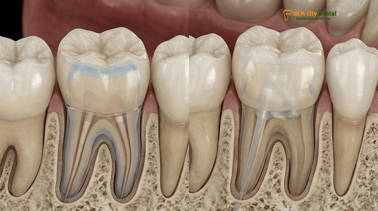
The anchoring mechanism of an endocrown is dual-faceted, relying on both macromechanical geometry and micromechanical adhesion. Macromechanically, the preparation utilizes the pulp chamber as a central retention cavity. The internal walls of the pulp chamber are prepared with a slight divergence (approximately 7 to 10 degrees of total occlusal convergence) to allow for the passive seating of the rigid ceramic block. The depth of this pulpal extension—often referred to as the pulp chamber macromechanical retention endocrown rule—is typically 2mm to 3mm. Extending deeper into the canal orifices does not significantly improve retention but drastically increases internal stress and complicates the manufacturing process. The cervical margin is prepared as a flat, circumferential butt-joint, ideally resting on sound enamel. This flat margin provides a broad surface area to resist compressive masticatory forces.
\n\n
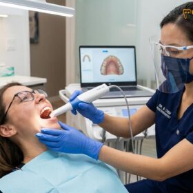
Micromechanically, the success of the endocrown is entirely dependent on flawless adhesive dentistry. Because the preparation lacks the traditional parallel walls and ferrule of a standard crown preparation, the resin cement must provide the primary retentive force. At HCMC Dental Clinic, this is achieved through rigorous biomimetic protocols. Following the excavation of the pulp chamber, the dentin is treated using Immediate Dentin Sealing (IDS). This involves applying a dentin bonding agent immediately after preparation, before taking the digital impression. IDS protects the freshly cut dentinal tubules from bacterial contamination, reduces postoperative sensitivity, and significantly increases the ultimate bond strength. [1]
\n\n
\n
“The endocrown represents the pinnacle of tissue-preserving prosthodontics for endodontically treated molars. By abandoning the radicular post, we respect the fragile anatomy of the root. The retention is no longer derived from friction deep within the canal, but from the profound chemical adhesion between etched lithium disilicate, dual-cure resin cement, and hybrid-layer dentin inside the pulp chamber.”
\n— Dr. Nguyen Van Cuong, DDS (Clinical Director, HCMC Dental Clinic)\n
\n\n
Furthermore, the bonding procedure is executed under a 100% latex-free nitrile rubber dam to ensure absolute moisture control. The internal surface of the pulp chamber is micro-roughened using 27-micron aluminum oxide air abrasion to remove the temporary cement and biofilm. To manage the high polymerization shrinkage stress inherent in deep cavity preparations—quantified by Feilzer’s Configuration Factor (C-Factor), which can reach up to 5.0 in a box-like pulp chamber—we utilize a 5-minute decoupling technique and, in specific cases, incorporate ultra-high-molecular-weight polyethylene fibers on the pulpal floor to absorb stress and prevent adhesive failure.
\n\n\n
\n\n\n
Why Drilling Post Spaces into Curved or Narrow Molar Roots Risks Strip Perforation and Catastrophic Root Fracture
\n\n
To understand why the endocrown has become the preferred restoration for molars, one must examine the inherent risks of traditional post-and-core procedures. Molars, particularly maxillary first molars and mandibular first molars, possess complex and highly variable root anatomies. The mesiobuccal roots of maxillary molars and the mesial roots of mandibular molars are frequently narrow, curved, and invaginated. The dentinal walls in these roots are notoriously thin, especially on the distal aspect of the mesial roots—an area endodontists refer to as the “danger zone.”
\n\n
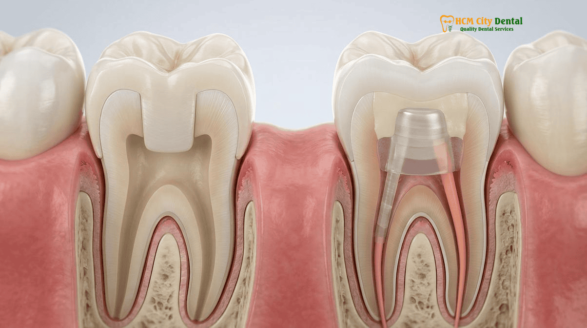
When a dentist prepares a tooth for a traditional post, they must use specialized drills (Peeso reamers or Gates Glidden drills) to remove gutta-percha and enlarge the canal space. This process inevitably removes sound radicular dentin. In curved or narrow roots, the rigid drill tends to straighten out, cutting aggressively into the inner curvature of the root. This can easily lead to a “strip perforation,” where the drill breaches the lateral wall of the root, communicating with the periodontal ligament. A strip perforation is a catastrophic complication that often necessitates the extraction of the tooth. Therefore, the ability to prevent root perforation curved molar canals is one of the most significant advantages of the endocrown technique.
\n\n
Even if a perforation does not occur, the removal of radicular dentin severely weakens the root. The root of an endodontically treated tooth is already compromised; hollowing it out further to accommodate a post creates a thin-walled tube that is highly susceptible to fracture. When masticatory forces are applied to a crown supported by a rigid post (particularly cast metal or thick zirconia posts), the post acts as a wedge. It concentrates stress at the apical end of the post or along the lateral walls of the root, frequently resulting in a vertical root fracture. Vertical root fractures are untreatable and invariably lead to tooth loss and the need for implant replacement. [2]
\n\n
While modern glass fiber posts have an elastic modulus closer to that of natural dentin—thereby distributing stress more evenly than metal posts—they still require the removal of radicular dentin for placement. For patients interested in learning more about when fiber posts are still indicated (such as in anterior teeth), we recommend reading our detailed guide on Glass Fiber Post & Core Buildup vs. Cast Metal Post Before a Dental Crown: Preventing Root Fracture. However, for molars, where the pulp chamber is large enough to provide adequate retention on its own, avoiding the root canals entirely via an endocrown is the most biologically sound approach.
\n\n\n
\n\n\n
Finite Element Stress Analysis: How a Monoblock IPS E.max Endocrown Distributes Chewing Forces Across the Cervical Butt-Joint Margin
\n\n
The biomechanical success of the endocrown is heavily reliant on the material chosen for its fabrication. In the debate of endocrown monolithic IPS E.max zirconia vs post and core molar root canal, material science plays a pivotal role. Finite Element Analysis (FEA)—a computerized method for predicting how a product reacts to real-world forces—has been extensively used to study stress distribution in endocrowns. FEA studies consistently show that a monoblock endocrown distributes compressive masticatory forces far more favorably than a post-and-core assembly.
\n\n
In a traditional crown, forces are directed along the axial walls of the preparation and down into the root via the post. In an endocrown, the forces are distributed across the broad, flat cervical butt-joint margin and the floor of the pulp chamber. This broad distribution prevents the dangerous stress concentrations that cause root fractures. However, for this stress distribution to function correctly, the ceramic material must be rigidly bonded to the underlying tooth structure, creating a unified biomechanical complex.
\n\n
This is why IPS E.max (lithium disilicate) is overwhelmingly preferred over monolithic zirconia for endocrowns. Lithium disilicate is a glass-ceramic. Its glass matrix can be selectively dissolved using 5% to 9.5% hydrofluoric (HF) acid. This etching process creates a highly porous, micro-retentive topography on the internal surface of the endocrown. Following acid etching, the ceramic is treated with a pure, heat-dried silane coupling agent. Silane is a bifunctional molecule; one end bonds chemically to the silica in the ceramic, and the other end copolymerizes with the methacrylate groups in the dual-cure resin cement. This creates an incredibly strong, predictable, and durable chemical bond. [3]
\n\n
Monolithic zirconia, on the other hand, is a polycrystalline ceramic with no glass matrix. It cannot be etched with hydrofluoric acid. To bond zirconia, clinicians must rely on the Blatz APC protocol: Air-abrasion (with aluminum oxide to create micromechanical roughness), Primer (using a specialized zirconia primer containing 10-MDP monomer, such as Ivoclean for decontamination followed by Monobond Plus), and Cementation (with a dual-cure resin cement). While the 10-MDP monomer does form a chemical bond with the zirconium oxide layer, this bond is generally less robust and more susceptible to hydrolytic degradation over time compared to the silane-lithium disilicate bond. Because an endocrown relies almost entirely on adhesion for retention, the superior bond strength of IPS E.max makes it the undisputed gold standard. For a deeper dive into the clinical performance of zirconia in more retentive preparations, see our Monolithic Zirconia vs PFM Clinical Study | HCMC Dental.
\n\n\n
\n\n\n
Strict Clinical Indications: Heavily Broken-Down Molars, Short Clinical Crowns (<3mm Interocclusal Space), and Calcified Canals
\n\n
While the endocrown is a remarkable restorative option, it is not a universal panacea. Its success depends on strict adherence to specific clinical indications. At HCMC Dental Clinic, Dr. Nguyen Van Cuong, DDS, meticulously evaluates each endodontically treated tooth using 3D CBCT imaging and digital intraoral scanning to determine if an endocrown is the optimal treatment.
\n\n
The primary indication for an endocrown is a heavily broken-down molar that has undergone root canal therapy. Molars are subjected primarily to vertical, compressive masticatory forces, which the flat butt-joint margin of the endocrown is perfectly designed to resist. The large pulp chamber of a molar provides ample surface area for adhesive bonding and macromechanical retention. Therefore, the monolithic ips IPS E.max endocrown survival rate in molars is exceptionally high, often exceeding 90% to 95% over a 10-year period in peer-reviewed literature. [4]
\n\n
A specific and highly critical indication for an endocrown is a molar with a short clinical crown. In traditional prosthodontics, a crown requires a “ferrule”—a band of sound tooth structure at least 1.5mm to 2mm high and 1mm thick, extending 360 degrees around the preparation. The ferrule acts like the metal band around a wooden barrel, preventing the root from splitting under the wedging forces of the post and core. However, in many heavily decayed or worn molars, achieving a 2mm ferrule is impossible without performing surgical crown lengthening, which removes supporting alveolar bone and compromises the crown-to-root ratio. Furthermore, if the patient has less than 3mm of interocclusal space (the vertical distance between the prepared tooth and the opposing tooth), there is simply not enough room to build a core and place a traditional crown of adequate thickness. The endocrown solves both problems simultaneously. It does not require a ferrule, and because it is a single monolithic piece, it requires less total vertical space than a separate core and crown assembly. This makes it the ideal solution to restore short broken down molar after root canal.
\n\n
Clinical Case Study: Restoring a Compromised Mandibular Molar in District 1
\n
Patient Profile: A 45-year-old Australian expatriate presented to HCMC Dental Clinic with a fractured, root-canal-treated mandibular right first molar (Tooth 46). The tooth had previously been restored with a large composite filling that had failed, leaving less than 1.5mm of coronal tooth structure above the gumline. The mesial root canals were highly curved and partially calcified.
\n
Clinical Challenge: Placing a traditional post was contraindicated due to the severe curvature and calcification of the mesial roots, which carried a high risk of strip perforation. Furthermore, the lack of coronal tooth structure meant a traditional ferrule could not be achieved without invasive crown lengthening surgery.
\n
Treatment Protocol: Dr. Nguyen Van Cuong, DDS, opted for a monolithic IPS E.max CAD endocrown. Under a rubber dam, the pulp chamber was cleaned and prepared to a depth of 2.5mm. Immediate Dentin Sealing (IDS) was performed. A digital scan was taken, and the endocrown was designed and milled in-house. The restoration was crystallized and bonded using hydrofluoric acid, pure silane, and a dual-cure resin cement.
\n
Outcome: The patient received a highly aesthetic, biomechanically sound restoration in just 48 hours, avoiding root canal retreatment, post placement, and periodontal surgery. At the 2-year follow-up, the endocrown demonstrated perfect marginal integrity and zero signs of occlusal trauma.
\n
\n\n
Another excellent indication for an endocrown is a molar with calcified or obliterated root canals. In older patients, or in teeth that have suffered chronic trauma, the root canals can become so narrow that placing a post is physically impossible. Because the endocrown anchors solely in the pulp chamber, the condition of the radicular canals (provided the endodontic seal is sound) is irrelevant to the retention of the restoration. For a comprehensive overview of all post-endodontic restorative options, patients can review our Crown After Root Canal: Clinical Guide & Options | HCMC Dental.
\n\n
When an Endocrown Is Contraindicated (Anterior Incisors, Premolars Without Enamel Borders, and Subgingival Pulp Chamber Floors)
\n\n
Despite its advantages in molars, the endocrown is contraindicated in several specific clinical scenarios. Understanding these limitations is crucial for long-term restorative success. The most absolute contraindication for an endocrown is its use in anterior teeth (incisors and canines). Anterior teeth have very small pulp chambers that do not provide sufficient surface area for macromechanical retention. More importantly, anterior teeth are subjected to high shear and lateral forces during the incision and tearing of food, as well as during excursive mandibular movements. These lateral forces create massive leverage on the restoration. Without the deep radicular anchorage provided by a fiber post, an anterior endocrown would rapidly debond or fracture. Therefore, for anterior teeth, a conservative glass fiber post and a biomimetic composite core remain the standard of care.
\n\n
Premolars represent a clinical gray zone. Maxillary premolars, in particular, have narrow pulp chambers and are subjected to a mix of compressive and lateral forces. While some clinical studies show acceptable survival rates for premolar endocrowns, the failure rate (primarily due to debonding) is statistically higher than in molars. At HCMC Dental Clinic, we evaluate premolars on a strict case-by-case basis. If the premolar has a deep, box-like pulp chamber and a broad cervical enamel border, an endocrown may be viable. If the pulp chamber is shallow or the tooth is subjected to heavy lateral guidance, a fiber post and core is preferred.
\n\n
Another significant contraindication is a preparation where the cervical margins or the floor of the pulp chamber are deeply subgingival. Adhesive dentistry requires absolute isolation from saliva, blood, and crevicular fluid. If the margins cannot be isolated with a rubber dam, the resin bond will fail, leading to microleakage, recurrent decay, and debonding. In cases where decay extends deep below the gumline, Coslet’s Altered Passive Eruption (APE) principles must be evaluated. Surgical crown lengthening or orthodontic forced eruption may be necessary to elevate the margins supragingivally before an endocrown can be considered. [5]
\n\n
Clinical Precaution: When to Consult a Specialist
\n
If you have a root-canal-treated tooth that has fractured below the gumline, or if you have been told that your tooth requires a post but your roots are highly curved, do not proceed with standard treatment without a specialist evaluation. Improper post placement in curved roots is a leading cause of irreversible tooth loss. Contact Dr. Nguyen Van Cuong, DDS (Clinical Director — MOH License 0003265/BL-CCHN) at HCMC Dental Clinic for a 3D CBCT scan and a comprehensive biomimetic assessment to determine if a tissue-preserving endocrown can save your tooth.
\n
\n\n\n
\n\n\n
2026 CAD/CAM IPS E.max Endocrown vs. Fiber Post + Zirconia Crown Fee Schedule at HCMC Dental Clinic (District 1)
\n\n
For international patients, expats, and dental travelers seeking world-class biomimetic dentistry, understanding the biomimetic endocrown cost Ho Chi Minh City is essential. At HCMC Dental Clinic, we maintain absolute transparency in our pricing. By utilizing in-house digital CAD/CAM workflows, we eliminate third-party laboratory delays and markups, allowing us to offer premium restorations at highly competitive rates. Our 2026 fee schedule features a specialized -40% Online Booking Pricing tier, making advanced restorative care highly accessible.
\n\n
The table below outlines the exact costs for restoring an endodontically treated tooth, comparing the monolithic IPS E.max endocrown protocol with the traditional fiber post and zirconia crown approach. For a complete overview of all our services, please visit our Official 2026 Dental Price List & -40% Online Booking Fee Schedule.
\n\n
| Restorative Procedure / Material | 2026 Online Booking Fee (-40% USD) | 2026 Online Booking Fee (VND) | Standard Walk-In Price (USD) |
|---|---|---|---|
| IPS E.max All-Ceramic Crown / Inlay / Onlay / Endocrown (Gold standard for adhesive monoblock restorations) | $231 USD / tooth | 6,000,000 VND | $385 USD |
| Full Zirconia Crown (Cercon Standard / HT Multilayer) (Highly durable, requires traditional core buildup) | $154 – $192 USD / tooth | 4,000,000 – 5,000,000 VND | $256 – $321 USD |
| Biomimetic Quartz/Glass Fiber Post & Core Buildup (Required if choosing a traditional Zirconia crown) | $38 – $58 USD / tooth | 988,000 – 1,508,000 VND | $63 – $96 USD |
| Microscopic Endodontic Root Canal Therapy (Complex Molar) (25x Carl Zeiss + Nitrile Dam) | $115 – $154 USD / tooth | 3,000,000 – 4,000,000 VND | $192 – $256 USD |
| 3D CBCT Scan (Essential for evaluating root curvature and pulp chamber depth) | $18 USD | 480,000 VND | $30 USD |
\n
\n\n
When comparing the total cost, an IPS E.max Endocrown ($231 USD) is often more cost-effective than the combined cost of a Glass Fiber Post & Core ($38 – $58 USD) plus a Full Zirconia Crown ($154 – $192 USD), which totals $192 to $250 USD. More importantly, the endocrown preserves vital root structure and can often be completed in a faster timeframe. For international patients on a tight schedule, our digital workflow ensures rapid delivery. Learn more about our express services by reading the Global Dental Lab Turnaround Benchmark 2026: How Saigon CAD/CAM Milling Delivers Crowns in 48 Hours vs. 3 Weeks, and explore our full range of prosthetic options at Dental Crowns, Fixed Bridges & Biomimetic Ceramic Restorations in Vietnam.
\n\n
\n
“The true value of an endocrown is not just in its competitive price, but in the biological cost it saves. By avoiding the root canals, we eliminate the risk of strip perforations and vertical root fractures, potentially saving the patient from needing a dental implant a decade down the line.”
\n— Dr. Nguyen Van Cuong, DDS\n
\n\n
Frequently Asked Questions
\n\n
Does an Endocrown hold onto a broken-down molar as strongly as a traditional post, core buildup, and full crown?
\n
Yes. Extensive finite element analysis and long-term clinical studies demonstrate that a properly bonded monolithic IPS E.max endocrown provides equivalent or superior fracture resistance compared to a traditional post-and-core crown on molars. By utilizing the pulp chamber for macromechanical retention and preserving the cervical enamel for micromechanical adhesion, the endocrown eliminates the need for a radicular post, thereby reducing the risk of catastrophic root fracture.
\n\n
Why is IPS E.max lithium disilicate preferred over monolithic zirconia for bonding an adhesive Endocrown?
\n
IPS E.max (lithium disilicate) is a glass-ceramic that can be etched with 5% to 9.5% hydrofluoric acid, creating a highly porous surface. When treated with a pure heat-dried silane coupling agent, it forms a profound chemical and micromechanical bond with dual-cure resin cements. Monolithic zirconia lacks a glass matrix, meaning it cannot be etched with acid. While zirconia can be bonded using the Blatz APC protocol (Air-abrasion, Ivoclean, 10-MDP monomer), the bond strength is generally lower than that of etched lithium disilicate, making E.max the gold standard for non-retentive, adhesion-dependent endocrown preparations.
\n\n
How deep into the pulp chamber does an Endocrown extend (the 2mm–3mm central endo-core rule)?
\n
A standard endocrown preparation extends approximately 2mm to 3mm into the central pulp chamber. Clinical research indicates that this depth provides optimal macromechanical retention and sufficient ceramic thickness for structural durability. Extending the ceramic deeper than 3mm into the canal orifices does not significantly increase retention but drastically complicates the manufacturing process, increases internal stress concentrations, and makes future endodontic retreatment exceedingly difficult.
\n\n
If the molar ever needs endodontic retreatment in the future, is it easier to access through an Endocrown or a metal post?
\n
Endodontic retreatment is significantly easier and safer through an endocrown. To access the root canals, an endodontist simply drills a conservative access hole through the occlusal surface of the ceramic endocrown, directly into the pulp chamber. In contrast, retreating a tooth with a cast metal post or a deeply cemented glass fiber post requires the perilous removal of the post itself, which carries a high risk of irreversible root fracture or lateral perforation.
\n\n
How much does a same-day/48h CAD/CAM IPS E.max Endocrown cost at HCMC Dental Clinic in District 1?
\n
Under the official 2026 fee schedule at HCMC Dental Clinic in District 1, Ho Chi Minh City, an IPS E.max All-Ceramic Crown, Inlay, Onlay, or Endocrown is priced at $231 USD per tooth when booked online (a 40% discount from the $385 USD walk-in rate). This fee includes the digital intraoral scanning, CAD/CAM milling, crystallization, and the strict biomimetic adhesive bonding protocol under a 100% latex-free nitrile rubber dam.
\n\n
References
\n
- \n
- Sedrez-Porto, J. A., Rosa, W. L., da Silva, A. F., Münchow, E. A., & Pereira-Cenci, T. (2016). Endocrown restorations: A systematic review and meta-analysis. Journal of Dentistry, 52, 8-14.
- Gresnigt, M. M., Özcan, M., van den Houten, M. L., Schipper, L., & Cune, M. S. (2016). Fracture strength, failure type and Weibull characteristics of lithium disilicate and multiphase resin composite endocrowns under axial and lateral forces. Dental Materials, 32(5), 607-614.
- Dartora, N. R., Maurício Moris, I. C., Poole, S. F., Bacchi, A., & Sousa-Neto, M. D. (2021). Mechanical behavior of endocrowns fabricated with different CAD-CAM ceramic systems. The Journal of Prosthetic Dentistry, 125(1), 117-125.
- Belleflamme, M. M., Geerts, S. O., Louwette, M. M., Grenade, C., Vanheusden, A. J., & Mainjot, A. K. (2017). No post-no core approach to restore severely damaged posterior teeth: An up to 10-year retrospective study of documented endocrown cases. Journal of Dentistry, 63, 1-7.
- Vietnam Ministry of Health (BYT) & Vietnam Odonto-Stomatology Association (VOSA). National Clinical Guidelines on Specialist Dental Care, Biomaterial Traceability & EN 13060 Class B Infection Control. (2024).
\n
\n
\n
\n
\n
\n\n