

The temporomandibular joint (TMJ) is arguably the most complex and biomechanically demanding articulation in the human body. For international patients, expatriates, and dental travelers seeking comprehensive restorative dentistry or orthodontics in Vietnam, the structural integrity of the TMJ must be the absolute foundation of any treatment plan. Attempting to execute full-mouth rehabilitations, porcelain veneers, or complex orthodontic movements on an unstable, actively degenerating, or displaced TMJ is a recipe for catastrophic clinical failure. At HCMC Dental Clinic in District 1, Ho Chi Minh City, our clinical protocol mandates rigorous 3D Cone Beam Computed Tomography (CBCT) diagnostics to evaluate the precise spatial relationship between the mandibular condyle, the articular disc, and the glenoid fossa. Understanding the critical nuances of CBCT tmj condylar resorption disc displacement with without reduction is paramount for distinguishing between a benign clicking jaw and a progressive, destructive joint pathology that threatens the entire occlusal scheme.
Clinical Summary:
Temporomandibular Joint (TMJ) disorders encompass a spectrum of intracapsular pathologies, ranging from Disc Displacement With Reduction (ADDwR—characterized by reciprocal clicking) to Disc Displacement Without Reduction (ADDwoR—characterized by an acute closed lock and limited mouth opening). Furthermore, Idiopathic Condylar Resorption (ICR) presents as an aggressive, progressive degeneration of the condylar head, leading to a sudden anterior open bite. At HCMC Dental Clinic, Dr. Nguyen Van Cuong, DDS, utilizes high-resolution bilateral 3D CBCT imaging to diagnose cortical erosion, joint space narrowing, and condylar positioning. Irreversible Phase II treatments (such as full-mouth zirconia crowns or skeletal orthodontics) must never commence until the TMJ is stabilized in an Adapted Centric Posture using Phase I conservative therapy, which includes custom CAD/CAM stabilization splints, manual distraction, and targeted therapeutic Botulinum Toxin protocols.
Key Takeaways:
- DC/TMD Classification: A clicking jaw usually indicates Disc Displacement With Reduction (ADDwR), whereas a sudden inability to open the mouth wider than 25mm signifies an acute closed lock, or Disc Displacement Without Reduction (ADDwoR).
- 3D CBCT Diagnostic Superiority: High-resolution CBCT is the gold standard for detecting hard-tissue TMJ pathologies, including condylar cortical erosion, osteophyte formation, subchondral cysts, and Idiopathic Condylar Resorption (ICR).
- The Danger of Premature Restorations: Executing irreversible full-mouth veneers, crowns, or orthodontics on an unstable TMJ will inevitably lead to occlusal collapse, porcelain fractures, and severe chronic orofacial pain.
- Phase I Stabilization: Conservative therapy utilizing a hard acrylic CAD/CAM Michigan stabilization splint is mandatory to unload the retrodiscal tissues and allow for fibrous pseudo-disc formation before any permanent dental work begins.
- Advanced Orthodontic Integration: Once the TMJ is stable, Phase II reconstruction may utilize advanced biomechanics, such as dual bucco-palatal TAD posterior molar intrusion, to conservatively close anterior open bites caused by condylar resorption.
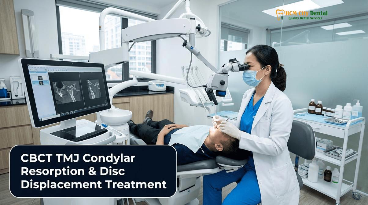
DC/TMD Clinical Classification: Distinguishing Reciprocal Clicking (Disc Displacement With Reduction — ADDwR) from Acute Closed Lock (Without Reduction — ADDwoR)
The Diagnostic Criteria for Temporomandibular Disorders (DC/TMD) provides a globally recognized, evidence-based framework for classifying intracapsular joint pathologies. The articular disc, a dense, avascular pad of fibrocartilage, normally sits interposed between the mandibular condyle and the articular eminence of the temporal bone. Its primary function is to absorb shock, distribute occlusal loads, and facilitate smooth hinge and translational movements of the mandible. However, due to microtrauma, macrotrauma, bruxism, or ligamentous laxity, the disc can become displaced—most commonly in an anteromedial direction due to the continuous traction of the superior belly of the lateral pterygoid muscle. Understanding the clinical presentation of this displacement is the first step in effective TMJ management.
Disc Displacement With Reduction (ADDwR): The Reciprocal Click
In Disc Displacement With Reduction (ADDwR), the articular disc rests anterior to the condylar head when the jaw is closed in Maximum Intercuspation (MI). As the patient begins to open their mouth, the condyle rotates and then translates forward. During this forward translation, the condyle forcefully snaps over the thickened posterior band of the displaced disc, recapturing its normal position on the intermediate zone. This sudden recapture produces a distinct, audible, and palpable “opening click.” As the patient closes their mouth, the condyle slides backward, and just before teeth touch, the disc slips anteriorly off the condyle again, producing a softer “closing click.” This phenomenon is known as reciprocal clicking. Patients with ADDwR typically maintain a normal range of motion (maximum incisal opening of 40mm to 50mm) and may experience only mild, intermittent discomfort. However, the presence of a reciprocal click indicates that the collateral ligaments tethering the disc to the condyle have been elongated and permanently stretched[1].
Disc Displacement Without Reduction (ADDwoR): The Acute Closed Lock
A critical clinical transition occurs when a patient reports, “My jaw used to click all the time, but the clicking suddenly stopped, and now I can’t open my mouth.” This is the hallmark of Disc Displacement Without Reduction (ADDwoR). In this acute phase, the disc remains permanently displaced anterior to the condyle. During mouth opening, the condyle attempts to translate forward but is physically blocked by the bunched-up, displaced disc. The disc acts as a mechanical obstacle, resulting in an acute “closed lock.” Clinically, the patient’s maximum unassisted opening is severely restricted, typically to less than 25mm to 30mm (often described as a “two-finger” opening). Furthermore, because the affected condyle cannot translate forward, the mandible will deflect toward the affected side upon opening. This condition is highly painful because the condyle is now articulating directly on the highly vascularized and innervated retrodiscal tissue (the bilaminar zone), leading to severe retrodiscitis, joint effusion, and muscle splinting.
To manage the acute muscular spasms associated with ADDwoR, our clinical protocol at HCMC Dental Clinic often integrates synergistic therapeutic Botulinum Toxin Type A injections. By carefully administering 20–30U into the Masseter muscle (strictly within the lower safety zone to avoid the marginal mandibular nerve and parotid gland) and 10–15U into the Temporalis muscle, we can significantly reduce elevator muscle hyperactivity. This pharmacological intervention, paired with a hard occlusal night guard, breaks the cycle of muscle trismus, allowing for manual mandibular distraction and unlocking of the joint.
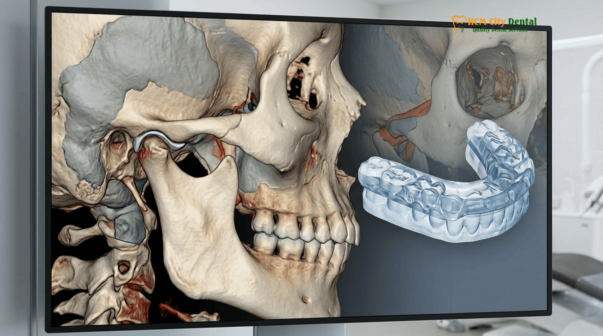
Bilateral 3D CBCT Temporomandibular Joint Imaging: Evaluating Condylar Cortical Erosion, Osteophytes, Joint Space Narrowing, and Idiopathic Condylar Resorption (ICR)
While Magnetic Resonance Imaging (MRI) remains the gold standard for visualizing the soft-tissue articular disc and joint effusion, high-resolution 3D Cone Beam Computed Tomography (CBCT) is the undisputed modality of choice for evaluating the osseous (bony) architecture of the TMJ. At HCMC Dental Clinic, a bilateral 3D CBCT TMJ diagnostic scan is a mandatory prerequisite for any patient presenting with signs of occlusal instability, facial asymmetry, or TMD symptoms prior to commencing major restorative or orthodontic therapy.
Radiographic Hallmarks of TMJ Osteoarthritis
When the articular disc is permanently displaced (ADDwoR), the protective fibrocartilage barrier is lost. The condyle begins to articulate directly against the glenoid fossa, separated only by the inflamed retrodiscal tissues. Over time, this abnormal mechanical loading triggers degenerative joint disease (osteoarthritis). A 3D CBCT scan allows our specialists to meticulously evaluate the condylar head in the sagittal, coronal, and axial planes. We specifically look for:
- Cortical Erosion: The loss of the continuous, radiopaque cortical outline of the condylar head, indicating active bone breakdown and acute inflammation.
- Osteophyte Formation: Bony outgrowths or “bone spurs,” typically forming on the anterior aspect of the condyle. This is a classic sign of the body attempting to increase the surface area of the joint to distribute abnormal loads.
- Subchondral Cysts (Ely’s Cysts): Radiolucent voids beneath the cortical bone, representing areas of localized bone necrosis due to micro-fractures and synovial fluid intrusion.
- Subchondral Sclerosis: Thickening and increased radiopacity of the bone beneath the articulating surface, representing a chronic, adaptive response to heavy loading.
- Joint Space Narrowing: By measuring the anterior, superior, and posterior joint spaces, we can infer condylar position. A significantly decreased posterior joint space strongly suggests posterior condylar displacement, which often correlates with anterior disc displacement[2].
Idiopathic Condylar Resorption (ICR): The Progressive Open Bite
One of the most devastating TMJ pathologies identifiable on a 3D CBCT is Idiopathic Condylar Resorption (ICR), historically referred to as “cheerleader syndrome.” ICR is an aggressive, progressive, and often bilateral resorption of the condylar heads, predominantly affecting females between the ages of 15 and 35. While the exact etiology remains multifactorial, it is strongly linked to hormonal imbalances (specifically estrogen receptor variations in the TMJ), systemic autoimmune conditions, and mechanical overloading.
As the condylar heads rapidly resorb and lose vertical height, the entire mandible undergoes a clockwise rotation. Clinically, this manifests as a sudden, progressive anterior open bite (where the front teeth no longer touch) and a rapidly developing retrognathic (recessed) chin profile. The posterior molars become the only point of contact, acting as a destructive fulcrum. Identifying ICR on a CBCT scan is critical; if a dentist fails to diagnose active ICR and attempts to close the open bite with traditional orthodontics or crowns, the increased mechanical load will simply accelerate the condylar destruction, leading to catastrophic failure.
“The 3D CBCT scan is the ultimate truth-teller in comprehensive dentistry. You cannot build a stable, long-lasting bite on a foundation of actively resorbing condyles. Diagnosing Idiopathic Condylar Resorption early allows us to halt the destruction, stabilize the joint, and prevent a lifetime of chronic pain and occlusal collapse.”
— Dr. Nguyen Van Cuong, DDS (Clinical Director — MOH License 0003265/BL-CCHN)
Why Irreversible Full-Mouth Veneers, Crowns, or Orthodontics Must Never Begin Until Condylar Position Is Stabilized in Adapted Centric Posture
A fundamental tenet of advanced prosthodontics and gnathology is that the occlusion (the bite) and the TMJ are intimately and inextricably linked. The teeth dictate the endpoint of jaw closure (Maximum Intercuspation – MI), while the TMJs dictate the trajectory of jaw movement. In a healthy system, MI coincides harmoniously with Centric Relation (CR)—the most superior, anterior position of the condyles in the glenoid fossa, resting against the posterior slopes of the articular eminences, with the discs properly interposed.
The Danger of Treating to a Pathological Bite
When a patient suffers from TMJ disc displacement or condylar resorption, their habitual bite (MI) is no longer in harmony with a stable joint position. The muscles of mastication are forced into a state of chronic hyperactivity (muscle engrams) to hold the jaw in a position that allows the teeth to mesh, often pulling the condyles down and forward out of the fossa to avoid compressing the inflamed retrodiscal tissues. This is a pathological, defensive posture.
If a dentist ignores the TMJ and proceeds to place irreversible full-mouth porcelain veneers, zirconia crowns, or initiates skeletal orthodontics based on this unstable, pathological bite, the results are disastrous. Once the restorations are cemented or the teeth are moved, the new occlusion locks the jaw into this strained position. Eventually, the muscles will fatigue, or the joint will undergo further structural breakdown. As the joint settles or degenerates, the entire occlusal scheme shifts. The patient will suddenly develop severe premature contacts on their new crowns, leading to porcelain fractures, mobility of the teeth, periodontal trauma, and excruciating orofacial pain.
Achieving Adapted Centric Posture
Before any irreversible Biomimetic Full-Mouth Reconstruction & VDO Rehabilitation in Vietnam can commence, the TMJ must be stabilized. In cases where the articular disc is permanently displaced (ADDwoR) and cannot be recaptured, the goal is not to force the disc back into place, but rather to achieve an “Adapted Centric Posture.” This concept, pioneered by Dawson and Okeson, involves unloading the joint to allow the highly vascularized retrodiscal tissues to undergo fibrosis and form a dense, avascular “pseudo-disc.” Once this pseudo-disc forms, the condyle can seat comfortably and stably in the fossa without pain, even in the absence of the original articular disc[3]. Only when this pain-free, repeatable, and structurally stable Adapted Centric Posture is achieved—and verified by serial 3D CBCT scans showing no active bone resorption—can Phase II restorative or orthodontic treatment safely begin.
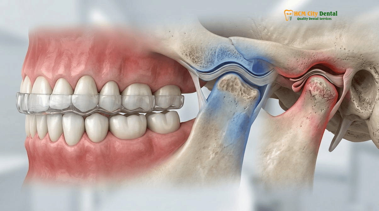
Conservative Reversible Phase I Therapy: CAD/CAM Stabilization Splints, Manual Mandibular Distraction Unlocking, and Physical Therapy
The management of TMJ disc displacement and condylar resorption must always follow a conservative, reversible, and phased approach. Phase I therapy is strictly diagnostic and therapeutic, aimed at eliminating pain, reducing inflammation, deprogramming hyperactive musculature, and establishing a stable condylar position without permanently altering the natural dentition.
The CAD/CAM Michigan Stabilization Splint
The cornerstone of Phase I therapy at HCMC Dental Clinic is the custom-milled CAD/CAM hard acrylic occlusal stabilization splint, often referred to as a Michigan splint. Unlike soft, over-the-counter night guards—which actually stimulate the masseter muscles and exacerbate bruxism by providing a “chewy” surface—a hard, flat-plane stabilization splint provides a rigid, frictionless surface. The splint is meticulously designed to provide simultaneous, even, bilateral pinpoint contacts for all posterior teeth in Centric Relation, while incorporating smooth anterior guidance (canine rise) to immediately disocclude the posterior teeth during any lateral or protrusive excursions.
By wearing the stabilization splint (typically at night, and sometimes 24/7 during acute flare-ups), the patient’s proprioceptive feedback from the teeth is temporarily erased. The lateral pterygoid muscles relax, the elevator muscles deprogram, and the condyles are allowed to seat superiorly and anteriorly into their most orthopedically stable position in the glenoid fossa. This unloads the inflamed retrodiscal tissues, facilitating the formation of the fibrous pseudo-disc required for Adapted Centric Posture.
Manual Mandibular Distraction and Physical Therapy
In cases of acute closed lock (ADDwoR), where the patient presents with a maximum opening of less than 25mm, immediate intervention is required to prevent permanent fibrous adhesions within the joint capsule. Following the administration of therapeutic Botox to relax the trismus, Dr. Cuong may perform manual mandibular distraction. This involves placing the thumbs on the patient’s mandibular molars and applying firm, controlled downward and forward pressure to distract the condyle away from the fossa, attempting to manipulate the condyle back beneath the displaced disc or, at minimum, stretch the joint capsule to restore a functional range of motion.
Adjunctive physical therapy is also critical. Patients are instructed in specific TMJ mobilization exercises, such as Rocabado’s 6×6 exercises, controlled hinge-axis opening, and isometric jaw resistance training. Furthermore, strict adherence to a soft-food diet, avoiding extreme jaw movements (like wide yawning), and the application of moist heat or cryotherapy are essential components of the Phase I protocol[4].
Clinical Case Study: Resolving Acute Closed Lock in an Expat Patient
Patient Profile: A 34-year-old Australian expatriate living in Thao Dien, District 2, presented to HCMC Dental Clinic with severe left pre-auricular pain and an inability to open her mouth wider than 22mm. She reported a history of a loud clicking jaw that had suddenly stopped two days prior.
Clinical Findings & CBCT Diagnosis: Clinical examination revealed severe deflection of the mandible to the left upon opening. A bilateral 3D CBCT scan ruled out active condylar resorption or fractures but showed severe posterior displacement of the left condyle with significant joint space narrowing. The diagnosis was acute Disc Displacement Without Reduction (ADDwoR).
Phase I Intervention: Dr. Nguyen Van Cuong, DDS, immediately administered 25U of Botulinum Toxin to the left masseter and 15U to the left temporalis to break the severe muscle trismus. A digital intraoral scan was taken, and a hard CAD/CAM stabilization splint was milled and delivered within 24 hours. Manual distraction therapy was performed, increasing her opening to 35mm.
Outcome: After 3 months of strict splint wear and physical therapy, the patient achieved a pain-free Adapted Centric Posture with a functional opening of 42mm. Serial CBCT confirmed joint stability, allowing her to safely proceed with her planned Invisalign therapy without risking further TMJ trauma.
Managing Progressive Condylar Resorption & Bite Collapse: Unloading the Retrodiscal Bilaminar Zone Before Phase II Prosthetic Reconstruction
When dealing with Idiopathic Condylar Resorption (ICR), the clinical stakes are exceptionally high. The primary objective is to arrest the active bone degradation. This requires absolute compliance with Phase I stabilization splint therapy to completely unload the retrodiscal bilaminar zone. In severe cases of ICR, systemic pharmacological management (such as the use of specific anti-inflammatory medications or consultation with a rheumatologist) may be necessary to control the underlying autoimmune or hormonal drivers of the resorption.
Transitioning to Phase II: Advanced Orthodontic Biomechanics
Once serial 3D CBCT scans (taken 6 to 12 months apart) confirm that the condylar cortical bone has re-corticated (formed a new, dense outer layer) and no further loss of vertical ramus height is occurring, the TMJ is deemed stable. Only then can Phase II reconstruction begin. The challenge in post-ICR patients is correcting the severe anterior open bite and retrognathic profile without re-loading the fragile, newly adapted TMJs.
Traditional orthodontics, which often involves extruding anterior teeth to close an open bite, is highly unstable and aesthetically poor. Instead, at HCMC Dental Clinic, we employ advanced Wave 4B skeletal TAD (Temporary Anchorage Device) orthodontics. By utilizing dual bucco-palatal TADs (such as extra-radicular Mandibular Buccal Shelf – MBS 2.0x12mm stainless steel TADs or Infra-Zygomatic Crest – IZC TADs), we can achieve true posterior molar intrusion. Biomechanically, for every 1mm of posterior molar intrusion achieved via TADs, the mandible undergoes a counterclockwise autorotation, resulting in 2–3mm of anterior open bite closure. This sophisticated technique closes the bite by rotating the jaw upward and forward, significantly improving the patient’s chin projection and facial profile, all while maintaining the condyles in their safe, Adapted Centric Posture.
In pediatric or adolescent patients presenting with early signs of TMJ dysfunction or transverse maxillary deficiency, early intervention is critical. Phase I pediatric Rapid Palatal Expansion (RPE or Invisalign First at ages 7–10, prior to midpalatal suture fusion) combined with Leeway Space (E-Space) preservation can correct skeletal crossbites, improve nasal airway volume, and provide the mandible with the necessary spatial freedom to grow downward and forward, potentially mitigating the risk of future disc displacement or condylar compression[5].
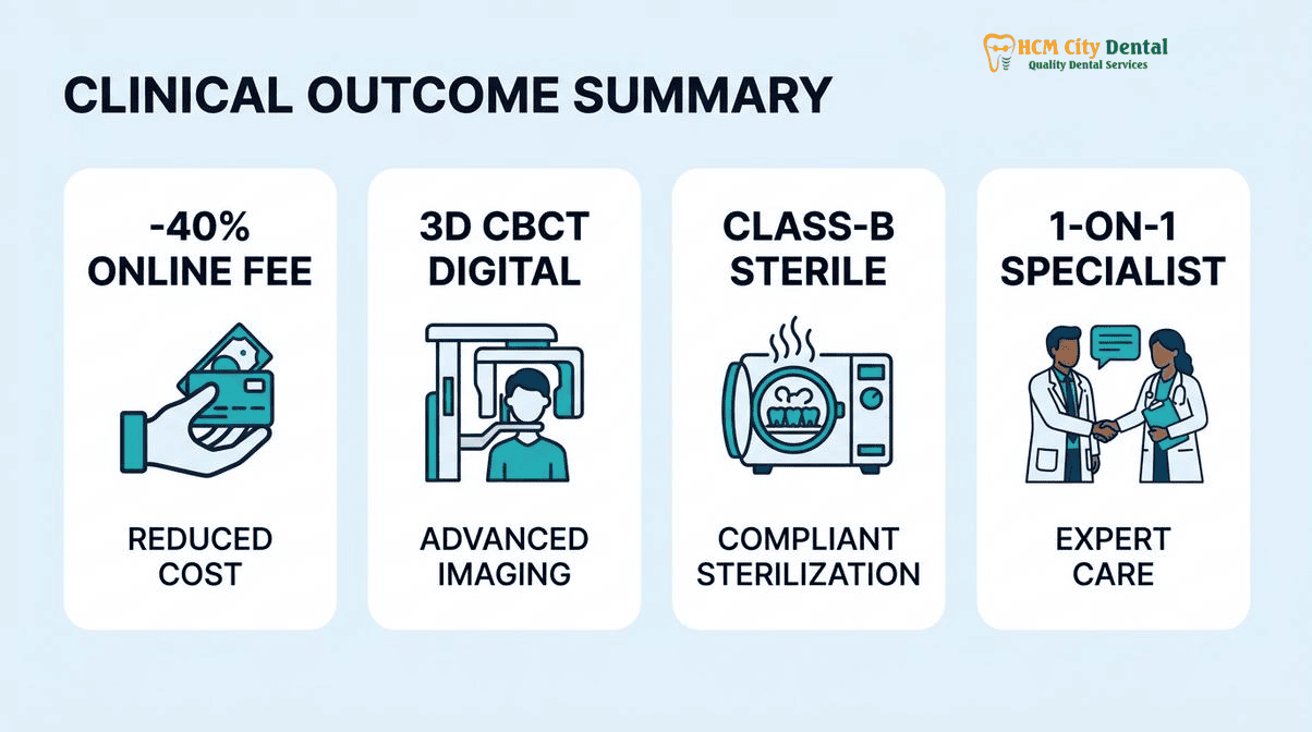
2026 Bilateral 3D CBCT TMJ Diagnostic Scan & Custom Occlusal Stabilization Splint Fee Schedule at HCMC Dental Clinic (District 1)
For international patients, expats, and dental travelers, accessing specialist-level TMJ diagnostics and CAD/CAM splint therapy in the US, Australia, or Europe can be prohibitively expensive, often costing thousands of dollars just for the initial Phase I stabilization. At HCMC Dental Clinic, we believe that foundational joint stability should be accessible. Our transparent Official 2026 Dental Price List & -40% Online Booking Fee Schedule ensures that patients receive world-class, evidence-based TMJ care utilizing the exact same Carl Zeiss optics, Dentsply Sirona CBCT imaging, and CAD/CAM milling technology found in top-tier Western clinics, but at a fraction of the cost.
| Diagnostic & Phase I TMJ Therapy Procedure | US / Australian Benchmark (USD) | HCMC Dental Clinic Walk-In Price | 2026 Online Booking Price (-40% USD) |
|---|---|---|---|
| Bilateral 3D CBCT TMJ Diagnostic Scan (High-Resolution) | $250 – $400 | $30 (480,000 VND) | $18 USD |
| Custom CAD/CAM Hard Acrylic Stabilization Splint (Michigan Splint) | $800 – $1,500 | $135 – $192 | $81 – $115 USD |
| Full Zirconia Crown (Cercon Standard / HT Multilayer – Phase II) | $1,200 – $2,000 | $256 – $321 | $154 – $192 USD / tooth |
| Orthodontic TAD Mini-Screw (IZC / Buccal Shelf for Molar Intrusion) | $400 – $800 | $128 – $192 | $77 – $115 USD / screw |
| Invisalign Comprehensive (Unlimited aligners for Phase II) | $6,000 – $8,500 | $4,231 (66,000,000 VND) | $2,538 USD |
By securing your consultation through our online booking portal, you guarantee access to these exclusive 2026 rates. Whether you require a simple diagnostic CBCT to investigate a clicking jaw, or comprehensive Phase I splint therapy followed by Phase II full-mouth rehabilitation, our clinical team is equipped to manage the most complex TMJ and occlusal challenges.
“Transparency in clinical protocols and pricing is the bedrock of patient trust. We utilize the exact same DC/TMD diagnostic criteria and CAD/CAM stabilization splint protocols as the leading orofacial pain centers in the United States, ensuring our international patients receive uncompromising care without the exorbitant financial burden.”
— Dr. Nguyen Van Cuong, DDS (Clinical Director — MOH License 0003265/BL-CCHN)
Clinical Red Flags: When to Seek Immediate TMJ Evaluation
Do not ignore the warning signs of progressive TMJ pathology. Contact Dr. Nguyen Van Cuong, DDS, immediately via WhatsApp at +84 853 020 003 if you experience any of the following:
- A sudden inability to open your mouth wider than two fingers (acute closed lock).
- A sudden change in your bite, where your front teeth no longer touch but your back teeth hit heavily (progressive anterior open bite).
- Severe, sharp pain directly in front of the ear that radiates to the temples or neck, especially during chewing.
- A history of a loud clicking jaw that has suddenly gone quiet, accompanied by restricted movement.
Frequently Asked Questions
Why did my jaw stop clicking right before I suddenly couldn’t open my mouth wider than 25mm (2 fingers)?
This clinical scenario represents the acute transition from TMJ Disc Displacement With Reduction (ADDwR) to Disc Displacement Without Reduction (ADDwoR). The clicking sound was the articular disc popping back onto the condyle during opening. When the clicking suddenly stops and mouth opening is restricted to less than 25mm, it means the disc has become permanently trapped anterior to the condyle, acting as a physical mechanical barrier that prevents the jaw from translating forward. This is an acute closed lock requiring immediate clinical intervention.
Can a 3D CBCT scan at HCMC Dental Clinic see the bony TMJ condyles, and when is an adjunct MRI indicated for soft-tissue disc position?
Yes, a high-resolution 3D CBCT scan is the gold standard for evaluating the osseous (bony) structures of the TMJ. It clearly reveals condylar cortical erosion, osteophytes, subchondral cysts, joint space narrowing, and signs of Idiopathic Condylar Resorption (ICR). However, CBCT cannot directly visualize soft tissue. If soft-tissue disc position, retrodiscal inflammation, or joint effusion must be definitively mapped—especially in complex cases of ADDwoR—an adjunct Magnetic Resonance Imaging (MRI) scan is indicated.
Why does Idiopathic Condylar Resorption (ICR) cause the back teeth to hit first and create a sudden adult anterior open bite?
Idiopathic Condylar Resorption (ICR) involves the progressive, aggressive breakdown of the condylar bone, effectively shortening the vertical height of the mandibular ramus. As the condyles resorb and lose height, the mandible rotates downward and backward (clockwise rotation). This skeletal shift causes the posterior molars to collide prematurely during closure, acting as a fulcrum that levers the front teeth apart, resulting in a progressive anterior open bite and a retrognathic (recessed) chin profile.
How long must I wear a Phase I stabilization splint before starting full-mouth zirconia crowns or orthodontics?
Phase I stabilization splint therapy typically lasts between 3 to 6 months, though complex cases of ICR may require 9 to 12 months of monitoring. The goal is to achieve an ‘Adapted Centric Posture’ where the condyles are seated comfortably in the glenoid fossa, the retrodiscal tissues have formed a fibrous pseudo-disc, and the patient is entirely pain-free with a stable, repeatable bite. Irreversible Phase II treatments (like full-mouth zirconia crowns or orthodontics) must never begin until serial 3D CBCT scans confirm that active condylar resorption has ceased and the joint is structurally stable.
How much does a comprehensive 3D CBCT TMJ evaluation and custom CAD/CAM stabilization splint cost in District 1?
At HCMC Dental Clinic in District 1, Ho Chi Minh City, our transparent 2026 online booking fee schedule offers a 3D CBCT scan for just $18 USD (480,000 VND). A custom-milled CAD/CAM hard acrylic occlusal stabilization splint (Michigan splint) costs between $81 and $115 USD. This represents a savings of over 70% compared to equivalent specialist TMJ diagnostic and splint therapy fees in the United States or Australia, without compromising on international clinical standards.
Before initiating irreversible prosthodontics, explore our TMJ Disorder Specialist Hub and Kois Deprogrammer vs. Michigan Stabilization Splint protocol in District 1.
References
- Okeson, J. P. (2019). Management of Temporomandibular Disorders and Occlusion (8th ed.). Elsevier. (Comprehensive guidelines on DC/TMD classification and stabilization splint therapy).
- Ahmad, M., & Schiffman, E. L. (2016). Temporomandibular Joint Disorders and Orofacial Pain. Dental Clinics of North America, 60(1), 105-124. (Analysis of 3D CBCT imaging for condylar cortical erosion and joint space narrowing).
- Wolford, L. M. (2001). Idiopathic condylar resorption of the temporomandibular joint in teenage girls (cheerleaders syndrome). Baylor University Medical Center Proceedings, 14(3), 246-252.
- Dawson, P. E. (2006). Functional Occlusion: From TMJ to Smile Design. Mosby. (Protocols for achieving adapted centric posture before irreversible prosthodontics).
- Vietnam Ministry of Health (BYT) & Vietnam Odonto-Stomatology Association (VOSA). National Clinical Guidelines on Specialist Dental Care, Biomaterial Traceability & EN 13060 Class B Infection Control. (2024).