

The maxillary canine is arguably the most critical tooth in the human dentition, serving as the cornerstone of the dental arch, the primary support for facial aesthetics, and the functional guide for the entire masticatory system. However, due to its long path of eruption from high within the maxilla, it is also the second most frequently impacted tooth, trailing only behind third molars. For international patients, expatriates, and dental travelers seeking advanced orthodontic reconstruction, the management of an unerupted “eye tooth” represents a complex intersection of oral surgery, periodontics, and biomechanical orthodontics. The contemporary gold standard for resolving this clinical challenge is impacted maxillary canine 3D CBCT surgical exposure gold chain traction, a highly specialized protocol that rescues the tooth from the alveolar bone and guides it into its rightful position without compromising the adjacent roots or the surrounding gingival architecture.
Historically, the diagnosis and treatment of impacted canines relied heavily on two-dimensional panoramic radiographs, which often led to surgical guesswork, unexpected root collisions, and devastating resorption of the adjacent lateral incisors. Today, the integration of ultra-high-resolution 0.075mm Cone-Beam Computed Tomography (CBCT) has revolutionized this field. By providing a flawless three-dimensional map of the maxilla, specialists can precisely pinpoint the canine’s crown, assess its proximity to vital structures, and design a targeted surgical approach—whether that involves a closed-eruption technique for deep palatal impactions or an apically positioned flap for labial variants. At HCMC Dental Clinic in District 1, Ho Chi Minh City, this multidisciplinary approach is executed with microscopic precision, utilizing state-of-the-art Temporary Anchorage Devices (TADs) and biomechanical cantilever springs to deliver the optimal 60g of light continuous force required for safe, predictable extrusion.
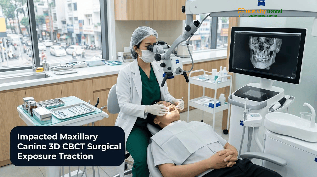
Clinical Summary:
The successful rescue of an impacted maxillary canine requires a meticulously planned sequence of 3D digital diagnostics, minimally invasive surgical exposure, and controlled orthodontic biomechanics. Utilizing 0.075mm 3D CBCT imaging, the exact spatial orientation of the impacted tooth is mapped to prevent collateral damage to adjacent incisor roots. Depending on the impaction vector, a closed-eruption mucoperiosteal flap or an apically positioned flap is utilized to expose the crown, followed by the bonding of a 24k gold traction chain under strict hemostatic isolation. Orthodontic extrusion is then achieved using TAD-supported cantilever springs delivering a precise 60g force, ensuring the canine is safely navigated away from adjacent roots before being integrated into the main archwire. This evidence-based protocol preserves the periodontium, restores canine-protected occlusion, and maintains vital midface aesthetic support.
Key Takeaways:
- 3D CBCT Localization: Essential for mapping palatal vs. labial impactions and detecting silent lateral incisor root resorption before initiating treatment.
- Periodontal Preservation: Closed-eruption techniques are mandatory for palatal impactions, while apically positioned flaps are required for labial impactions to ensure a healthy zone of keratinized tissue.
- Biomechanical Vector Control: Initial traction must pull the canine away from adjacent roots using cantilever springs; tying directly to the main archwire causes catastrophic root collision.
- Optimal Force Application: A light, continuous force of exactly 60g is utilized to stimulate osteoclastic activity without causing hyalinization or root ankylosis.
- Advanced Anchorage: Extra-radicular TADs (mini-implants) provide absolute anchorage, preventing the unwanted intrusion or tipping of adjacent anchor teeth during canine extrusion.
Why the Maxillary Canine Is the Cornerstone of the Dental Arch (Canine Guidance & Facial Support) and Must Rarely Be Extracted
In the architectural framework of the human skull, the maxillary canine occupies a position of unparalleled importance. Anatomically, it possesses the longest and most robust root of any human tooth, deeply embedded within the canine eminence of the maxilla. This dense ridge of bone not only anchors the tooth but also provides critical structural support for the alar base of the nose and the upper lip. When a maxillary canine is missing or extracted, the canine eminence inevitably resorbs, leading to a sunken, prematurely aged appearance in the midface—a phenomenon often referred to as midface collapse. Therefore, the decision to extract an impacted canine is never taken lightly and is generally reserved only for cases of severe ankylosis, untreatable external root resorption, or pathological cystic degeneration.
Beyond aesthetics, the maxillary canine is the functional linchpin of the masticatory system. It is uniquely designed to absorb and dissipate the heavy lateral forces generated during chewing and bruxism. This is achieved through a biomechanical concept known as “canine-protected occlusion” or “canine guidance.” During lateral excursions of the mandible, the upper and lower canines engage, causing the posterior teeth (premolars and molars) to disclude (separate) immediately. This separation protects the multi-rooted posterior teeth from destructive non-axial forces that can lead to enamel fractures, abfractions, and temporomandibular joint (TMJ) dysfunction. The canine’s extensive periodontal ligament (PDL) is also densely packed with proprioceptive nerve endings, which send rapid feedback to the brain to regulate muscle contraction and prevent excessive bite force.
Attempting to substitute a premolar for a missing canine—a compromise sometimes suggested in outdated orthodontic protocols—is fundamentally flawed. A premolar, with its smaller root surface area and different crown morphology, cannot adequately support canine guidance or maintain the volume of the canine eminence. Consequently, modern specialist orthodontics prioritizes the salvage and alignment of the impacted canine whenever biologically possible. This requires a profound understanding of the tooth’s ectopic position and the implementation of advanced surgical and orthodontic techniques to guide it into the arch. For patients seeking to save their impacted eye tooth with braces in Ho Chi Minh City, the clinical team at HCMC Dental Clinic employs a rigorous, preservation-first philosophy.
“The maxillary canine is not just another tooth; it is the primary guardian of the occlusion and the structural pillar of the midface. Extracting it for convenience compromises the patient’s functional longevity and facial aesthetics. With modern 3D CBCT and targeted biomechanics, our primary mandate is always to rescue, erupt, and restore the impacted canine to its rightful place in the arch.”
— Dr. Nguyen Van Cuong, DDS (Clinical Director — MOH License 0003265/BL-CCHN)
0.075mm 3D CBCT Localization (Ericson & Kurol Sector Analysis): Mapping Palatal vs. Labial Impaction and Detecting Adjacent Lateral Incisor Root Resorption
The successful management of an impacted canine begins with flawless diagnostic imaging. Traditional two-dimensional panoramic radiographs are notoriously inadequate for this task. They suffer from magnification errors, distortion, and superimposition of anatomical structures, making it nearly impossible to accurately determine whether a canine is located palatally (toward the roof of the mouth) or labially (toward the lip). Furthermore, 2D imaging fails to detect early-stage root resorption of the adjacent lateral incisors in over 50% of cases. To eliminate these diagnostic blind spots, the implementation of ultra-high-resolution 0.075mm Cone-Beam Computed Tomography (CBCT) is an absolute clinical necessity.
CBCT technology captures a volumetric dataset of the maxilla, allowing the specialist to view the impacted tooth in the axial, coronal, and sagittal planes. This 3D visualization is critical for applying the Ericson and Kurol sector analysis, a standardized method for quantifying the severity of the impaction. The analysis divides the anterior maxilla into five distinct sectors based on the position of the canine cusp tip relative to the long axis of the adjacent lateral and central incisors. A canine located in Sector 1 (distal to the lateral incisor root) has a highly favorable prognosis for eruption, whereas a canine in Sector 5 (mesial to the central incisor root) represents a severe, complex impaction requiring extensive biomechanical intervention. Additionally, the alpha angle—the angle formed by the long axis of the impacted canine and the mid-sagittal plane—is measured to predict the difficulty of the required rotational and tipping movements[1].
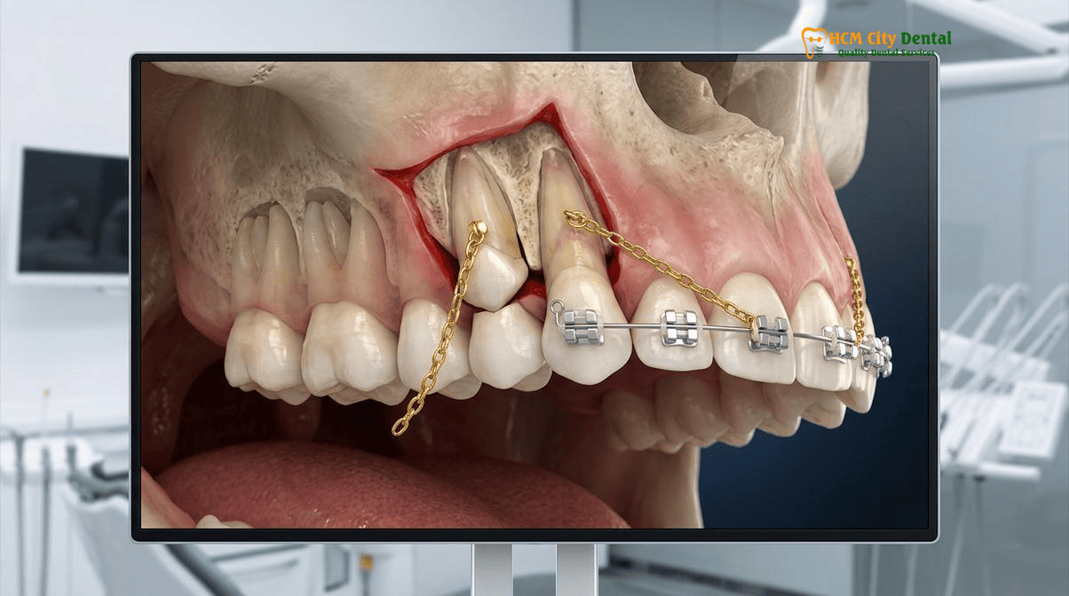
Perhaps the most critical function of 3D CBCT in these cases is the early detection of lateral incisor root resorption. As the ectopic canine follicle enlarges and the crown attempts to erupt, it can exert pressure on the adjacent roots, triggering a localized inflammatory response and osteoclastic clastic activity. This silent, asymptomatic process can rapidly dissolve the root of the lateral incisor, leading to tooth mobility and eventual loss. High-resolution CBCT allows the clinician to detect microscopic resorption lacunae on the palatal or distal aspects of the incisor roots long before they become visible on conventional X-rays. If resorption is detected, the orthodontic traction vector must be immediately designed to pull the canine crown away from the affected root, halting the clastic process and allowing the periodontium to repair.
Furthermore, CBCT imaging is essential for evaluating the morphology of the impacted canine itself. It reveals the presence of root dilacerations (severe bends in the root), hypercementosis, or cystic changes within the dental follicle. Most importantly, it allows the clinician to assess the periodontal ligament space. A continuous, uniform radiolucent PDL space indicates a healthy, movable tooth. Conversely, an obliterated PDL space where the root dentin appears fused directly to the surrounding alveolar bone is a definitive sign of ankylosis, a condition that renders orthodontic movement impossible and necessitates alternative surgical strategies[2].
Periodontal Flap Selection: Closed-Eruption Gold-Chain Technique for Deep Palatal Canines vs. Apically Positioned Flap for Labial Impactions
Once the exact 3D coordinates of the impacted canine are established, the next critical phase is the surgical exposure. The choice of surgical technique is not arbitrary; it is dictated entirely by the anatomical position of the tooth (palatal vs. labial) and the necessity to preserve a healthy, functional periodontium. The overarching goal is not merely to uncover the tooth, but to ensure that when it finally erupts into the dental arch, it is surrounded by a robust collar of keratinized, attached gingiva, free from mucosal inflammation or recession.
For palatally impacted canines, which account for approximately 85% of all cases, the closed-eruption technique is the undisputed gold standard. The palatal mucosa is entirely composed of thick, keratinized masticatory mucosa, which provides an excellent environment for healing. The procedure begins with the elevation of a full-thickness mucoperiosteal flap to expose the overlying palatal bone. Using a piezoelectric surgical handpiece or a high-speed surgical bur under copious sterile saline irrigation, the bone covering the canine crown is carefully removed. Strict hemostasis is then achieved using epinephrine-soaked pellets to ensure a perfectly dry field. The enamel is etched, primed, and a specialized orthodontic eyelet with a pre-attached 24k gold traction chain is bonded to the crown using a dual-cure composite resin. The critical step in the closed-eruption technique is that the mucoperiosteal flap is then sutured back into its original position, completely covering the tooth and the bonded attachment, with only the gold chain exiting through a small surgical window. This technique mimics the natural eruption process, allowing the canine to migrate through the bone and emerge through the crestal tissue with a perfectly formed periodontal attachment[3].
Clinical Warning: Avoiding Open Exposure in the Palate
Performing an “open exposure” (excising a large window of palatal tissue and leaving the crown uncovered) for deep palatal impactions is strongly discouraged. This outdated approach leads to delayed healing, severe post-operative discomfort, and the formation of thick, fibrotic scar tissue that can physically impede the orthodontic traction of the canine. The closed-eruption technique ensures primary intention healing and optimal biomechanical efficiency.
Conversely, labially impacted canines present a completely different periodontal challenge. These teeth are often located high in the buccal vestibule, covered only by thin, fragile, unattached alveolar mucosa. If a simple window is cut through this mucosa to expose the tooth (a “window excision” or laser gingivectomy), the canine will erupt without any protective keratinized tissue. This inevitably leads to chronic marginal inflammation, severe gingival recession, and alveolar bone loss on the facial aspect of the tooth. To prevent this, an apically positioned flap (APF) must be utilized. The surgeon creates a split-thickness or full-thickness flap that includes the band of keratinized tissue from the crest of the ridge. After exposing the canine crown and bonding the attachment, the flap is sutured apically (higher up on the root), physically relocating the keratinized tissue over the erupting crown. As the tooth is pulled downward into the arch, it brings this healthy band of attached gingiva with it, ensuring long-term periodontal stability and a highly aesthetic gingival margin.
Testing for Ankylosis & Controlled Biomechanical Traction: Using Ballista Loops and Mini-Implant (TAD) Cantilever Springs (60g Light Continuous Force)
Following successful surgical exposure and bonding, the clinical focus shifts to the biomechanical phase: the controlled orthodontic traction of the impacted canine. Before active force is applied, the surgeon must definitively rule out ankylosis. While CBCT provides excellent predictive value, clinical confirmation is essential. During the surgical exposure, the clinician may gently apply a luxating force to the canine crown. A slight, microscopic mobility confirms the presence of an intact periodontal ligament. Additionally, a metallic, high-pitched sound upon percussion is a classic clinical indicator of ankylosis. If the tooth is ankylosed, no amount of orthodontic force will move it; attempting to do so will only result in the severe intrusion of the adjacent anchor teeth.
Once mobility is confirmed, the traction protocol begins. The fundamental principle of moving an impacted tooth through dense alveolar bone is the application of light, continuous force. The optimal force magnitude for canine extrusion is approximately 60 grams (about 2 ounces). This specific force level is critical because it slightly exceeds the capillary blood pressure within the periodontal ligament, inducing a localized, controlled ischemia that triggers osteoclastic bone resorption ahead of the moving tooth. If heavy forces (e.g., >150g) are applied, the blood supply to the PDL is completely crushed, leading to sterile necrosis and hyalinization. The bone remodeling process halts entirely, and the tooth stops moving—a phenomenon known as “orthodontic lock-up.”
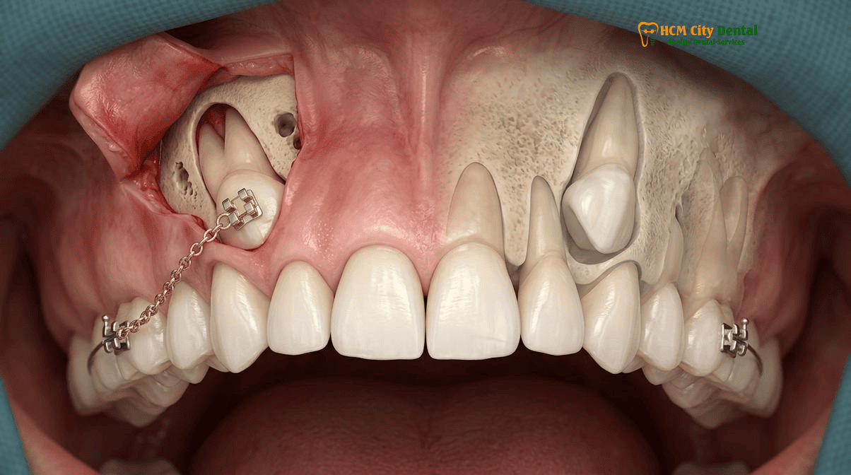
To deliver this precise, continuous force, specialists utilize advanced biomechanical auxiliaries, most notably cantilever springs and ballista loops fabricated from Titanium Molybdenum Alloy (TMA) or stainless steel. A cantilever spring is a single-ended wire engaged in the auxiliary tube of the molar band, extending forward to attach to the gold chain. Because of its long span and the specific properties of the TMA alloy, the cantilever delivers a near-constant, light force over a long range of activation, requiring fewer adjustments and minimizing patient discomfort[4].
However, Newton’s Third Law of Motion dictates that for every action, there is an equal and opposite reaction. When a 60g extrusive force is applied to pull the canine down, an equal 60g intrusive force is exerted on the anchor teeth (usually the molars and premolars). In patients with compromised periodontal support or when multiple teeth are impacted, this reactive force can cause the posterior teeth to intrude, leading to a lateral open bite. To neutralize this unwanted side effect, modern protocols integrate Temporary Anchorage Devices (TADs). These titanium mini-implants are placed directly into the dense cortical bone of the Infra-Zygomatic Crest (IZC) or the palatal vault. By anchoring the cantilever spring directly to the TAD rather than the adjacent teeth, the clinician achieves “absolute anchorage.” The canine is extruded efficiently, while the rest of the dental arch remains perfectly stable and undisturbed.
Why Never to Pull Directly Into the Main Archwire Initially: Vector Control to Move the Canine Crown Away from Lateral Incisor Roots Before Arch Alignment
One of the most catastrophic errors in the management of impacted canines is the premature engagement of the tooth into the continuous main archwire. When a deeply impacted palatal canine is tied directly to a flexible Nickel-Titanium (NiTi) archwire spanning the incisors and premolars, the biomechanical vectors are entirely uncontrolled. The archwire will attempt to take the shortest path to alignment, which often pulls the bulky crown of the canine directly mesially and labially—straight into the delicate roots of the adjacent lateral and central incisors. This “collision course” is the primary cause of severe, iatrogenic root resorption, often resulting in the tragic loss of healthy anterior teeth.
To prevent this, specialist orthodontists employ a strict two-vector traction protocol. The initial phase of movement is never about aligning the tooth; it is solely about spatial clearance. The first vector of force must be directed vertically (downward toward the occlusal plane) and distally (backward toward the premolars), intentionally moving the canine crown away from the incisor roots. This is achieved using the aforementioned TAD-supported cantilever springs or a heavy, rigid base archwire (e.g., 0.019 x 0.025 stainless steel) equipped with a soldered directional hook.
Clinical Case Study: Vector Control in District 1, HCMC
A 24-year-old Australian expat presented to HCMC Dental Clinic with a retained deciduous upper right canine and a deeply impacted permanent canine (Sector 4, palatal). 3D CBCT revealed the canine crown was resting less than 0.5mm from the apical third of the lateral incisor root. Instead of immediate archwire engagement, Dr. Cuong designed a custom TMA cantilever spring anchored to a palatal TAD. For the first 4 months, a purely distal-occlusal force of 60g was applied, safely navigating the canine crown away from the incisor root. Only after CBCT confirmation showed complete spatial clearance was the gold chain attached to the main archwire for final labial torque and alignment. The result: perfect canine guidance restored with zero root resorption to adjacent teeth.
During this initial clearance phase, the patient’s main archwire serves only as a rigid stabilizing unit for the rest of the dentition. The gold chain is activated every 3 to 4 weeks, and the progress is monitored clinically and radiographically. Only when the crown of the canine has safely bypassed the roots of the incisors and has emerged through the palatal or alveolar mucosa is the second vector applied. At this stage, an orthodontic bracket is bonded to the newly exposed facial surface of the canine, and it is finally engaged into a flexible NiTi archwire to initiate the labial movement, rotation, and final detailing required to integrate it seamlessly into the dental arch[5].
“Vector control is the essence of safe canine traction. We must think in three dimensions. Pulling an impacted tooth blindly toward the archwire is akin to driving a car through a building to get to the street. We must first navigate the tooth out of the bony vault, clear the adjacent roots, and only then steer it into its final aesthetic position.”
— Specialist Orthodontics, TADs & Adult Palatal Expanders in Ho Chi Minh City
2026 Impacted Canine 3D CBCT Surgical Exposure & Orthodontic Traction Fee Schedule at HCMC Dental Clinic (District 1)
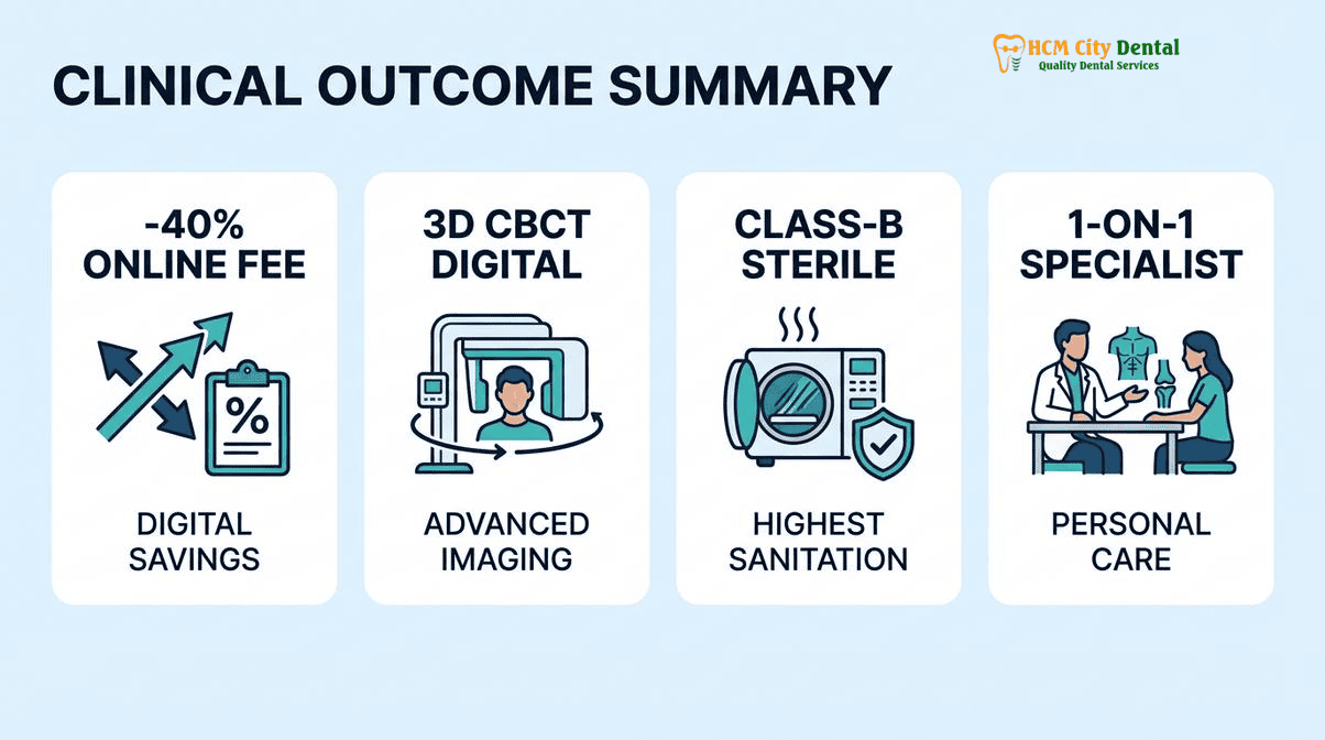
For international patients, expats, and dental travelers, the cost of complex interdisciplinary treatments like the surgical exposure and orthodontic traction of an impacted canine can be prohibitively expensive in countries like Australia, the US, or the UK. At HCMC Dental Clinic, located in the heart of District 1, Ho Chi Minh City, patients receive world-class, evidence-based care utilizing the exact same 3D CBCT technology, Carl Zeiss microscopes, and biocompatible materials (TMA wire, 24k gold chains, titanium TADs) at a fraction of the Western cost. The clinic’s transparent Official 2026 Dental Price List & -40% Online Booking Fee Schedule ensures that patients can plan their medical travel with absolute financial clarity.
The management of an impacted canine is typically integrated into a comprehensive orthodontic treatment plan, with the surgical exposure and 3D imaging billed as specific adjunctive procedures. Below is the detailed 2026 fee schedule for the diagnostic, surgical, and orthodontic components required for this specialized protocol.
| Clinical Procedure / Biomaterial Component | 2026 Online Booking Fee (-40% USD) | Walk-In / Standard List Price (USD) | Estimated US / AUD Benchmark |
|---|---|---|---|
| High-Resolution 3D CBCT Scan (0.075mm Voxel, Ericson & Kurol Analysis) | $18 USD (480,000 VND) | $30 USD | $250 – $400 USD |
| Surgical Exposure & Gold Chain Bonding (Closed Eruption or Apically Positioned Flap) | $58 – $115 USD (1,500,000 – 3,000,000 VND) | $96 – $192 USD | $800 – $1,500 USD |
| Orthodontic TAD Mini-Screw (IZC / Palatal Anchorage for Cantilever Mechanics) | $77 – $115 USD / screw | $128 – $192 USD | $400 – $800 USD |
| Invisalign Comprehensive (Unlimited Aligners, includes complex traction planning) | $2,538 USD (66,000,000 VND) | $4,231 USD | $6,500 – $9,000 USD |
| Invisalign Vivera Retainers (Genuine Align Technology SmartTrack – 3 Sets) | $308 USD (8,000,000 VND) | $513 USD | $800 – $1,200 USD |
| Single European / Swiss Implant (Straumann BLT-SLA / BLA-Active – if canine is ankylosed and extracted) | $962 – $1,346 USD (25,000,000 – 35,000,000 VND) | $1,603 – $2,244 USD | $4,500 – $6,500 USD |
| Full Zirconia Crown (Cercon Standard / HT Multilayer – for implant restoration) | $154 – $192 USD / tooth (4,000,000 – 5,000,000 VND) | $256 – $321 USD | $1,200 – $2,000 USD |
By leveraging the Official 2026 Dental Price List & -40% Online Booking Fee Schedule, patients can access elite-level surgical orthodontics without compromising on safety, sterilization (EN 13060 Class B), or clinical expertise. Dr. Nguyen Van Cuong and his multidisciplinary team ensure that every impacted canine is treated with the utmost respect for periodontal biology and biomechanical precision, restoring both the functional integrity of the bite and the aesthetic harmony of the smile.
Frequently Asked Questions
How can 3D CBCT predict whether an impacted upper canine in an adult is fused to the bone (ankylosed) before starting braces?
High-resolution 0.075mm 3D CBCT imaging allows specialists to evaluate the periodontal ligament (PDL) space surrounding the impacted canine. If the dark radiolucent line representing the PDL is obliterated and the tooth root appears continuous with the surrounding alveolar bone, ankylosis is highly suspected. This critical diagnostic step prevents futile orthodontic traction attempts and allows the clinical team to plan alternative treatments, such as surgical luxation or extraction followed by implant placement.
Why does a labially impacted canine require an apically positioned mucogingival flap rather than simple laser gingivectomy through unattached mucosa?
A labially impacted canine must erupt through a zone of keratinized, attached gingiva to ensure long-term periodontal health. If a simple laser gingivectomy is performed through unattached alveolar mucosa, the tooth will erupt with a mucosal margin, leading to chronic inflammation, severe gingival recession, and bone loss. An apically positioned flap surgically relocates the keratinized tissue over the erupting crown, creating a robust, healthy biological width and preventing future mucogingival defects.
How long does it take to bring a palatally impacted canine into the dental arch using a gold chain and cantilever spring?
The duration of orthodontic traction depends on the severity of the impaction, the patient’s age, and bone density. On average, moving a palatally impacted canine into the dental arch takes between 8 to 14 months. The process utilizes light, continuous forces (approximately 60g) via a cantilever spring and gold chain to move the tooth at a biological rate of about 1mm per month, ensuring the preservation of the root structure and surrounding alveolar bone.
What if my baby canine (deciduous tooth) is still in place at age 25 or 35—should I extract it for an implant or pull down the impacted adult canine?
The decision depends on the condition of the deciduous canine, the position of the impacted adult canine, and the presence of ankylosis. If the adult canine is in a favorable position (low Ericson & Kurol sector) and not ankylosed, surgical exposure and orthodontic traction is the gold standard, even in adults. However, if the adult canine is deeply impacted, ankylosed, or causing severe root resorption to adjacent teeth, it may be extracted. If the baby tooth has good root length, it can be retained; otherwise, it is extracted and replaced with a dental implant.
How much does surgical exposure and bonding of a 24k gold traction chain cost at HCMC Dental Clinic in District 1?
At HCMC Dental Clinic, the comprehensive management of an impacted canine is highly affordable. A high-resolution 3D CBCT scan costs just $18 USD online. The surgical exposure and bonding of the gold chain typically falls under advanced minimally invasive procedures or surgical extractions, ranging from $58 to $115 USD depending on complexity. The subsequent orthodontic traction is integrated into the comprehensive braces or Invisalign treatment plan, offering world-class specialist care at a fraction of Western prices.
Patients can also review our Piezosurgery ultrasonic bone window protocol and 2026 orthodontic & Invisalign fee schedule prior to scheduling 3D CBCT impacted canine localization in District 1.
References
- American Journal of Orthodontics and Dentofacial Orthopedics. “Three-dimensional localization of maxillary canines with cone-beam computed tomography and assessment of lateral incisor root resorption.” (2021).
- Journal of Oral and Maxillofacial Surgery. “Periodontal outcomes of closed eruption versus open surgical exposure of palatally impacted maxillary canines: A systematic review.” (2022).
- The Angle Orthodontist. “Biomechanical considerations in the orthodontic traction of impacted maxillary canines using temporary anchorage devices (TADs) and cantilever springs.” (2020).
- International Journal of Periodontics & Restorative Dentistry. “Mucogingival considerations and apically positioned flaps in the management of labially impacted canines.” (2019).
- Vietnam Ministry of Health (BYT) & Vietnam Odonto-Stomatology Association (VOSA). National Clinical Guidelines on Specialist Dental Care, Biomaterial Traceability & EN 13060 Class B Infection Control. (2024).