Yes, bruxism frequently causes ear pain and tinnitus. The temporomandibular joint (TMJ) shares complex nerve pathways and ligaments with the middle ear. Chronic teeth clenching inflames these structures, leading to referred earaches, a sensation of fullness, and persistent ringing without any underlying auditory disease.
For many patients, the journey to resolving unexplained auditory symptoms involves multiple visits to medical specialists, only to discover that the root cause lies within their dental health. Understanding the intricate biomechanical and neurological relationship between the jaw and the ear is the first step toward achieving lasting relief from these debilitating symptoms.
Clinical Summary:
Bruxism-induced otalgia and somatosensory tinnitus arise from the anatomical proximity of the temporomandibular joint to the auditory system. Hyperactivity of the masticatory muscles, particularly the lateral pterygoid, can trigger spasms in the tensor tympani and tensor veli palatini muscles via the trigeminal nerve. This neuromuscular tension manifests as ear pain, aural fullness, and ringing. Clinical management requires a multidisciplinary approach, often utilizing custom occlusal splints to decompress the joint, alongside physical therapy to restore normal neuromuscular function and alleviate the aberrant sensory signals causing the auditory distress.
Key Takeaways:
- The TMJ and middle ear share the trigeminal nerve pathway, allowing jaw muscle tension to radiate directly into the ear canal.
- Secondary otalgia from teeth grinding typically presents without fever, fluid discharge, or acute hearing loss, distinguishing it from an ear infection.
- Somatosensory tinnitus can fluctuate in pitch or volume based on jaw movements, chewing, or clenching intensity.
- Custom occlusal appliances are the primary conservative intervention to reduce joint loading and alleviate auditory symptoms.
- A multidisciplinary diagnostic approach involving both dental and ENT specialists is crucial for ruling out primary otologic pathology.
The TMJ-Ear Anatomy Connection
The temporomandibular joint and the middle ear are anatomically linked by shared embryonic origins, the trigeminal nerve, and the malleomandibular ligament, allowing jaw tension to directly affect auditory function.
To comprehend why a dental condition like bruxism can cause profound auditory symptoms, one must examine the complex embryological and anatomical relationship between the jaw and the ear. During human embryonic development, the structures of the middle ear (specifically the malleus and incus bones) and the mandible both originate from the first branchial arch, known as Meckel’s cartilage. Because they share this common developmental origin, they remain intimately connected throughout life via shared neural pathways, vascular supplies, and ligamentous attachments [1].
The physical proximity of these structures is striking. The temporomandibular joint is located mere millimeters from the external auditory meatus (the ear canal) and the middle ear cavity. The posterior aspect of the TMJ capsule is separated from the ear canal only by a thin plate of bone known as the tympanic portion of the temporal bone. When the jaw joint undergoes trauma, inflammation, or displacement due to chronic clinical definition of bruxism, the resulting edema and pressure can easily transfer to the adjacent auditory structures.
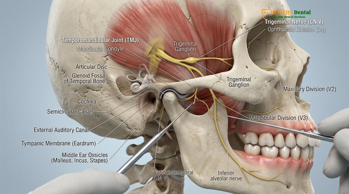
Furthermore, there is a direct physical connection between the jaw and the ear known as the malleomandibular ligament, or Pinto’s ligament. This tiny ligament connects the neck of the malleus (one of the tiny hearing bones in the middle ear) directly to the capsule and meniscus of the temporomandibular joint. When a patient clenches or grinds their teeth, the extreme forces placed on the TMJ can cause tension along Pinto’s ligament, physically tugging on the middle ear bones and altering their movement. This mechanical interference can result in a sensation of ear fullness, muffled hearing, and TMJ ear symptoms.
Neurologically, the connection is even more profound. Both the masticatory muscles (which control chewing) and two critical muscles in the middle ear (the tensor tympani and the tensor veli palatini) are innervated by the mandibular branch of the trigeminal nerve (Cranial Nerve V3). The tensor tympani dampens loud sounds, while the tensor veli palatini opens the Eustachian tube to equalize pressure. When bruxism causes the jaw muscles to become hyperactive and spasm, this neurological hyperactivity can spill over to the ear muscles, causing them to spasm as well. Dr. Nguyen Van Cuong frequently notes in clinical practice that patients are often surprised to learn that their ear fullness is actually a muscle spasm triggered by their jaw, rather than fluid in the ear.
How Bruxism Affects Your Ears
Chronic teeth grinding overworks the masticatory muscles, causing severe inflammation that radiates to the ear canal and disrupts the Eustachian tube, leading to referred pain and aural fullness.
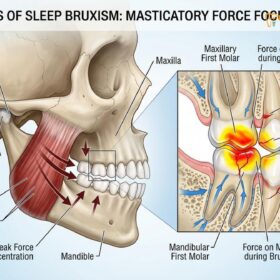
Bruxism is a parafunctional habit characterized by the involuntary clenching, grinding, or gnashing of teeth. It can occur during the day (awake bruxism), often as a subconscious reaction to stress, or during sleep (sleep bruxism), which is considered a sleep-related movement disorder. The human jaw is capable of exerting tremendous force—up to 600 pounds per square inch during nocturnal bruxism episodes. When this force is applied continuously over hours, it leads to profound biomechanical consequences for the entire craniomandibular system [2].
The primary mechanism by which bruxism affects the ears is through referred pain. Referred pain occurs when pain signals originating in one area of the body are perceived by the brain as coming from a different, adjacent area. Because the muscles of mastication—specifically the masseter, temporalis, and medial/lateral pterygoids—are heavily innervated by the trigeminal nerve, chronic overuse leads to the accumulation of lactic acid and inflammatory cytokines within the muscle tissue. This creates myofascial trigger points. A trigger point in the deep masseter or the lateral pterygoid muscle frequently refers a deep, aching pain directly into the ear canal, a condition known as secondary otalgia.
“Secondary otalgia is one of the most common misdiagnoses in primary care. Patients present with severe ear pain, yet otoscopic examination reveals a perfectly healthy tympanic membrane. In these cases, the source of the pain is almost always myofascial, originating from the temporomandibular joint and the surrounding masticatory musculature due to parafunctional habits.”
In addition to referred pain, bruxism directly impacts the function of the Eustachian tube. As previously mentioned, the tensor veli palatini muscle is responsible for opening the Eustachian tube during swallowing or yawning to equalize pressure between the middle ear and the outside environment. Because this muscle shares its nerve supply with the jaw muscles, the chronic hyperactivity seen in bruxism can cause the tensor veli palatini to become hypertonic or go into spasm. When this occurs, the Eustachian tube fails to open properly, leading to negative pressure in the middle ear. Patients experience this as a frustrating sensation of their ears feeling “clogged,” “full,” or “underwater,” which is a hallmark of bruxism ear pain.
Furthermore, the physical displacement of the TMJ articular disc can compress the retrodiscal tissues. The retrodiscal tissue is a highly vascular and heavily innervated pad of tissue located just behind the jaw joint, directly in front of the ear canal. When a patient grinds their teeth, the condyle of the mandible can be pushed backward, crushing this sensitive tissue. The resulting severe inflammation (retrodiscitis) causes acute pain that is almost indistinguishable from a severe earache. Understanding these nocturnal bruxism patterns is essential for accurate diagnosis and effective intervention.
Ear Pain vs Ear Infection: Key Differences
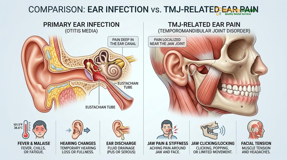
While an ear infection typically presents with fever, fluid discharge, and acute hearing loss, bruxism-induced ear pain is characterized by a dull, constant ache that fluctuates with jaw movement.
Distinguishing between primary otalgia (pain originating from the ear itself, such as an infection) and secondary otalgia (pain referred to the ear from another source, such as the TMJ) is a critical diagnostic step. Because the symptoms can feel remarkably similar to the patient, misdiagnosis is common, leading to unnecessary courses of antibiotics and delayed treatment of the actual underlying dental issue [3].
Primary ear infections, such as acute otitis media (middle ear infection) or otitis externa (swimmer’s ear), are typically caused by bacterial or viral pathogens. They are characterized by acute inflammatory signs. Patients will often present with a fever, purulent (pus-like) discharge from the ear canal, and a sudden, measurable decrease in hearing acuity. The pain is usually sharp, throbbing, and constant, regardless of facial movement. Upon clinical examination with an otoscope, a physician will observe a red, bulging, or inflamed tympanic membrane (eardrum), or severe swelling of the external ear canal.
Conversely, secondary otalgia caused by bruxism and temporomandibular disorders presents quite differently upon careful clinical investigation. The pain is typically described as a deep, dull, continuous ache that may radiate down the neck or up into the temples. Crucially, this pain is heavily influenced by jaw function. Patients will often report that their earache worsens significantly when chewing tough foods, yawning widely, or upon waking in the morning (a classic sign of sleep bruxism). There is no fever, no discharge, and standard audiometric testing usually reveals normal hearing.
| Clinical Feature | Primary Ear Infection (Otitis Media/Externa) | Bruxism-Induced Ear Pain (Secondary Otalgia) |
|---|---|---|
| Pain Characteristics | Sharp, throbbing, acute onset | Dull, aching, chronic, fluctuates in intensity |
| Aggravating Factors | Lying down, pulling on the earlobe | Chewing, yawning, talking, morning waking |
| Associated Symptoms | Fever, fluid discharge, acute hearing loss | Jaw stiffness, facial fatigue, clicking/popping TMJ |
| Otoscopic Exam | Red, bulging eardrum, fluid behind eardrum | Normal, healthy eardrum and clear ear canal |
| Palpation Findings | Tenderness around the ear itself | Severe tenderness in masseter, temporalis, and TMJ |
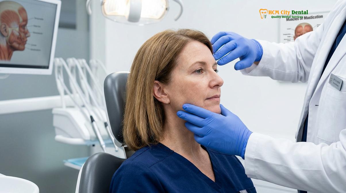
During a clinical evaluation, a dentist will perform specific provocation tests to confirm the source of the pain. Palpating the preauricular area (just in front of the ear) while the patient opens and closes their mouth can often reproduce the exact ear pain the patient is experiencing. Additionally, examining the oral cavity for signs of long-term structural damage to the dentition, such as flattened enamel, abfractions, and scalloped tongue borders, provides strong corroborating evidence that bruxism is the true culprit behind the auditory distress.
Tinnitus and Teeth Grinding
Jaw clenching can trigger somatosensory tinnitus by sending aberrant sensory signals through the trigeminal nerve to the dorsal cochlear nucleus, altering how the brain perceives sound.
Tinnitus is the perception of sound—often described as ringing, buzzing, hissing, or roaring—in the absence of an external acoustic stimulus. While tinnitus is frequently associated with noise-induced hearing loss or age-related auditory decline, a significant subset of patients suffers from what is known as somatosensory (or somatic) tinnitus. This specific subtype of tinnitus is directly influenced by the somatosensory system of the cervical spine and the temporomandibular region [4].
The neurophysiological link between teeth grinding tinnitus and the auditory system lies in the brainstem. The trigeminal nerve, which carries sensory information from the jaw, teeth, and masticatory muscles, projects into the brainstem. Research has demonstrated that there are direct neural connections between the medullary somatosensory nuclei (where trigeminal signals arrive) and the dorsal cochlear nucleus (the first relay station for auditory processing in the brain). When a patient chronically grinds their teeth, the constant barrage of sensory input from the overworked jaw muscles hyper-excites the trigeminal nerve.
This hyperactivity spills over into the dorsal cochlear nucleus, essentially “cross-wiring” the sensory and auditory pathways. The brain misinterprets this excessive muscle tension as auditory signals, resulting in the perception of ringing in the ears. This is why jaw grinding ear ringing is a highly documented clinical phenomenon. A defining characteristic of somatosensory tinnitus is its ability to be modulated by physical movement. Patients often report that the pitch, volume, or intensity of their tinnitus changes when they clench their teeth, protrude their jaw, or press on their facial muscles.
Clinical Case Review: Somatosensory Tinnitus Management
A 42-year-old patient visited HCMC Dental Clinic in Ho Chi Minh City complaining of persistent, high-pitched tinnitus in the right ear that had not responded to traditional ENT treatments. The patient also reported morning jaw stiffness. Clinical examination by Dr. Nguyen Van Cuong revealed severe masseter hypertrophy and a right TMJ click. Provocation testing (asking the patient to clench forcefully) immediately increased the volume of the tinnitus. The patient was diagnosed with sleep bruxism and somatosensory tinnitus. Following the fabrication and consistent use of a custom rigid occlusal splint, combined with targeted physical therapy, the patient reported a 80% reduction in tinnitus volume within six weeks, confirming the somatogenic origin of the auditory symptoms.
It is crucial to differentiate somatosensory tinnitus from objective tinnitus (which can be heard by an examiner and is often vascular in origin, such as pulsatile tinnitus) and otologic tinnitus (caused by inner ear damage). If the tinnitus is accompanied by a heartbeat-like pulsing sound, sudden profound hearing loss, or severe dizziness, immediate medical evaluation is required to rule out vascular anomalies or neurological conditions. However, if the ringing is a steady tone that fluctuates with stress levels and jaw tension, bruxism is a highly probable contributing factor.
Treatment Approaches
Managing jaw-related ear symptoms requires decompressing the TMJ and relaxing masticatory muscles through custom occlusal appliances, physical therapy, and behavioral modifications.
Effectively treating bruxism ear pain and somatosensory tinnitus requires a comprehensive, multidisciplinary approach aimed at reducing the mechanical load on the temporomandibular joint, relaxing the hyperactive musculature, and addressing the underlying parafunctional habits. Because the auditory symptoms are secondary to the dental condition, treating the jaw will inherently alleviate the ear pain and ringing [5].
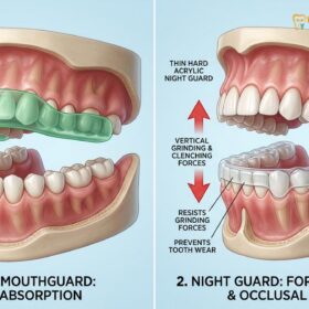
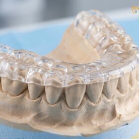
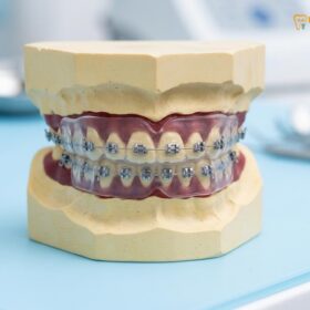
The cornerstone of conservative management is the use of custom occlusal appliances, commonly known as nightguards or occlusal splints. These devices are meticulously fabricated from precise impressions of the patient’s dentition. Unlike over-the-counter boil-and-bite guards, which are soft and can actually encourage more chewing and clenching, professionally fabricated night guards are typically made of hard, durable acrylic. A rigid splint provides a smooth, flat surface that prevents the upper and lower teeth from locking together. This disocclusion reduces the activation of the masseter and temporalis muscles, effectively decompressing the TMJ and allowing the inflamed retrodiscal tissues and associated ear structures to heal.
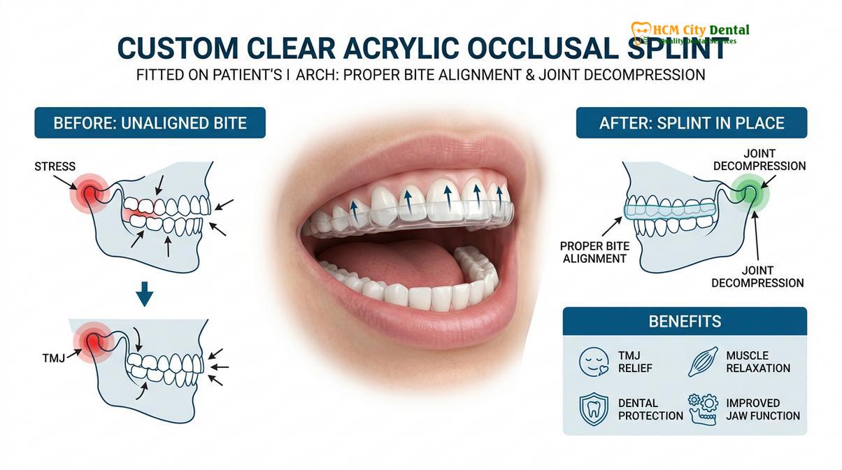
When selecting an appliance, the choice between a rigid acrylic splints and a dual-laminate design depends on the severity of the bruxism and the specific TMJ pathology. A permissive splint allows the mandible to glide freely, seeking its most relaxed, neurologically neutral position, which is highly effective for reducing the muscle spasms that cause Eustachian tube dysfunction and referred ear pain.
“The application of a properly adjusted, hard acrylic occlusal splint remains the gold standard for managing TMD-related auditory symptoms. By establishing a mutually protected occlusion on the appliance, we immediately reduce the proprioceptive feedback that drives nocturnal muscle hyperactivity, thereby breaking the cycle of referred otalgia and somatosensory tinnitus.”
Beyond oral appliances, comprehensive strategies to stop grinding include physical therapy and pharmacological interventions. Physical therapy for the TMJ involves targeted exercises to stretch and strengthen the jaw and neck muscles, improving range of motion and correcting postural imbalances that contribute to cervical-trigeminal tension. Techniques such as myofascial release, trigger point massage, and ultrasound therapy can significantly reduce the localized muscle knots that refer pain to the ear.
In cases of severe, refractory bruxism, pharmacological management may be necessary. Short-term use of muscle relaxants (such as cyclobenzaprine) taken before bed can help break the cycle of nocturnal muscle spasms. Additionally, targeted injections of Botulinum Toxin (Botox) into the masseter and temporalis muscles have shown high success rates in reducing the force of muscle contractions without affecting normal chewing function. By chemically relaxing the muscles, the pressure on the TMJ and the adjacent auditory structures is profoundly reduced, offering significant relief from both ear pain and tinnitus.
When to See an ENT vs Dentist
Navigating the healthcare system when experiencing ear pain and tinnitus can be confusing. Because the symptoms overlap significantly between otologic and dental conditions, knowing which specialist to see—and when—is crucial for timely and accurate diagnosis. A systematic triage approach ensures that serious medical conditions are ruled out before focusing on functional dental management.
As a general clinical guideline, any new onset of ear pain, hearing loss, or tinnitus should first be evaluated by an Ear, Nose, and Throat (ENT) specialist or an otolaryngologist. The primary goal of this initial medical evaluation is to rule out acute infections, structural ear damage, or serious neurological conditions. An ENT will perform a thorough otoscopic examination, an audiogram to assess hearing acuity, and potentially tympanometry to measure middle ear pressure.
Red Flag Symptoms Requiring Immediate Medical Attention:
- Pulsatile Tinnitus: Ringing that pulses in time with your heartbeat (may indicate vascular issues).
- Unilateral Hearing Loss: Sudden or gradual loss of hearing in only one ear (requires evaluation for acoustic neuroma).
- Severe Vertigo: Intense dizziness or a spinning sensation accompanied by nausea (potential Meniere’s disease or labyrinthitis).
- Purulent Discharge: Fluid, blood, or pus draining from the ear canal.
- Facial Weakness: Numbness, tingling, or paralysis on one side of the face.
If the ENT specialist conducts these examinations and concludes that your ears are structurally healthy, your eardrums are clear, and your hearing is normal, the diagnosis of secondary otalgia becomes highly probable. At this juncture, the patient should be referred to a qualified dentist or a specialist in Orofacial Pain and Temporomandibular Disorders.
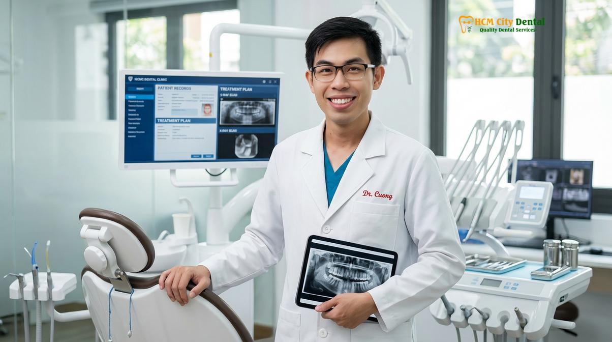
During the dental consultation, the focus shifts from the ear itself to the craniomandibular system. At HCMC Dental Clinic in Ho Chi Minh City, the clinical evaluation involves a comprehensive assessment of the patient’s bite (occlusion), an examination of the teeth for wear facets indicative of bruxism, and precise palpation of the TMJ and masticatory muscles. Advanced imaging, such as a Cone Beam Computed Tomography (CBCT) scan, may be utilized to evaluate the bony structures of the jaw joint and the condylar position. By working collaboratively, ENTs and dental professionals can ensure that patients suffering from bruxism ear pain and tinnitus receive the targeted, effective care they need to restore their quality of life.
Frequently Asked Questions
Can teeth grinding cause ear pain?
Yes, teeth grinding frequently causes ear pain. Because the temporomandibular joint is located just millimeters from the ear canal, inflammation and muscle spasms from chronic clenching radiate directly into the ear, creating a deep, aching sensation known as secondary otalgia. This referred pain is often misdiagnosed as a standard ear infection.
Does bruxism cause tinnitus?
Bruxism can trigger a specific type of ringing known as somatosensory tinnitus. The trigeminal nerve, which innervates the jaw muscles, shares neural pathways with the auditory system in the brainstem, allowing jaw tension to alter how the brain perceives sound. This results in a ringing sensation that often fluctuates with jaw movement.
Why does my ear feel full when I clench?
Ear fullness during clenching occurs due to spasms in the tensor veli palatini muscle. This muscle, controlled by the same nerve as your jaw, is responsible for opening the Eustachian tube. When it spasms due to bruxism, the tube cannot equalize pressure properly, creating a clogged or underwater sensation in the ear.
Will treating bruxism stop ear ringing?
Treating bruxism can significantly reduce or eliminate ear ringing if the tinnitus is somatosensory in origin. By using occlusal splints and muscle relaxation techniques to decompress the jaw, the aberrant nerve signals causing the auditory symptoms are often minimized, leading to a profound decrease in tinnitus volume and frequency.
Should I see an ENT or dentist for ear pain?
You should initially see an ENT to rule out primary ear infections or hearing loss. If the ENT finds your ears are healthy and structural issues are ruled out, you should then consult a dentist or TMJ specialist to evaluate for bruxism, temporomandibular joint dysfunction, and referred secondary otalgia.
References
- Journal of Oral Rehabilitation. Anatomical relationship between the TMJ and middle ear. (2021).
- American Journal of Otolaryngology. Secondary otalgia and temporomandibular disorders. (2020).
- Journal of Craniomandibular & Sleep Practice. Somatosensory tinnitus and bruxism management. (2019).
- International Journal of Prosthodontics. Efficacy of occlusal splints in TMD-related tinnitus. (2022).
- European Archives of Oto-Rhino-Laryngology. Eustachian tube dysfunction in bruxism patients. (2018).