Bruxism effects on teeth include severe enamel erosion, micro-fractures, and compromised dental restorations due to excessive occlusal forces. Left untreated, chronic teeth grinding accelerates tooth wear, triggers temporomandibular joint (TMJ) disorders, and causes gum recession, requiring comprehensive clinical intervention to restore oral function and alleviate chronic facial pain.
Clinical Summary:
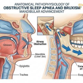
Bruxism, clinically defined as the involuntary clenching and grinding of teeth, is a destructive parafunctional habit that predominantly occurs during sleep. The masticatory forces generated during a bruxism episode can exceed 250 pounds per square inch—exponentially higher than the forces used during normal chewing. This relentless mechanical stress initiates a cascade of oral health deterioration. The primary bruxism effects on teeth manifest as accelerated enamel attrition, exposing the highly sensitive underlying dentin. As the condition progresses, the structural integrity of the dentition is compromised, leading to craze lines, cusp fractures, and the catastrophic failure of dental restorations such as crowns and implants. Furthermore, the hyperactive muscle contractions overload the temporomandibular joint (TMJ) and the periodontium, resulting in chronic facial pain, disc displacement, and non-carious cervical lesions (abfractions). Effective management requires early diagnostic recognition and the implementation of protective occlusal splints to mitigate force distribution and preserve the remaining dentition.
Key Takeaways:
- Sleep bruxism generates occlusal forces up to ten times greater than normal mastication, causing rapid structural degradation.
- Enamel erosion from grinding exposes dentin, leading to severe thermal hypersensitivity and an increased risk of decay.
- Excessive lateral forces cause teeth to flex, contributing to gum recession and non-carious cervical lesions at the gumline.
- Dental restorations, including porcelain crowns and implants, are highly susceptible to fracture and failure under bruxism loads.
- Early intervention with custom-fitted occlusal splints is critical to preventing irreversible damage and complex full-mouth rehabilitation.
Enamel Erosion and Tooth Wear
Chronic grinding strips away the protective enamel layer, exposing the underlying dentin and significantly increasing tooth sensitivity while altering the natural bite.
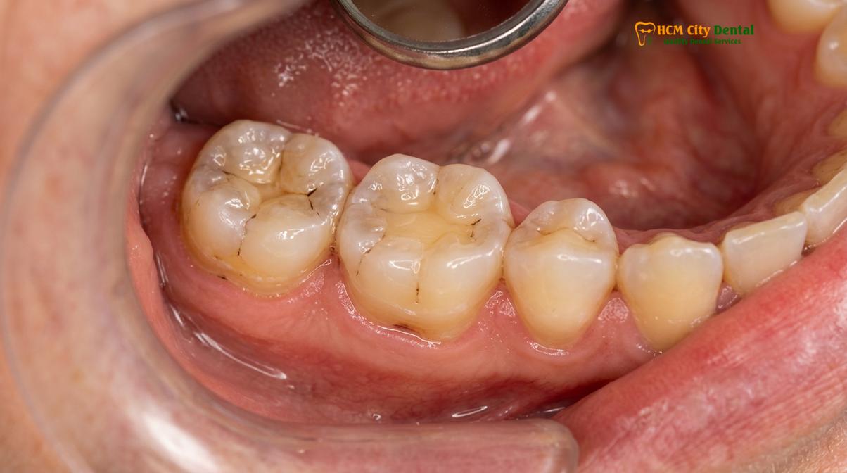
The most immediate and visually apparent consequence of chronic teeth grinding is the accelerated loss of tooth structure. Enamel, the outermost layer of the tooth, is the hardest substance in the human body, composed primarily of highly mineralized hydroxyapatite crystals. However, despite its remarkable hardness, enamel is highly brittle and susceptible to mechanical wear when subjected to the relentless, repetitive friction characteristic of bruxism. This specific type of wear, caused by tooth-to-tooth contact, is clinically referred to as attrition.
During normal mastication (chewing), the upper and lower teeth rarely make direct, forceful contact; food acts as a buffer, and neuromuscular feedback mechanisms prevent excessive force. In contrast, sleep bruxism bypasses these protective reflexes. The continuous grinding motion acts like a millstone, gradually flattening the natural cusps and anatomical ridges of the molars and premolars. Over time, the incisal edges of the front teeth may also become chipped, thin, and translucent. [1]
The Pathophysiology of Dentin Exposure
As the enamel layer is systematically worn away, the underlying dentin is exposed. Dentin is significantly softer than enamel and wears down at a much faster rate once the protective enamel shell is breached. This creates a compounding problem: the rate of tooth destruction accelerates exponentially once dentin is involved. Furthermore, dentin contains thousands of microscopic tubules that communicate directly with the dental pulp (the nerve center of the tooth). When these tubules are exposed to the oral environment, patients typically experience severe dentin hypersensitivity, reacting sharply to hot, cold, sweet, or acidic stimuli.
Dr. Nguyen Van Cuong, a leading specialist at HCMC Dental Clinic in Ho Chi Minh City, emphasizes the importance of early detection: “Many patients are entirely unaware of their nocturnal grinding until they present with unexplained, severe thermal sensitivity. By the time dentin is exposed, the structural compromise is significant, and the treatment shifts from simple prevention to complex restoration.”
Loss of Vertical Dimension of Occlusion (VDO)
The cumulative enamel wear from grinding does not merely affect individual teeth; it alters the entire architecture of the lower face. As the teeth become shorter due to severe attrition, the vertical distance between the upper and lower jaws decreases. This is known as the loss of the Vertical Dimension of Occlusion (VDO). A collapsed VDO can lead to a prematurely aged facial appearance, characterized by deep creases at the corners of the mouth (angular cheilitis) and a protruding lower jaw. Restoring a collapsed bite is one of the most complex procedures in prosthodontics, often requiring full-mouth rehabilitation with multiple crowns to re-establish the proper anatomical height.
“The mechanical degradation of enamel due to parafunctional habits represents a progressive, irreversible loss of the stomatognathic system’s primary protective barrier, necessitating immediate interceptive therapy.”
Cracked and Fractured Teeth
The immense pressure generated during sleep bruxism creates micro-fractures in the tooth structure, which can propagate into catastrophic vertical root fractures if left unmanaged.
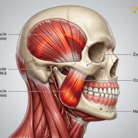
While enamel erosion is a gradual process, the extreme compressive and shear forces generated by bruxism can also cause acute, catastrophic structural failures. The human jaw muscles, particularly the masseter and temporalis muscles, are incredibly powerful. During a sleep bruxism episode, these muscles can contract with maximum intensity, generating forces that far exceed the structural tolerance of natural teeth. This immense pressure is a primary catalyst for cracked tooth syndrome and various forms of dental fractures. [2]
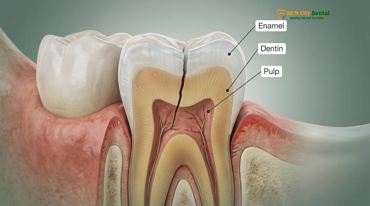
The initial signs of structural stress often manifest as craze lines—superficial, microscopic cracks in the enamel that do not extend into the dentin. While craze lines are generally asymptomatic and common in adult dentition, they serve as a clinical indicator of heavy occlusal loading. However, under the sustained pressure of chronic bruxism, these superficial lines can propagate deeper into the tooth structure.
Crack Propagation Mechanics
When a tooth is subjected to heavy, off-axis forces (shear forces), the stress concentrates at the base of the cusps or around existing dental restorations. A crack can initiate and slowly propagate through the dentin toward the dental pulp. This is known as a fractured cusp or a cracked tooth. Patients with a cracked tooth often experience a very specific type of pain: a sharp, shooting pain that occurs precisely upon the release of biting pressure. This happens because the crack opens slightly when biting down, allowing fluid to move within the dentinal tubules, and then snaps shut when the pressure is released, stimulating the nerve.
Clinical Warning: Cracked Tooth Syndrome
If you experience sharp, localized pain when chewing certain foods, or pain that occurs immediately upon releasing a bite, you may have a cracked tooth. Unlike a cavity, a crack will not heal and will progressively worsen under the continuous strain of bruxism. Immediate clinical evaluation is required to prevent the crack from splitting the tooth entirely, which would necessitate extraction.
Catastrophic Failures: Split Teeth and Vertical Root Fractures
If the bruxism complications are not addressed, a cracked tooth can progress into a split tooth, where the crack extends completely from the occlusal surface down below the gumline, separating the tooth into two distinct segments. At this stage, the tooth is generally unsalvageable and must be extracted. Even more insidious is the vertical root fracture, which originates in the root of the tooth and propagates upward toward the crown. These fractures are notoriously difficult to diagnose in their early stages, as they often do not appear on standard two-dimensional X-rays until significant surrounding bone loss has occurred.
Damage to Dental Restorations
Restorative materials like porcelain crowns, composite fillings, and veneers are highly susceptible to chipping, debonding, or catastrophic failure under the extreme shear forces of bruxism.
Dental restorations are engineered to withstand the normal forces of mastication, but they are frequently pushed beyond their material limits by the pathological forces of teeth grinding. The longevity of fillings, crowns, bridges, and veneers is drastically reduced in patients with unmanaged bruxism. The failure of these restorations is not only frustrating for the patient but also represents a significant recurring financial burden.
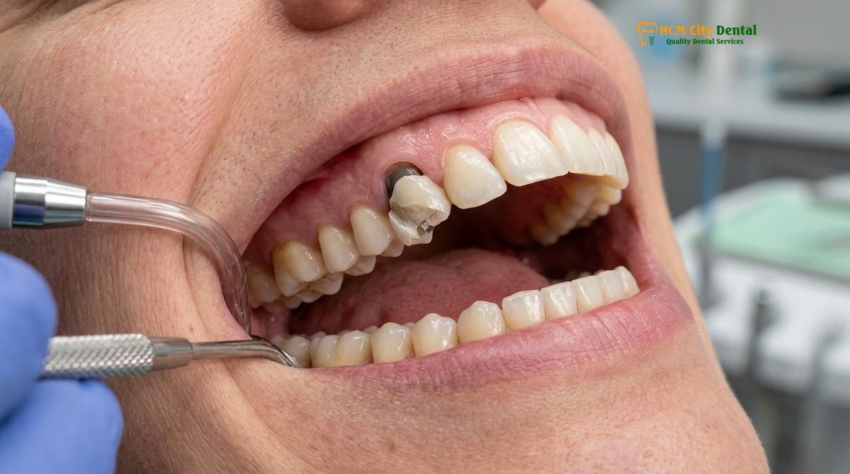
Different restorative materials respond differently to bruxism forces. Composite resin (tooth-colored filling material) is relatively soft compared to enamel and will wear down rapidly under heavy grinding. Amalgam (silver fillings), while highly durable under compressive forces, can act as a wedge when subjected to the lateral forces of bruxism, potentially causing the surrounding natural tooth structure to fracture. [3]
Porcelain Fracture and Debonding
Porcelain and ceramic restorations, such as veneers and traditional crowns, are highly aesthetic but inherently brittle. When a patient grinds their teeth, the lateral shear forces can cause the porcelain to chip or fracture entirely. This is particularly common with porcelain-fused-to-metal (PFM) crowns, where the aesthetic porcelain layer can shear off the underlying metal substructure. Even modern, high-strength ceramics like lithium disilicate or monolithic zirconia can fail if the occlusal forces are severe enough or if the bite is not perfectly balanced.
Furthermore, the continuous flexing of the tooth under heavy loads can degrade the cement seal holding the restoration in place. This micro-leakage allows bacteria to infiltrate beneath the crown or filling, leading to recurrent decay that can destroy the underlying tooth structure without the patient realizing it until the restoration completely debonds and falls out.
Clinical Case Review: Restoration Failure
A 45-year-old patient presented to HCMC Dental Clinic in Ho Chi Minh City with a history of repeatedly fracturing porcelain crowns on their lower molars. Clinical examination revealed severe masseter muscle hypertrophy and flattened anterior teeth, confirming a diagnosis of severe sleep bruxism. The treatment protocol involved replacing the fractured restorations with high-strength monolithic zirconia crowns and fabricating a custom-fitted hard acrylic occlusal splint. By addressing the underlying parafunctional habit, the new restorations have remained stable and intact for over five years.
Implant Biomechanics Under Parafunction
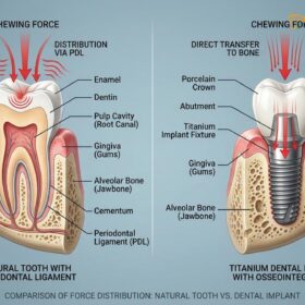
The impact of bruxism on dental implants is a critical clinical concern. Natural teeth are suspended in the jawbone by the periodontal ligament (PDL), a fibrous connective tissue that acts as a shock absorber, allowing the tooth to move slightly and dissipate heavy forces. Dental implants, however, are osseointegrated—they are fused directly to the bone without a PDL. Consequently, the massive forces generated by grinding are transferred directly to the implant components and the surrounding crestal bone. This occlusal overload can lead to the loosening or fracture of the abutment screw, fracture of the implant fixture itself, or progressive bone loss around the implant, ultimately resulting in implant failure. [4]
TMJ Disorder Development
The continuous hyperactive contraction of masticatory muscles during grinding overloads the temporomandibular joint, leading to disc displacement, chronic facial pain, and restricted jaw movement.
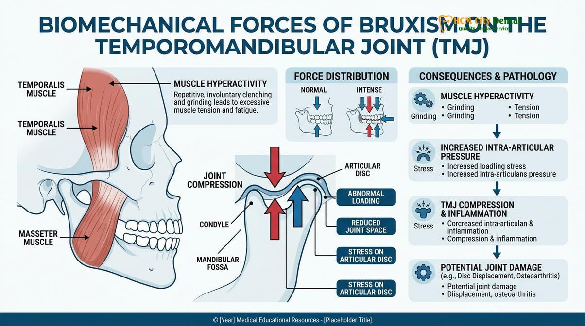
The teeth grinding consequences extend far beyond the teeth themselves; the entire stomatognathic system, particularly the temporomandibular joint (TMJ) and the associated musculature, bears the brunt of the parafunctional activity. The TMJ is a highly complex, bilateral joint that connects the lower jaw (mandible) to the skull (temporal bone). It allows for the intricate movements required for speaking, chewing, and yawning. However, the relentless, high-intensity muscle contractions associated with bruxism place extraordinary stress on this delicate joint complex.
During a sleep bruxism episode, the masseter, temporalis, and medial pterygoid muscles contract forcefully and continuously. This hyperactivity leads to muscle fatigue, lactic acid buildup, and the development of myofascial trigger points. Patients frequently wake up with a dull, aching pain radiating through their jaw, cheeks, and temples. This muscle tension is also a primary trigger for tension-type headaches, which are commonly misdiagnosed as migraines.
Intra-articular Derangement
Beyond muscular pain, the excessive compressive forces can cause structural damage within the TMJ itself. The joint contains a cartilaginous articular disc that acts as a cushion between the condyle of the mandible and the glenoid fossa of the skull. Chronic bruxism can cause this disc to become displaced, typically anteriorly (forward). When the disc is out of position, patients may experience a clicking or popping sound when opening or closing their mouth. [5]
If the disc displacement progresses, it can lead to a condition known as “closed lock,” where the patient is unable to fully open their mouth. Furthermore, the continuous friction within the joint can degrade the protective cartilage, leading to osteoarthritis of the TMJ, characterized by crepitus (a grinding, gravel-like sound in the joint) and severe, chronic pain.
| Clinical Parameter | Normal Masticatory Function | Bruxism / Parafunctional Activity |
|---|---|---|
| Force Exerted | 20 – 40 lbs per square inch | Up to 250 lbs per square inch |
| Duration of Contact | Minutes per day (during meals) | Hours per night (sustained clenching) |
| Muscle Status | Relaxed at rest, normal tone | Hypertrophic, chronic fatigue, spasms |
| Joint Loading | Intermittent, well-tolerated | Continuous compression, disc displacement risk |
| Direction of Force | Primarily vertical (axial) | Heavy lateral and shear forces |
Gum Recession from Grinding
Excessive occlusal loading causes the teeth to flex at the cervical margin, leading to non-carious cervical lesions (abfractions) and subsequent gingival recession.
While gum recession is commonly associated with periodontal disease or aggressive brushing, the mechanical forces of bruxism play a highly significant, yet often overlooked, role in the degradation of gingival tissues. The relationship between occlusal stress and gum recession is rooted in the biomechanical response of the tooth structure to heavy lateral forces.
When a tooth is subjected to the intense, side-to-side grinding motions of bruxism, it acts like a lever. The crown of the tooth is pushed laterally, causing the entire tooth to flex microscopically. The fulcrum of this flexion occurs at the cervical margin—the neck of the tooth, right at the gumline. This area is where the enamel is at its thinnest, tapering off to meet the root surface.
The Abfraction Mechanism
The continuous flexing of the tooth concentrates immense stress at the cervical margin, causing the crystalline structure of the enamel to break down. Micro-fractures develop, and small, wedge-shaped pieces of enamel and dentin eventually chip away. This specific type of tooth wear is clinically known as an abfraction lesion. These non-carious cervical lesions create deep notches at the gumline, which are highly susceptible to plaque accumulation and severe dentin hypersensitivity.
Furthermore, the trauma from the excessive occlusal forces is transmitted down the root of the tooth into the periodontal ligament (PDL) and the surrounding alveolar bone. The body responds to this trauma by widening the PDL space, which can cause the tooth to become mobile (loose). The chronic inflammation and stress in the periodontium signal the delicate gingival tissues to pull away from the site of trauma, resulting in progressive gum recession. This bruxism damage exposes the root surface, which is covered only by cementum—a tissue much softer than enamel and highly vulnerable to rapid decay.
“The interplay between occlusal trauma and periodontal attachment loss highlights the necessity of managing parafunctional forces to maintain the integrity of the gingival architecture.”
Estimated Cost of Not Treating Bruxism
Delaying treatment for chronic teeth grinding exponentially increases long-term dental expenses, as patients transition from needing simple preventive mouthguards to requiring complex full-mouth rehabilitations.
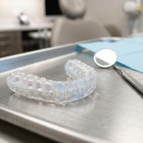
From a clinical and economic perspective, bruxism is a progressive condition that becomes exponentially more expensive to treat the longer it is ignored. The initial intervention for teeth grinding is highly conservative and cost-effective. A custom-fabricated occlusal splint, designed by a dental professional, acts as a protective barrier, absorbing the destructive forces and preventing tooth wear. The cost of a high-quality nightguard is a minor investment compared to the extensive restorative work required once the dentition is compromised.
If the condition is left unmanaged, the financial trajectory shifts dramatically. As enamel is worn away and teeth begin to fracture, the patient will require multiple restorative procedures. A single fractured cusp may necessitate a porcelain crown. If the crack propagates into the pulp, endodontic therapy (root canal treatment) will be required before the crown can be placed. If the tooth splits entirely, it must be extracted and replaced with a dental implant and an implant-supported crown.
The Economics of Full Mouth Rehabilitation
In severe cases of chronic bruxism, where the patient has lost significant vertical dimension of occlusion (VDO) due to widespread attrition, a few isolated crowns will not suffice. The entire bite must be reconstructed to restore proper function, aesthetics, and joint health. This process, known as full mouth rehabilitation, involves placing crowns or veneers on nearly every tooth in the mouth. The estimated cost of such comprehensive treatment is substantial, requiring extensive planning, multiple clinical phases, and significant laboratory fees.
Dr. Cuong frequently advises patients that preventive dentistry is the most economically sound approach to oral health. By diagnosing parafunctional habits early and implementing protective strategies, such as custom dental splints, patients can preserve their natural dentition and avoid the profound financial and biological costs associated with advanced bruxism complications.
When to See a Doctor
Recognizing the early warning signs of bruxism is critical for preventing irreversible damage to your teeth and jaw joints. Because sleep bruxism occurs unconsciously, many patients rely on secondary symptoms or the observations of a sleep partner to identify the problem. You should schedule a comprehensive clinical evaluation if you experience any of the following symptoms:
- Unexplained Facial Pain: Waking up with a dull, aching pain in your jaw, cheeks, or temples, or experiencing frequent morning headaches.
- Tooth Sensitivity: A sudden or progressive increase in sensitivity to hot, cold, or sweet foods, indicating potential enamel loss or dentin exposure.
- Visible Tooth Wear: Noticing that your teeth appear shorter, flatter, or have chipped edges, particularly on the front teeth.
- Jaw Dysfunction: Experiencing a clicking, popping, or grating sound in your jaw joint, or having difficulty opening your mouth fully.
- Partner Observations: If your sleep partner reports hearing loud grinding, clenching, or gnashing sounds during the night.
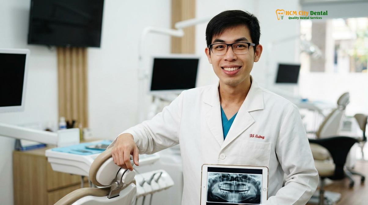
If you suspect you are suffering from parafunctional habits, seeking professional care is essential. A thorough examination, including an assessment of your bite, muscle palpation, and a review of your dental restorations, can confirm the diagnosis. For personalized diagnostic advice and to explore protective options like Nightguards, consulting with the experienced team at HCMC Dental Clinic ensures that your smile is safeguarded against the destructive forces of bruxism.
Frequently Asked Questions
Can bruxism break dental crowns?
Yes, the extreme forces of bruxism can easily fracture or dislodge dental crowns. While natural chewing generates around 20 to 40 pounds of force, sleep bruxism can exert up to 250 pounds per square inch. This immense pressure can cause porcelain to chip, shear off, or cause the underlying cement seal to fail, necessitating premature crown replacement.
Does grinding cause gum recession?
Yes, excessive grinding forces cause teeth to flex at the gumline, leading to abfraction and subsequent gum recession. The lateral forces generated during parafunctional habits concentrate stress at the cervical margin (the neck of the tooth). This stress causes micro-chipping of the enamel and widening of the periodontal ligament, which ultimately triggers the gingival tissue to recede.
Can bruxism damage dental implants?
Yes, uncontrolled bruxism can cause occlusal overload on dental implants, leading to screw loosening or even implant failure. Unlike natural teeth, dental implants lack a periodontal ligament to act as a shock absorber. Consequently, the massive shear forces of teeth grinding are transferred directly to the implant-bone interface, which can cause crestal bone loss or mechanical fracture.
How fast does bruxism destroy teeth?
The rate of destruction varies, but severe sleep bruxism can cause significant enamel loss and micro-fractures within just a few months. Factors such as the intensity of the grinding, the pH level of the patient’s saliva, and the presence of acid reflux can exponentially accelerate tooth wear. Without intervention, patients can lose millimeters of tooth structure annually.
Is bruxism damage reversible?
The structural damage to teeth is not naturally reversible, but it can be clinically restored using crowns, bonding, or veneers. Once enamel is worn away or a tooth is fractured, the body cannot regenerate it. However, comprehensive restorative dentistry can rebuild the lost vertical dimension, restore aesthetics, and protect the remaining tooth structure from further parafunctional damage.
References
- Journal of Prosthetic Dentistry. Biomechanics of occlusal wear and bruxism. (2021).
- American Academy of Sleep Medicine. Sleep-related bruxism pathophysiology. (2020).
- Journal of Clinical Periodontology. Non-carious cervical lesions and occlusal stress. (2019).
- International Journal of Prosthodontics. Implant complications in bruxing patients. (2022).
- Journal of Oral Rehabilitation. Temporomandibular disorders and parafunctional habits. (2018).