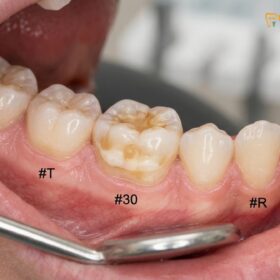
A pediatric root canal in HCMC is a specialized dental procedure designed to save a severely decayed or infected baby tooth. By removing the diseased pulp tissue, pediatric dentists eliminate pain and preserve the tooth to guide the proper eruption of future permanent teeth.
Clinical Summary:
Pediatric endodontic therapy, commonly known as a baby root canal, encompasses pulpotomy and pulpectomy procedures. These treatments are essential for managing deep caries that have penetrated the pulp chamber of primary teeth. Preserving the primary dentition maintains arch space, supports proper mastication, and prevents speech impediments. At HCMC Dental Clinic, advanced diagnostic tools and child-friendly behavioral guidance ensure that these procedures are performed safely, effectively, and with minimal discomfort, utilizing biocompatible materials and durable pediatric crowns.
Key Takeaways:
- Primary teeth act as natural space maintainers, guiding the eruption of permanent teeth.
- A pulpotomy removes only the infected coronal pulp, leaving healthy root nerves intact.
- A pulpectomy is required when bacterial infection extends deep into the root canals of a baby tooth.
- Post-treatment restoration typically requires a stainless steel or zirconia crown to prevent tooth fracture.
- Modern pediatric dentistry utilizes behavioral guidance and precise anesthetics for a comfortable experience.
When a child complains of severe toothache, especially at night, or when parents notice a deep cavity in a baby tooth, immediate clinical intervention is required. Unlike adult teeth, primary teeth have thinner enamel and relatively larger pulp chambers, meaning that tooth decay can progress rapidly from the surface into the nerve tissue. Understanding the nuances of pediatric pulp therapy is essential for parents navigating dental care options in Ho Chi Minh City.
Why Saving Primary (Baby) Teeth is Vital for Child Development
Preserving primary teeth is crucial because they serve as natural space maintainers, facilitate proper chewing and speech development, and guide the eruption of permanent teeth.
A common misconception among parents is that primary teeth are expendable because they will eventually exfoliate (fall out). However, from a clinical perspective, saving primary baby teeth is one of the most critical aspects of pediatric dentistry. The primary dentition plays a multifaceted role in the physiological and anatomical development of a growing child.
First and foremost, primary teeth act as biological space maintainers. Each baby tooth holds a specific place in the dental arch, preserving the exact amount of room required for the underlying permanent tooth to erupt correctly. If a primary molar is lost prematurely due to untreated decay or infection, the adjacent teeth will naturally drift into the empty space. This mesial drift leads to a loss of arch length, causing the permanent tooth to become impacted or erupt out of alignment, which inevitably necessitates complex and costly orthodontic treatment later in life[1].
Furthermore, primary teeth are essential for proper mastication (chewing) and nutrition. A child suffering from dental pain or missing multiple teeth will naturally avoid hard, fibrous, or nutrient-dense foods, potentially leading to nutritional deficiencies and impaired physical growth. The ability to chew comfortably is directly linked to a child’s overall systemic health and well-being.
Speech development is another critical factor. The anterior primary teeth (incisors and canines) are vital for the correct articulation of consonants such as “s,” “z,” “t,” and “th.” Premature loss of these teeth can result in speech impediments that may require speech therapy to correct. Additionally, a healthy, complete smile contributes significantly to a child’s self-esteem and psychosocial development, preventing the self-consciousness that often accompanies visible dental decay or missing teeth.
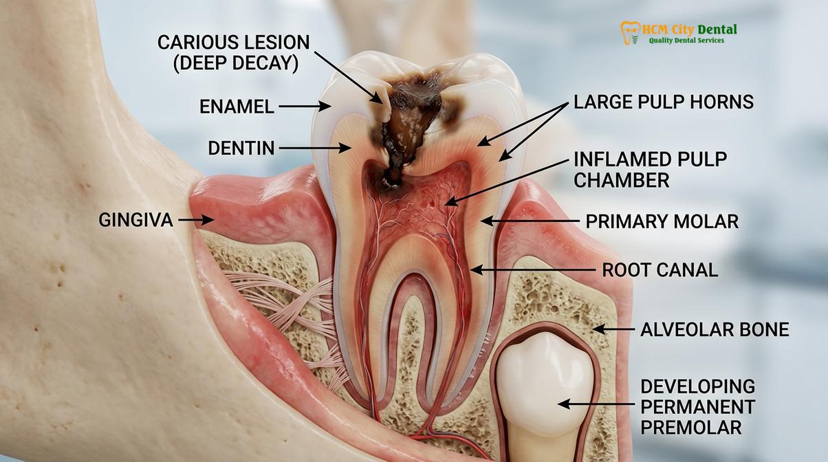
When decay reaches the dental pulp—the innermost part of the tooth containing nerves and blood vessels—the resulting inflammation (pulpitis) causes severe pain. In these cases, a pediatric root canal is the standard of care to eliminate the infection while retaining the functional tooth structure until natural exfoliation occurs.
Pulpotomy: The Partial Nerve Removal (Baby Tooth Root Canal)
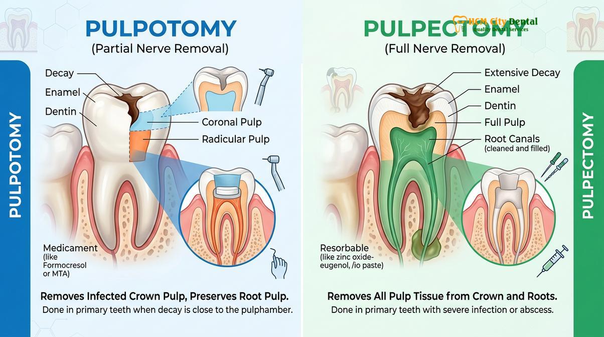
A pulpotomy involves removing only the infected upper portion of the dental pulp while preserving the healthy, vital nerve tissue inside the tooth’s roots.
The most common endodontic procedure performed on primary teeth is the pulpotomy. Often referred to colloquially as a “baby root canal,” a pulpotomy is indicated when dental caries have penetrated the enamel and dentin to reach the coronal pulp (the nerve tissue located in the crown of the tooth), but the radicular pulp (the nerve tissue inside the roots) remains healthy and uninfected.
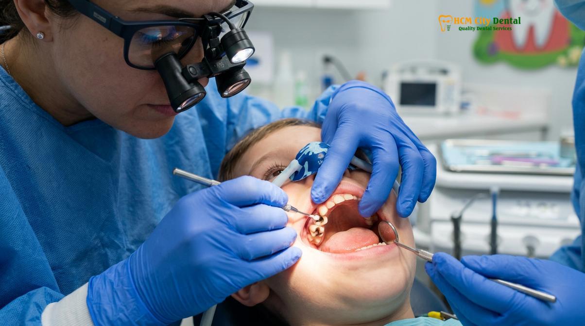
The clinical workflow for a pulpotomy is precise and highly successful when performed by an experienced pediatric dentist Saigon. The procedure begins with the administration of profound local anesthesia to ensure the child is completely comfortable. A dental rubber dam is often placed to isolate the tooth, keeping the operating field sterile and protecting the child’s airway from water and debris.
The dentist then carefully excavates the decayed tooth structure to gain access to the pulp chamber. The infected coronal pulp is amputated (removed) using specialized rotary instruments or sharp hand excavators. Once the diseased tissue is removed, achieving hemostasis (stopping the bleeding) is a critical diagnostic step. If the bleeding from the root orifices stops within a few minutes, it indicates that the remaining radicular pulp is healthy and vital. If the bleeding cannot be controlled, it suggests that the infection has spread into the roots, necessitating a more extensive procedure (pulpectomy)[2].
“The success of a pulpotomy relies heavily on accurate clinical diagnosis and the meticulous handling of the remaining vital pulp tissue. By utilizing advanced biocompatible materials, we can effectively seal the nerve and promote long-term healing.”
After hemostasis is achieved, a medicament is applied to the remaining pulp stumps to fixate the tissue, eliminate residual bacteria, and promote healing. Historically, Formocresol was the standard medicament; however, modern pediatric dentistry has largely shifted toward more biocompatible materials such as Mineral Trioxide Aggregate (MTA) or Ferric Sulfate. MTA, in particular, is highly regarded for its excellent sealing ability, biocompatibility, and capacity to stimulate the formation of a dentinal bridge, protecting the underlying nerve.
Once the pulp chamber is treated and sealed with a base material, the tooth must be restored. Because a pulpotomy removes the internal blood supply to the crown, the remaining tooth structure becomes brittle and highly susceptible to fracture. Therefore, a full-coverage restoration, such as a stainless steel or zirconia crown, is universally recommended to protect the tooth and ensure it survives until it naturally falls out.
Pulpectomy: Full Nerve Removal for Severe Infections
When bacterial infection irreversibly damages the entire nerve, a pulpectomy completely removes the diseased pulp from both the crown and the roots of the baby tooth.
In cases where tooth decay has been left untreated for an extended period, the bacterial infection will progress beyond the coronal pulp and invade the radicular pulp inside the root canals. This condition, known as irreversible pulpitis or pulpal necrosis, often presents with severe spontaneous pain, sensitivity to hot stimuli, or the presence of a dental abscess (a pimple-like swelling on the gums). In these scenarios, a pulpotomy is insufficient, and a full pulpectomy is required to save the tooth.
A pulpectomy is the pediatric equivalent of a traditional adult root canal, but with crucial anatomical and material differences. The primary objective is to completely debride (clean out) the infected nerve tissue from the entire length of the root canals, disinfect the canal system, and seal it to prevent reinfection.
The procedure begins similarly to a pulpotomy, with anesthesia, isolation, and access to the pulp chamber. However, the dentist then uses specialized endodontic files to carefully navigate and clean the delicate, often curved root canals of the primary tooth. Copious irrigation with antimicrobial solutions is used to flush out bacteria and necrotic debris.
The most significant difference between a pediatric pulpectomy and an adult root canal lies in the obturation (filling) material. In adult teeth, root canals are filled with gutta-percha, a solid, non-resorbable rubber material. However, primary teeth must eventually undergo root resorption—a natural process where the body dissolves the baby tooth’s roots to make way for the erupting permanent tooth. If a non-resorbable material were used, it would block the permanent tooth from erupting[3].
Therefore, pediatric dentists use specialized resorbable pastes to fill the root canals of baby teeth. Common materials include Zinc Oxide Eugenol (ZOE) paste or iodoform-based pastes (such as Vitapex or Metapex). These materials are designed to resorb at the exact same rate as the natural tooth root, ensuring that the permanent tooth can erupt without interference.
| Clinical Parameter | Pulpotomy (Partial) | Pulpectomy (Full) |
|---|---|---|
| Indication | Infection limited to the coronal pulp (crown). | Infection extends into the radicular pulp (roots). |
| Pulp Status | Radicular pulp is vital and healthy. | Entire pulp is necrotic or irreversibly inflamed. |
| Procedure Scope | Removal of upper nerve tissue only. | Removal of nerve tissue from crown and root canals. |
| Filling Material | MTA, Ferric Sulfate, or Biodentine. | Resorbable paste (ZOE or Iodoform-based). |
| Clinical Signs | Pain provoked by eating/drinking; no swelling. | Spontaneous pain, night pain, or visible gum abscess. |
Following a successful pulpectomy, the tooth is structurally compromised and must be protected with a durable dental crown. Parents often inquire about the root canal recovery process for children; fortunately, because the source of the infection is completely removed, children typically experience rapid relief from pain and return to normal activities within 24 hours.
Restoring Pediatric Teeth: Stainless Steel Crowns and White Zirconia Crowns
Following pulp therapy, baby teeth become brittle and require full-coverage restorations like stainless steel or zirconia crowns to withstand chewing forces until they naturally fall out.
A critical component of pediatric endodontic therapy is the final restoration of the treated tooth. Parents frequently ask why a simple composite (white) filling cannot be used after a pulpotomy or pulpectomy. The clinical reality is that once the pulp chamber is accessed and the internal blood supply is altered, the remaining enamel and dentin become desiccated and significantly weaker. Furthermore, the cavities that necessitate pulp therapy are usually very large, leaving little sound tooth structure to support a traditional filling.
Placing a large filling on a pulpotomized primary molar carries a high risk of catastrophic tooth fracture under the heavy forces of mastication. If the tooth fractures below the gumline, it becomes unsalvageable and must be extracted, defeating the entire purpose of the pulp therapy. Therefore, full-coverage crowns are the gold standard for restoring primary teeth following endodontic treatment.
There are two primary options for pediatric crowns: Stainless Steel Crowns (SSCs) and Zirconia Crowns.
Stainless Steel Crowns (SSCs): For decades, SSCs have been the workhorse of pediatric dentistry. They are highly durable, cost-effective, and require minimal tooth preparation. Because the metal is somewhat flexible, the dentist can crimp and contour the edges of the crown to snap securely over the tooth, providing an excellent seal against future bacterial leakage. While they are silver in color and less aesthetic, they are exceptionally reliable for posterior (back) molars where aesthetics are less of a concern. Finding a reliable provider for a stainless steel crown child HCMC is a common priority for parents seeking durable, functional restorations.
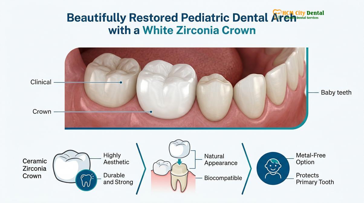
Zirconia Crowns: With advancements in dental materials, white Zirconia crowns have become increasingly popular for parents seeking a highly aesthetic, metal-free alternative. Zirconia is a biocompatible ceramic that is incredibly strong and tooth-colored, blending seamlessly with the child’s natural dentition. These crowns are pre-fabricated in various sizes and cemented into place. While they provide superior aesthetics, they require more precise tooth preparation and are technique-sensitive, as they cannot be crimped or bent like stainless steel[4]. For parents interested in aesthetic options, understanding the benefits of zirconia crowns is essential.
Clinical Case Study: Pediatric Pulp Therapy
Patient Profile: A 5-year-old patient presented to HCMC Dental Clinic in Ho Chi Minh City with severe pain in the lower right primary molar, preventing sleep.
Clinical Findings: Radiographs revealed deep occlusal caries penetrating the pulp chamber, but no periapical radiolucency (no root infection). Diagnosis: Reversible pulpitis.
Treatment Executed: Dr. Nguyen Van Cuong performed a vital pulpotomy using MTA to preserve the healthy root nerves. To ensure long-term durability and aesthetics, the tooth was restored with a pre-fabricated white Zirconia crown.
Outcome: The patient experienced immediate pain relief. At the 6-month follow-up, the tooth was fully functional, asymptomatic, and the surrounding gingival tissues were perfectly healthy, maintaining the vital space for the future permanent premolar.
Regardless of the material chosen, the placement of a dental crown after root canal therapy in children is a non-negotiable step to ensure the long-term success of the treatment and the preservation of the dental arch.
Space Maintainers: What Happens If a Baby Tooth Must Be Extracted Early
If a primary tooth is unsalvageable and requires extraction, a custom space maintainer is placed to prevent adjacent teeth from shifting and blocking the permanent tooth.
Despite the best efforts of pediatric dentists, there are clinical scenarios where a primary tooth is deemed unsalvageable. This typically occurs when tooth decay has destroyed so much of the clinical crown that a restoration cannot be supported, when a severe infection has caused extensive root resorption, or when a dental abscess threatens the developing permanent tooth bud beneath it. In these cases, extraction is the only viable medical option.
However, extracting a primary molar without subsequent intervention leads to severe orthodontic complications. As previously mentioned, teeth have a natural tendency to drift forward (mesial drift). If a primary molar is removed years before the permanent tooth is ready to erupt, the adjacent teeth will tilt and drift into the extraction site. This closes the space, leaving no room for the permanent tooth, which may then become impacted inside the jawbone or erupt completely out of alignment.
To prevent this, pediatric dentists utilize devices called Space Maintainers. These are custom-made, passive orthodontic appliances designed to hold the space open until the permanent tooth is ready to emerge. There are several types of space maintainers, chosen based on the specific tooth lost and the child’s dental development:
- Band and Loop: The most common unilateral space maintainer. A stainless steel band is cemented onto the tooth adjacent to the empty space, and a wire loop extends across the gap to rest against the next tooth, holding the space open.
- Distal Shoe: Used when the second primary molar is lost before the first permanent molar has erupted. A metal guide plane extends into the gum tissue to guide the erupting permanent molar into its correct position.
- Lower Lingual Holding Arch (LLHA): A bilateral appliance used when multiple lower primary teeth are lost. Bands are placed on the permanent molars, and a heavy wire runs along the inside (lingual) of the lower teeth to prevent forward shifting.
- Nance Appliance: Similar to the LLHA but used for the upper arch, utilizing an acrylic button that rests on the palate to provide anchorage.
Space maintainers require diligent oral hygiene and regular monitoring by the dentist to ensure they remain intact and are removed at the precise moment the permanent tooth begins to erupt. While space maintainers are highly effective, the primary goal always remains saving the natural baby tooth whenever clinically possible.
Behavioral Guidance: Keeping Your Child Calm and Pain-Free During Treatment
Pediatric dental specialists employ techniques like Tell-Show-Do, positive reinforcement, and safe sedation to ensure children remain comfortable and cooperative during endodontic procedures.
One of the most significant concerns parents have regarding pediatric root canals is how their child will tolerate the procedure. Dental anxiety is common among children, and managing this anxiety is just as important as the technical execution of the dental work. Pediatric dentists are extensively trained in behavioral guidance techniques to create a positive, non-threatening environment.
The foundation of pediatric behavioral management is the “Tell-Show-Do” technique. Before any instrument is used, the dentist explains the procedure in child-friendly language (Tell), demonstrates how the instrument works in a non-threatening way, such as on the child’s fingernail (Show), and then proceeds with the treatment (Do). This demystifies the process and builds trust between the child and the clinician[5].
“Our primary goal is not just to fix the tooth, but to ensure the child leaves the clinic with a positive attitude toward dental care. By using gentle communication and modern pain management, we can turn a potentially scary procedure into a routine, comfortable experience.” — Dr. Nguyen Van Cuong
Pain management is paramount. Modern local anesthetics are highly effective, and pediatric dentists use specialized techniques to deliver them painlessly. Topical anesthetic gels are applied to numb the gums before any injection, and the use of computer-assisted anesthesia delivery systems can make the process virtually imperceptible.
For children who are highly anxious, very young, or require extensive treatment, pharmacological behavioral guidance may be recommended. Nitrous Oxide (laughing gas) is a safe, mild sedative inhaled through a small nose mask. It helps the child relax, reduces the gag reflex, and provides a mild analgesic effect, all while the child remains fully conscious and responsive. The effects of nitrous oxide wear off completely within minutes of stopping the gas, allowing the child to return to normal activities immediately.
In cases of severe anxiety, special needs, or when extensive full-mouth rehabilitation is required, treatment under deep sedation or general anesthesia in a hospital setting may be the safest and most humane option. A thorough consultation at HCMC Dental Clinic will determine the most appropriate behavioral guidance strategy for your child’s specific needs.
When to See a Pediatric Dentist
Early detection of dental issues in children can prevent the need for complex endodontic therapy. Parents should be vigilant and schedule a consultation with a pediatric dentist if they observe any of the following signs:
- Spontaneous Pain: The child complains of toothache without any provocation, especially pain that wakes them up at night.
- Prolonged Sensitivity: Severe pain when consuming hot, cold, or sweet foods that lingers after the stimulus is removed.
- Visible Decay: Dark brown or black spots on the teeth, or visible holes (cavities) in the tooth structure.
- Gum Swelling: Red, swollen gums around a specific tooth, or the presence of a “gum boil” (parulis) which indicates a draining dental abscess.
- Facial Swelling: Any swelling of the cheek or jaw, which is a medical emergency indicating a spreading infection.
Important Clinical Warning
Never ignore a dental abscess or facial swelling in a child. An untreated dental infection can rapidly spread to surrounding facial spaces, posing a serious risk to the child’s systemic health and potentially causing irreversible damage to the developing permanent tooth bud located directly beneath the infected baby tooth. Seek immediate professional dental care.
Following a procedure, parents often wonder what to eat after a root canal. A soft diet is recommended for the first 24 to 48 hours to allow the surrounding tissues to heal. Additionally, if your family has travel plans, it is generally safe, but reviewing guidelines on flying after a root canal with your dentist is advised to ensure pressure changes do not cause discomfort.
Frequently Asked Questions
Why do dentists do root canals on baby teeth if they will fall out anyway?
Baby teeth act as essential space maintainers for permanent teeth and are vital for chewing and speech. Extracting them prematurely can cause adjacent teeth to shift, leading to severe orthodontic crowding and impaction of developing adult teeth. Preserving the baby tooth through pulp therapy maintains the integrity of the dental arch until natural exfoliation occurs.
Does a child feel pain during a pulpotomy?
No, a child should not feel pain during a pulpotomy. Pediatric dentists use precise local anesthesia to completely numb the tooth and surrounding tissues, often combined with behavioral guidance or nitrous oxide to ensure a comfortable, anxiety-free experience. The goal is to eliminate the pain caused by the infection, not cause further discomfort.
Are dental X-rays safe for toddlers and young children?
Yes, modern digital dental X-rays are highly safe for children. They emit an extremely low dose of radiation, and pediatric clinics utilize lead aprons with thyroid collars to provide maximum protection during the diagnostic imaging process. Radiographs are essential for diagnosing decay between teeth and assessing the health of the tooth roots and developing permanent teeth.
How long does a pediatric root canal take?
A pediatric root canal, such as a pulpotomy, typically takes between 30 to 45 minutes per tooth. This timeframe includes the administration of local anesthesia, the removal of infected pulp tissue, the application of medicaments, and the placement of a protective dental crown. Pediatric dentists work efficiently to minimize the time the child spends in the dental chair.
What is the recovery process like for a child?
Recovery is generally swift, with most children returning to normal activities the next day. Mild tissue sensitivity is common but can be managed with over-the-counter pediatric analgesics and by following a soft diet for the first 24 to 48 hours. The child should avoid chewing hard or sticky foods on the treated tooth to protect the new crown.
References
- American Academy of Pediatric Dentistry (AAPD). Pulp Therapy for Primary and Immature Permanent Teeth. (2022).
- Journal of Endodontics. Mineral Trioxide Aggregate vs. Formocresol in Primary Molar Pulpotomies. (2020).
- International Journal of Paediatric Dentistry. Resorbable Root Canal Fillings in Primary Teeth. (2021).
- Pediatric Dentistry Journal. Clinical Success of Zirconia vs Stainless Steel Crowns in Children. (2019).
- Journal of the American Dental Association. Behavioral Management Techniques in Pediatric Dentistry. (2023).
For professional children’s dental care, preventive sealants, or to schedule a check-up, visit our Children & Pediatric Dentistry service page at HCMC Dental Clinic in Ho Chi Minh City.