Chalky teeth in kids, clinically known as Molar Incisor Hypomineralisation (MIH), is a developmental defect where tooth enamel fails to calcify properly. This results in porous, discolored, and hypersensitive teeth that are highly susceptible to rapid decay and require specialized pediatric dental intervention.
Clinical Summary:
Molar Incisor Hypomineralisation (MIH) is a prevalent structural anomaly affecting the first permanent molars and frequently the incisors. Originating from systemic disruptions during early childhood enamel maturation, the condition leaves teeth with a significantly reduced mineral density. These “chalky teeth” are prone to post-eruptive breakdown, severe thermal sensitivity, and accelerated caries progression. Effective clinical management requires early diagnosis, aggressive remineralization protocols using fluoride and CPP-ACP, and the strategic use of moisture-tolerant restorative materials like Glass Ionomer Cements (GIC) or Stainless Steel Crowns (SSCs). Furthermore, specialized behavioral management techniques are essential, as the chronic subclinical pulpal inflammation associated with MIH often makes these teeth challenging to anesthetize using standard protocols.
Key Takeaways:
- MIH primarily affects the 6-year permanent molars and incisors, presenting as white, yellow, or brown opacities.
- The condition stems from developmental disruptions during the first three years of life, not from poor oral hygiene.
- Chalky teeth suffer from severe hypersensitivity and are highly vulnerable to rapid structural breakdown and decay.
- Treatment prioritizes remineralization, cavity arrest with Silver Diamine Fluoride (SDF), and moisture-tolerant GIC fillings.
- Advanced pediatric behavioral management, such as the Tell-Show-Do technique, is crucial for comfortable treatment.
What is Molar Incisor Hypomineralisation?
Molar Incisor Hypomineralisation (MIH) is a developmental defect characterized by a severe lack of mineral content in the enamel of first permanent molars and incisors, leading to porous, fragile teeth.
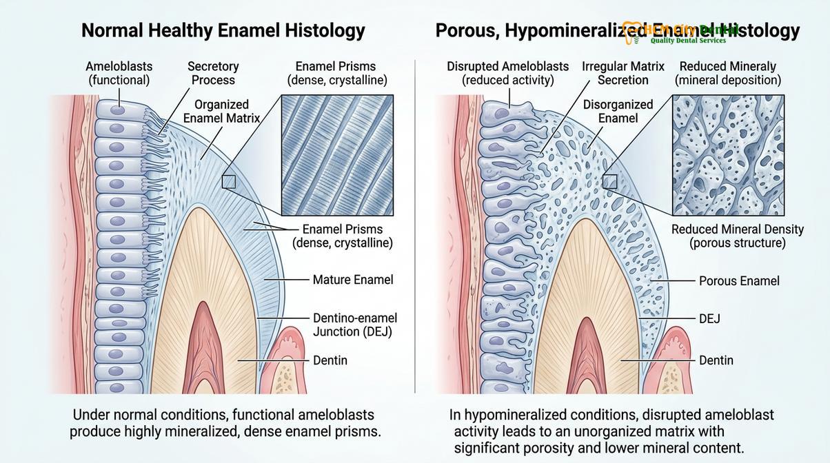
To understand the clinical significance of chalky teeth, it is essential to examine the biological process of tooth formation, known as amelogenesis. Enamel development occurs in two primary phases: the secretory phase, where a protein matrix is laid down, and the maturation phase, where these proteins are degraded and replaced by calcium and phosphate crystals. When a child develops Children & Pediatric Dentistry conditions like MIH, the disruption occurs specifically during the maturation phase. The enamel matrix fails to fully mineralize, leaving residual serum proteins (such as albumin) trapped within the tooth structure[1].
This incomplete calcification results in enamel that is structurally compromised. While normal enamel is the hardest substance in the human body, the enamel of an MIH-affected tooth can be up to ten times softer. This condition predominantly affects the first permanent molars—often referred to as the 6-year molars—because their crowns are mineralizing during the critical period from birth to three years of age. In many cases, the permanent incisors (front teeth) are also affected, presenting significant aesthetic and functional challenges.
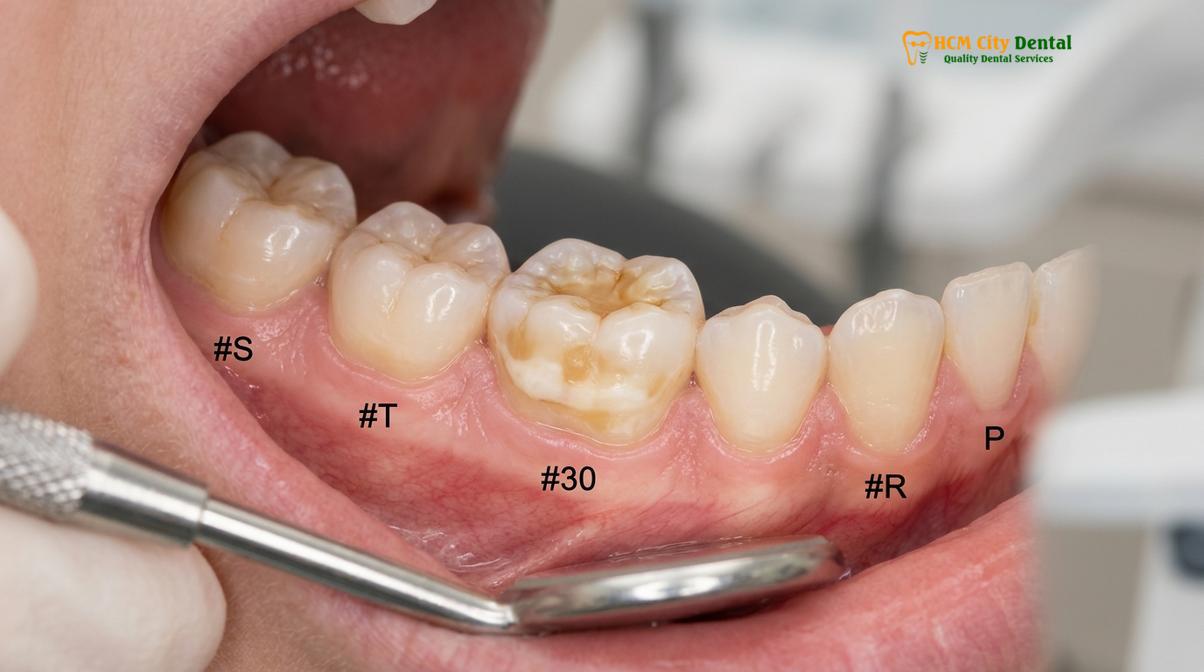
It is crucial for parents to understand that this specific type of enamel hypoplasia children experience is entirely developmental. It is not caused by a high-sugar diet or poor brushing habits, although inadequate oral hygiene will rapidly accelerate the decay of these already vulnerable teeth. Because the underlying dentin is less protected, the pulp (nerve) of the tooth is subjected to constant micro-trauma from temperature changes and chewing forces, leading to chronic subclinical inflammation.
Recognizing Chalky Enamel
Chalky enamel is visually identified by demarcated opacities ranging from creamy white to dark brown, accompanied by severe hypersensitivity and a high risk of the tooth crumbling under normal chewing forces.
The clinical presentation of MIH is highly variable, ranging from mild aesthetic concerns to severe structural failure. The hallmark visual sign is the presence of demarcated opacities. Unlike the diffuse, cloudy white spots associated with early decay or mild fluorosis, MIH opacities have clear, distinct boundaries. The color of the opacity directly correlates with the severity of the mineral deficiency: creamy white spots indicate a milder defect, while yellow or dark brown lesions signify highly porous, severely hypomineralized enamel that is at imminent risk of fracturing.
Clinical Warning: Post-Eruptive Breakdown (PEB)
Teeth with severe yellow-brown MIH opacities are at high risk for Post-Eruptive Breakdown. This occurs when the fragile enamel simply crumbles away under normal chewing forces shortly after the tooth erupts, rapidly exposing the sensitive underlying dentin and accelerating deep cavity formation.
Beyond visual signs, the most distressing symptom for the child is profound hypersensitivity. This tooth sensitivity in children can be so severe that the child avoids eating on the affected side, refuses cold drinks, or cries during routine tooth brushing. The porosity of the chalky enamel allows thermal and chemical stimuli to penetrate directly to the dentinal tubules, triggering sharp pain responses. Furthermore, because the enamel surface is rough and porous, it acts as a magnet for plaque accumulation, creating a vicious cycle of hypersensitivity, poor hygiene, and rapid caries progression[2].
During a comprehensive diagnostic evaluation, a pediatric dentist must differentiate MIH from other enamel defects. For instance, dental fluorosis typically affects all teeth symmetrically and presents with diffuse white striations, whereas MIH is usually asymmetrical and localized to specific molars and incisors. Amelogenesis Imperfecta, a rare genetic disorder, affects both primary and permanent dentitions globally, whereas MIH is primarily an acquired defect of specific permanent teeth.
Causes of Poor Enamel Calcification
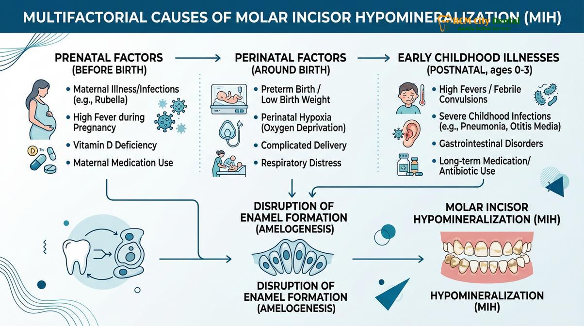
The exact cause of MIH is multifactorial, typically resulting from systemic health disturbances, illnesses, or environmental factors occurring during the child’s first three years of life when enamel is maturing.
The etiology of Molar Incisor Hypomineralisation is one of the most extensively researched topics in modern pediatric dentistry. Current clinical consensus indicates that there is no single causative agent; rather, it is a multifactorial condition linked to systemic disturbances that interrupt the delicate function of ameloblasts (enamel-forming cells) during the critical window of tooth maturation.
These systemic disruptions can be categorized into three distinct developmental periods:
- Prenatal Factors: Complications during the last trimester of pregnancy, such as maternal hypoxia, severe vitamin D deficiency, or maternal infections, can impact the initial stages of molar development.
- Perinatal Factors: Premature birth, low birth weight, prolonged labor, and neonatal hypoxia are strongly correlated with an increased risk of developing weak tooth enamel kids. The physiological stress of a difficult birth can temporarily halt enamel calcification.
- Postnatal Factors (Ages 0-3): This is the most critical period. Frequent early childhood illnesses, particularly respiratory diseases (like asthma or severe bronchitis), recurrent high fevers, and childhood infections (such as chickenpox or measles) are significant risk factors. Additionally, the frequent administration of certain antibiotics, particularly amoxicillin, during this developmental window has been heavily scrutinized as a potential contributing factor, though it is often difficult to separate the effect of the antibiotic from the effect of the underlying illness itself[3].
“Molar Incisor Hypomineralisation serves as a permanent biological archive of early childhood systemic stress. Because ameloblasts are highly sensitive to metabolic changes, any significant illness or prolonged fever during the first three years of life can leave an indelible mark on the maturing enamel of the permanent molars.”
While genetic predispositions are being studied, the overwhelming evidence points to environmental and systemic health factors. It is vital for parents to understand that they are not to blame for this condition; the developmental timeline of the 6-year molars makes them uniquely vulnerable to the common illnesses that many infants and toddlers experience.
Strengthening Weak Teeth: CPP-ACP & Fluoride
Early intervention for chalky teeth relies on aggressive remineralization therapies using topical fluoride, CPP-ACP complexes, and cavity-arresting agents to harden the enamel and eliminate hypersensitivity.
When a child is diagnosed with mild to moderate MIH, the immediate clinical objective is to strengthen the existing tooth structure, seal the porous surfaces, and manage hypersensitivity before structural breakdown occurs. Because the enamel is chemically deficient in calcium and phosphate, we must introduce these minerals externally through targeted therapies.
Topical fluoride varnishes are the first line of defense. Applied professionally in the clinic, these high-concentration varnishes create a protective fluorapatite layer over the chalky enamel, which is significantly more resistant to bacterial acids than natural hydroxyapatite. For home care, pediatric dentists often prescribe Casein Phosphopeptide-Amorphous Calcium Phosphate (CPP-ACP) creams, commonly known as Tooth Mousse. When applied nightly, CPP-ACP delivers bioavailable calcium and phosphate directly into the porous enamel matrix, promoting deep remineralization and significantly reducing thermal sensitivity.
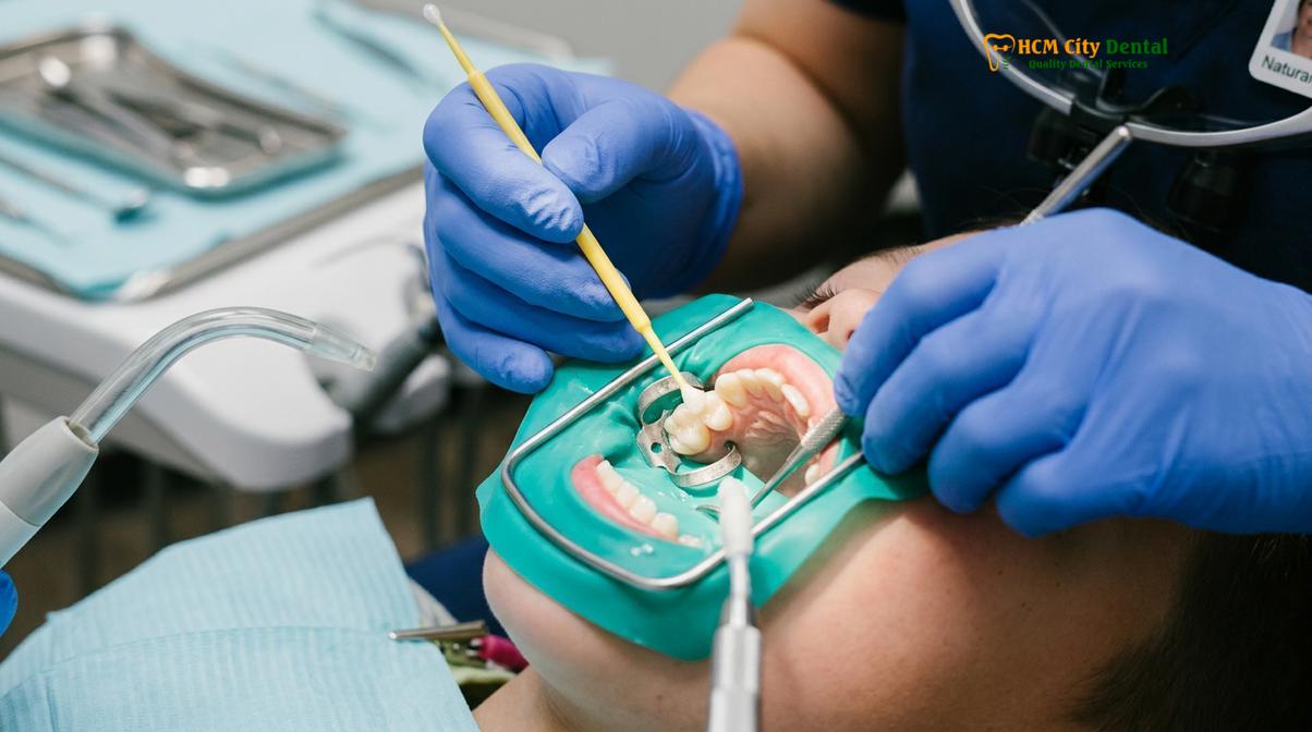
In cases where early decay has already begun but the child is too young or anxious for a traditional filling, Silver Diamine Fluoride (SDF) is a revolutionary intervention. SDF provides non-invasive cavity arrest; the silver ions act as a powerful antimicrobial agent, while the high-concentration fluoride promotes profound remineralization. While SDF is highly effective and completely painless, parents must be informed that it leaves a permanent, localized black stain on the active decay area. This is often an acceptable trade-off to halt disease progression without drilling.
| Treatment Modality | Primary Clinical Indication | Mechanism of Action | Aesthetic Outcome |
|---|---|---|---|
| Fluoride Varnish | Mild MIH, intact enamel, hypersensitivity | Forms acid-resistant fluorapatite layer | Clear/Invisible |
| CPP-ACP (Tooth Mousse) | Daily home remineralization | Delivers bioavailable calcium & phosphate | Clear/Invisible |
| Silver Diamine Fluoride (SDF) | Active decay, extreme anxiety, pre-cooperative child | Silver kills bacteria; Fluoride hardens dentin | Turns decayed areas black |
| Fissure Sealants | Deep grooves in intact MIH molars | Physical barrier against plaque and acids | Tooth-colored/White |
For molars that are fully erupted but structurally intact, the application of fissure sealants is critical. We often utilize glass ionomer-based sealants (such as GC Fuji Triage) for MIH teeth because they tolerate the moisture of a pediatric oral environment better than resin sealants and provide a continuous release of fluoride into the vulnerable fissures.
Restorations for MIH Molars
Restoring chalky teeth requires specialized materials like Glass Ionomer Cements (GIC) or Stainless Steel Crowns (SSCs) that can bond to weak enamel and withstand chewing forces without failing.
When post-eruptive breakdown occurs or caries develop, restorative intervention becomes mandatory. Restoring an MIH-affected tooth is notoriously challenging. Traditional composite resin fillings, which are the standard for healthy teeth, often fail prematurely on chalky teeth. Composite resins are hydrophobic (intolerant to moisture) and rely on micromechanical retention achieved by acid-etching the enamel. Because MIH enamel is already porous and structurally disorganized, the acid etching process is unpredictable, leading to poor bond strength and frequent filling dislodgement.
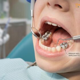
Instead, pediatric dentists prioritize glass ionomer fillings chalky teeth protocols. Glass Ionomer Cement (GIC) is uniquely suited for MIH for several reasons. First, it chemically bonds to both dentin and enamel without the need for aggressive acid etching. Second, it is hydrophilic, meaning it can bond effectively even in a slightly moist environment—a crucial advantage when treating a hypersensitive, salivating child. Most importantly, GIC acts as a fluoride reservoir; it continuously releases fluoride into the surrounding weak tooth structure and can be “recharged” by daily fluoride toothpaste use, preventing recurrent decay at the margins of the filling[4].
Clinical Case Study: Managing Severe MIH
An 8-year-old patient presented to HCMC Dental Clinic in Ho Chi Minh City with severe pain in the lower right 6-year molar. Clinical examination revealed severe yellow-brown MIH with post-eruptive breakdown and early pulpal involvement. Due to the extensive loss of structure, a standard filling was contraindicated. The clinical team performed a pediatric pulpotomy (nerve treatment) to eliminate the pain and restored the tooth with a bespoke Stainless Steel Crown (SSC). The SSC provided full-coverage protection, preventing further fracture and preserving the tooth for proper jaw development.
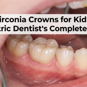
In cases of severe structural loss, full-coverage restorations are required. Preformed Stainless Steel Crowns (SSCs) are the gold standard for severely broken-down MIH molars. They encapsulate the entire tooth, instantly resolving hypersensitivity, preventing further enamel crumbling, and restoring full chewing function. For older children or adults where aesthetics are a primary concern, zirconia crowns or indirect composite onlays may be considered once the dental pulp has stabilized.
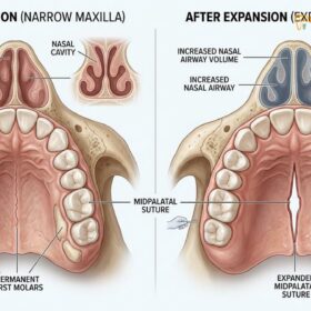
If an MIH molar is deemed unsalvageable and requires extraction, space management becomes the immediate priority. The premature loss of a 6-year molar can lead to severe orthodontic crowding as adjacent teeth drift into the extraction site. In such cases, a fixed space maintainer (like a band-and-loop) is placed. Furthermore, this triggers a comprehensive pediatric interceptive orthodontics evaluation. We assess the child’s overall jaw development, looking for signs of mouth breathing, tongue-ties, or pediatric sleep apnea, ensuring that the loss of the molar does not compromise their long-term airway and facial growth.
When to Consult a Pediatric Dentist
Early diagnosis is the most critical factor in successfully managing chalky teeth; parents should schedule a pediatric dental evaluation as soon as the first permanent molars begin to erupt around age six.
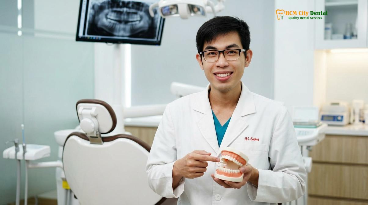
Because MIH can lead to rapid tooth deterioration, waiting for a child to complain of pain is often too late for conservative treatments. Dr. Nguyen Van Cuong, a leading specialist in pediatric care, emphasizes that early intervention is critical for managing MIH effectively. Dr. Cuong and his clinical team utilize advanced moisture-tolerant materials to protect fragile pediatric enamel, ensuring children can chew comfortably without pain. Comprehensive Children & Pediatric Dentistry protocols are designed to catch these developmental defects before post-eruptive breakdown occurs.
Parents should be particularly vigilant if their child experienced significant health issues, premature birth, or frequent high fevers during their first three years of life. According to guidelines supported by the Vietnam Odonto-Stomatology Association (VOSA), children with a history of early childhood systemic illnesses should be closely monitored for enamel defects during the mixed dentition phase[5]. If you notice any creamy white, yellow, or brown spots on your child’s newly erupting back teeth or front incisors, immediate professional assessment is warranted.
“Proactive management of Molar Incisor Hypomineralisation shifts the clinical focus from reactive pain control to preventive structural preservation. By applying remineralizing agents and sealants the moment a vulnerable tooth erupts, we can often prevent the need for invasive restorations entirely.”
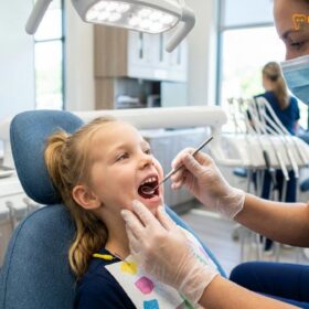
Furthermore, if your child exhibits signs of severe dental anxiety or behavioral challenges stemming from chronic tooth sensitivity, specialized pediatric care is essential. MIH teeth are notoriously difficult to numb with standard local anesthesia due to chronic subclinical pulpal inflammation. Pediatric specialists employ advanced behavioral management techniques, such as Tell-Show-Do, alongside specialized anesthetic protocols to ensure the child remains comfortable and cooperative throughout the procedure.
Whether your child requires preventive fissure sealants, interceptive orthodontics like Phase 1 vs Phase 2 Braces Children, or comprehensive management of chalky teeth, expert care is available. For families seeking specialized pediatric dental services, including Braces for Expat Children, scheduling a consultation at HCMC Dental Clinic in Ho Chi Minh City ensures your child receives the highest standard of compassionate, evidence-based treatment to protect their developing smile.
References
- European Academy of Paediatric Dentistry. Policy document on best clinical practice for Molar Incisor Hypomineralisation.
- International Journal of Paediatric Dentistry. Aetiology, pathogenesis and management of MIH.
- Journal of the American Dental Association. Clinical efficacy of Silver Diamine Fluoride in pediatric dentistry.
- Pediatric Dentistry. Behavioral management techniques and local anesthesia efficacy in hypomineralized teeth.
- Vietnam Odonto-Stomatology Association (VOSA). National guidelines on pediatric enamel defects and early childhood caries management.