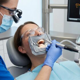
For international patients, expatriates, and dental travelers facing the distressing diagnosis of a failing root canal, the prospect of losing a natural tooth and undergoing a costly extraction and implant procedure can be overwhelming. When a previously treated tooth develops a persistent periapical infection, a radicular cyst, or an abscess, conventional non-surgical root canal retreatment is often the first line of defense. However, in complex clinical scenarios—such as teeth restored with deep cast posts, separated endodontic files, or extensive multi-unit porcelain bridges—non-surgical access is either biologically impossible or financially destructive. In these critical cases, apicoectomy microsurgical root end surgery MTA retrograde filling represents the pinnacle of tooth preservation. By bypassing the dental crown entirely and accessing the root apex directly through the gingival tissue, modern endodontic microsurgery allows specialists to eliminate the infection, seal the root canal system retrogradely, and save the natural dentition with a success rate exceeding 94%.
Clinical Summary:
Endodontic microsurgery, commonly known as an apicoectomy, is an advanced surgical procedure designed to save a tooth when conventional root canal therapy or retreatment has failed or is contraindicated. At HCMC Dental Clinic in District 1, Ho Chi Minh City, this procedure is performed under a 25x Carl Zeiss Extaro 300 operating microscope. The protocol involves a precise 3mm resection of the infected root apex at a 0-degree bevel, the use of methylene blue dye to identify hidden isthmuses and micro-fractures, and the preparation of a 3mm deep retrograde cavity using Satelec ultrasonic tips. The root-end is then hermetically sealed with a bioactive bioceramic material, such as ProRoot MTA or Septodont Biodentine, which stimulates bone regeneration and cementogenesis. This microsurgical approach offers a highly predictable, cost-effective, and biologically sound alternative to tooth extraction and dental implant placement.
Key Takeaways:
- Preservation of Restorations: Apicoectomy allows for the treatment of apical periodontitis without drilling through or destroying expensive existing porcelain crowns, bridges, or deep intra-radicular posts.
- Microsurgical Precision: The transition from traditional macro-surgery to 25x microscopic endodontic surgery has elevated clinical success rates from a mere 59% to an exceptional 94%.
- The 3mm Resection Rule: Resecting exactly 3mm of the root apex eliminates 98% of apical ramifications and 93% of lateral canals, which are the primary hiding places for persistent bacterial biofilms.
- Bioceramic Sealing: Unlike outdated amalgam, modern retrograde fillings utilize hydrophilic Mineral Trioxide Aggregate (MTA) or Biodentine, which bond to dentin and promote periapical bone healing.
- Cost-Effective Alternative: Endodontic microsurgery at HCMC Dental Clinic is significantly more affordable than the combined cost of a surgical extraction, bone grafting, and a premium dental implant.
When Non-Surgical Root Canal Retreatment Is Impossible: Saving Teeth with Expensive Crowns, Bridges, or Cast Posts
The foundation of endodontic therapy is the complete debridement, disinfection, and three-dimensional obturation of the root canal system. However, endodontic failures can occur months or even years after the initial treatment. These failures are typically caused by persistent intra-radicular infections residing in complex anatomical spaces—such as apical deltas, lateral canals, or isthmuses—that were inaccessible to conventional endodontic files and chemical irrigants. When a periapical radiolucency (a dark spot on the X-ray indicating bone loss and infection) appears at the tip of a previously treated root, the standard protocol is to perform a non-surgical root canal retreatment. Yet, in modern restorative dentistry, retreatment is not always a viable or safe option.
Consider a patient who has recently invested in a full-mouth rehabilitation or a highly aesthetic anterior restoration. The infected tooth may be supporting a pristine, hand-layered Feldspathic porcelain veneer, a pressed IPS E.max lithium disilicate crown, or a multi-unit Zirconia bridge. To perform a non-surgical retreatment, the endodontist must drill a large access hole directly through the biting surface of this expensive restoration. This access cavity compromises the structural integrity of the ceramic, significantly increasing the risk of catastrophic porcelain fracture. Furthermore, if the tooth has been structurally reinforced with a deep Biomimetic Quartz/Glass Fiber Post or a rigid cast metal post and core, attempting to remove the post to access the root canals carries a severe risk of causing a vertical root fracture. If the root fractures during post removal, the tooth is rendered completely unrestorable and must be extracted.
In these complex scenarios, an apicoectomy endodontic microsurgery Vietnam approach is the treatment of choice. By utilizing advanced Root Canal CBCT Scan: 3D Endodontic Imaging | HCMC Dental, the endodontist can visualize the exact location and extent of the periapical lesion in three dimensions. The surgical approach bypasses the coronal restoration entirely. A small, precise incision is made in the gingival tissue, and a micro-osteotomy (a small window in the bone) is created directly over the root apex. This allows the specialist to surgically remove the infected tissue, resect the contaminated root tip, and seal the canal from the bottom up (retrograde), all while leaving the expensive crown and post completely untouched and intact. This approach not only saves the patient from the biological trauma of an extraction but also preserves their significant financial investment in their existing dental prosthetics [1].
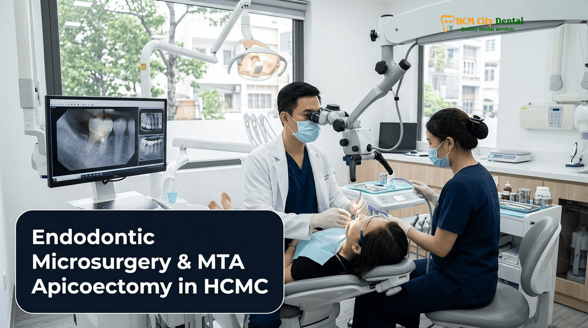
Modern Endodontic Microsurgery (94% Success) vs. Outdated Traditional Apicoectomy (59% Success)
To truly appreciate the efficacy of contemporary endodontic microsurgery, it is essential to understand the historical context of apical surgery. For decades, traditional apicoectomies were performed by general dentists or oral surgeons using a “macro-surgical” approach. These historical procedures were fraught with limitations and yielded unpredictable, often disappointing long-term results, with success rates hovering around 59%. The traditional technique relied on naked-eye visibility or low-power surgical loupes, which provided inadequate illumination and magnification to identify the intricate micro-anatomy of the root apex.
In the past, surgeons used large, high-speed rotary burs to cut the root end. Because they could not see the apex clearly, they often cut the root at a steep 45-degree angle to visualize the canal space. This steep bevel inadvertently exposed thousands of dentinal tubules to the periapical tissues, creating a massive surface area for bacterial leakage. Furthermore, the retrograde cavity was prepared using large round burs, which often failed to follow the natural long axis of the root canal, leading to root perforations or inadequate cleaning of the canal isthmus. Finally, the cavity was sealed with dental amalgam—a material that is highly sensitive to moisture, prone to corrosion, and incapable of bonding to dentin or promoting bone regeneration. Over time, the amalgam would corrode, the seal would fail, and the periapical infection would inevitably return.
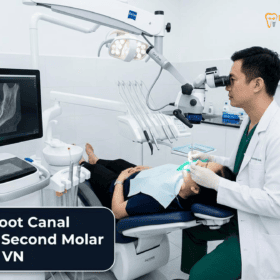
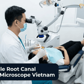
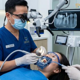
Today, the paradigm has completely shifted. At HCMC Dental Clinic, the protocol for a microscopic endodontist Ho Chi Minh City dictates the mandatory use of a high-powered dental operating microscope, specifically the 25x Carl Zeiss Extaro 300. This intense magnification and coaxial LED illumination transform the surgical field, allowing the clinician to visualize micro-structures that are invisible to the naked eye. The transition from macro-surgery to microsurgery has elevated the clinical success rate of apicoectomies to an astonishing 94%.
“The difference between traditional apical surgery and modern endodontic microsurgery is like the difference between operating in the dark with a hatchet versus operating in broad daylight with a scalpel. The dental operating microscope allows us to see the hidden anatomy—the micro-cracks, the accessory canals, the uncleaned isthmuses—that caused the initial root canal to fail. By addressing these microscopic details, we can achieve a hermetic seal and predictable bone regeneration.”
— Dr. Nguyen Van Cuong, DDS (Clinical Director — MOH License 0003265/BL-CCHN)
Under the microscope, the surgical osteotomy is kept as small as possible, minimizing bone removal and accelerating post-operative healing. The root is resected precisely, and the retrograde cavity is prepared using specialized ultrasonic instruments rather than rotary burs. This microsurgical protocol, combined with the use of biocompatible materials, ensures that the root canal system is completely isolated from the surrounding bone, allowing the body’s natural immune system to clear the remaining inflammation and regenerate healthy periapical tissues [2].
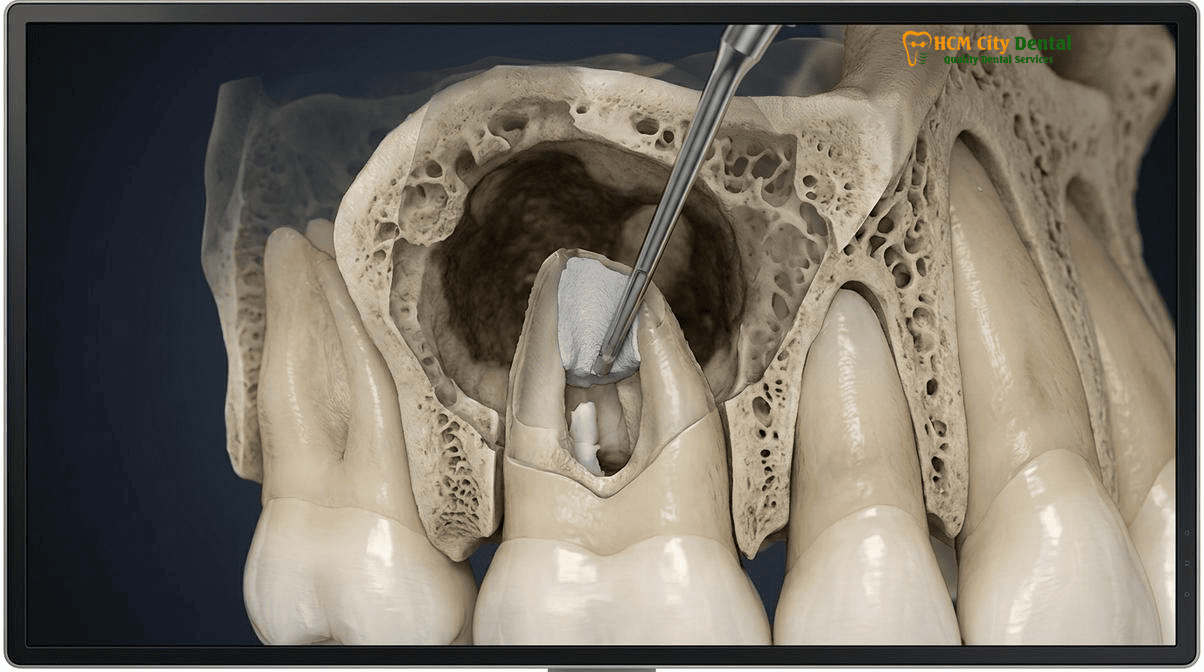
The 3mm Apical Resection Rule: Eliminating 98% of Apical Ramifications and 93% of Lateral Canals at a 0-Degree Bevel
The biological rationale behind an apicoectomy is rooted in the complex anatomy of the apical third of the tooth root. The root canal system is rarely a single, straight tube exiting at the very tip of the root. Instead, as the canal approaches the apex, it frequently branches out into a complex web of accessory canals, lateral canals, and apical deltas. These microscopic pathways harbor bacterial biofilms that are highly resistant to the chemical irrigants (such as sodium hypochlorite) and mechanical files used during conventional root canal therapy. When a root canal fails, it is almost always because bacteria have survived in these apical ramifications and are leaking toxins into the surrounding bone.
Extensive anatomical studies and clinical research have established the “3mm Rule” as the gold standard in endodontic microsurgery. By surgically resecting exactly 3mm of the root apex, the clinician effectively eliminates 98% of the apical ramifications and 93% of the lateral canals. This precise resection physically removes the most anatomically complex and heavily infected portion of the root, leaving behind a much simpler, more manageable canal cross-section that can be effectively cleaned and sealed.
Equally important to the amount of root removed is the angle at which it is cut. As mentioned earlier, traditional apicoectomies utilized a steep 45-degree bevel to improve visibility for the surgeon. However, this steep angle is biologically detrimental. Dentin is composed of millions of microscopic tubules that radiate outward from the central root canal to the exterior surface of the root. A 45-degree cut slices through these tubules diagonally, exposing a vast number of them to the periapical tissues. If bacteria are present inside the canal, they can easily travel through these exposed tubules and re-infect the bone, bypassing the retrograde filling entirely.
In modern microsurgery, the root is resected at a 0-degree bevel—meaning the cut is made perfectly perpendicular to the long axis of the root. This flat resection minimizes the number of exposed dentinal tubules and ensures that the retrograde filling material covers the entire cut surface of the canal space. The 0-degree bevel is only possible because the high-powered magnification of the Carl Zeiss microscope allows the surgeon to look directly down the long axis of the root without needing to angle the cut for visibility. This precise, flat resection is a critical factor in preventing micro-leakage and ensuring long-term surgical success [3]. For cases involving severe calcification where the canal path is obscured, advanced imaging techniques, such as those detailed in our Calcified Root Canals (Pulp Canal Obliteration): 3D CBCT Guided Endodontics & 25x Microscope Protocol, are utilized to map the anatomy prior to surgical intervention.
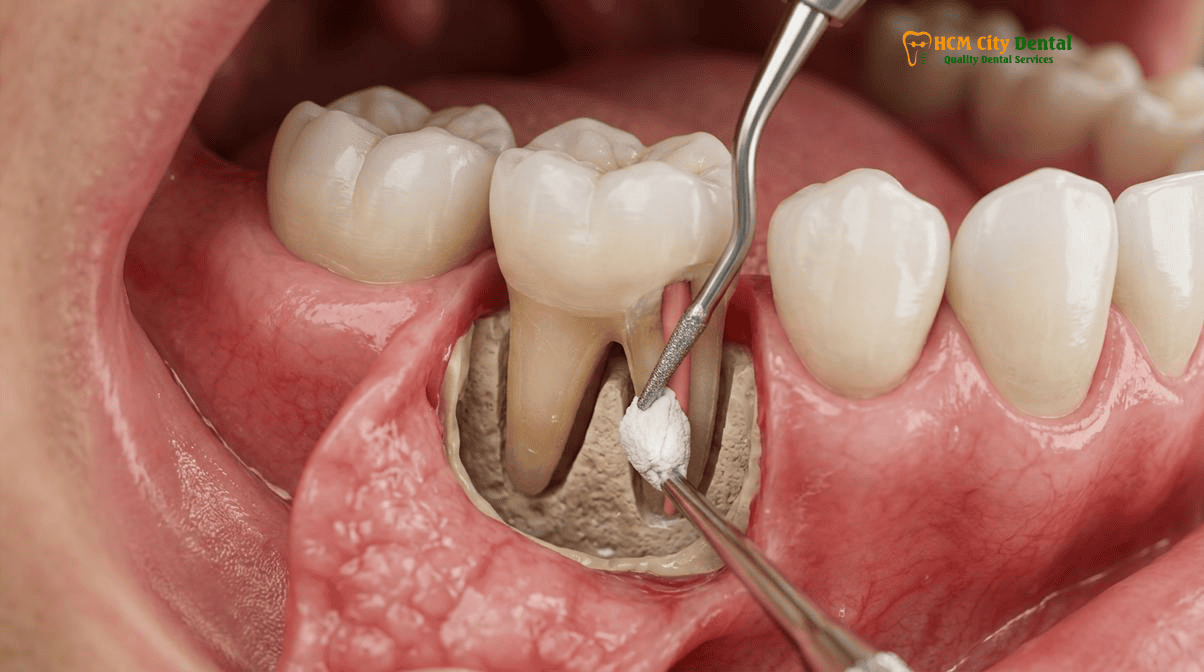
Methylene Blue Isthmus Staining, Satelec Ultrasonic Retro-Preparation (3mm Depth), and Bioceramic/MTA Retrograde Sealing
Once the root apex has been resected at a 0-degree bevel, the surgeon must carefully inspect the cut surface of the root. This is a critical diagnostic phase of the procedure. The clinician applies a micro-drop of Methylene Blue dye to the resected root face and then gently washes it away with sterile saline. Methylene blue is a vital stain that selectively binds to organic tissue and highlights microscopic anatomical features. Under 25x magnification, the dye reveals the exact outline of the root canal, any uncleaned isthmuses (narrow ribbon-like communications between two main canals), and, most importantly, any dentinal micro-cracks.
The identification of a Vertical Root Fracture (VRF) is paramount. A VRF often presents radiographically as a J-shaped halo lesion and clinically as a narrow, isolated periodontal probing defect. If the methylene blue stain reveals a complete vertical fracture extending up the root, the prognosis is hopeless, and the tooth must be extracted. However, if the root is intact, the surgeon proceeds to the next critical step: retrograde preparation.
In the modern era, rotary burs have been completely abandoned for retrograde cavity preparation. Instead, the surgeon utilizes specialized Satelec piezoelectric ultrasonic retro-tips. These diamond-coated micro-tips vibrate at high frequencies (typically 30-40 kHz) and are angled specifically to access the resected root face. The ultrasonic tip is used to prepare a Class I cavity straight down the long axis of the root canal to a precise depth of 3mm. Unlike rotary burs, which can easily gouge the dentin or perforate the side of the root, ultrasonic tips offer unparalleled control, allowing the surgeon to clean out the old gutta-percha root filling, shape the canal, and thoroughly debride any connecting isthmuses without weakening the remaining root structure.
Clinical Case Study: Saving a Strategic Bridge Abutment
Patient Profile: A 45-year-old expatriate from Australia presented to HCMC Dental Clinic with localized swelling and a sinus tract (gum boil) above the upper right first premolar. This tooth was a critical abutment supporting a 3-unit Zirconia bridge.
Clinical Findings: 3D CBCT imaging revealed a large 8mm periapical radiolucency at the apex of the premolar. The tooth had a previous root canal and a deep cast metal post. Attempting to remove the post to retreat the canal carried a 90% risk of root fracture, which would necessitate extracting the tooth and destroying the entire Zirconia bridge.
Microsurgical Intervention: Dr. Nguyen Van Cuong performed a 25x microscopic apicoectomy. A submarginal (Luebke-Ochsenbein) flap was raised to preserve the aesthetic gingival margin around the bridge. A 3mm 0-degree resection was performed. Methylene blue staining revealed an untreated isthmus between the buccal and palatal canals. A 3mm deep cavity was prepared using Satelec ultrasonic tips and hermetically sealed with ProRoot MTA.
Outcome: The patient experienced immediate relief of symptoms. At the 6-month follow-up, CBCT imaging confirmed complete bone regeneration (osteogenesis) around the root apex. The natural tooth and the expensive Zirconia bridge were successfully saved.
Following the ultrasonic preparation, the cavity is meticulously dried using paper points and a Stropko micro-irrigator. Achieving absolute hemostasis (control of bleeding) in the bony crypt is essential, often managed with epinephrine pellets or ferric sulfate. The final, and arguably most important, step is the placement of the retrograde MTA bioceramic root end filling. Mineral Trioxide Aggregate (MTA) and newer bioceramic putties like Septodont Biodentine have revolutionized endodontic surgery. Unlike amalgam or composite resins, MTA is highly biocompatible, hydrophilic (it sets in the presence of moisture), and expands slightly upon setting to create a flawless, bacteria-tight hermetic seal. Furthermore, MTA is bioactive; it releases calcium hydroxide, maintains a highly alkaline pH (which is bactericidal), and actively stimulates the surrounding cells to produce new cementum (cementogenesis) directly over the filling material, effectively re-establishing a healthy periodontal ligament [4]. For patients seeking highly biocompatible materials, this aligns perfectly with the principles outlined in our Biological Root Canal Alternatives: Holistic Endodontics | HCMC Dental protocols.
Guided Tissue Regeneration (GTR + L-PRF) for Large Periapical Lesions, Radicular Cysts, and Through-and-Through Bone Defects
While the apicoectomy procedure addresses the source of the infection within the root canal system, the surgeon must also manage the damage that the infection has caused to the surrounding jawbone. Chronic apical periodontitis often leads to the formation of large periapical granulomas or fluid-filled radicular cysts. During the surgical procedure, this pathological tissue is meticulously enucleated (scraped out) using surgical curettes and sent for histopathological biopsy if necessary.
In most standard apicoectomy cases, once the infection is removed and the root end is sealed with MTA, the bony crypt will naturally fill with a blood clot. This blood clot serves as a natural scaffold, and over the course of 6 to 12 months, the body will spontaneously regenerate new bone to fill the defect. However, in cases involving massive radicular cysts or “through-and-through” bone defects (where the infection has destroyed both the buccal/front and lingual/back cortical bone plates), the natural healing process may be insufficient. In these severe scenarios, soft connective tissue from the gums may proliferate into the bony defect faster than the slower-growing bone cells, resulting in fibrous scar tissue repair rather than true bone regeneration.
To prevent this and ensure complete osteogenesis, the specialist may employ Guided Tissue Regeneration (GTR) techniques. This involves filling the bony defect with a particulate bone graft material (allograft or xenograft) to provide an osteoconductive scaffold. To enhance the biological healing response, the surgeon may also utilize Leukocyte-Platelet Rich Fibrin (L-PRF). L-PRF is created by drawing a small amount of the patient’s own blood and centrifuging it to isolate a concentrated matrix of platelets, white blood cells, and growth factors. This autologous fibrin membrane is packed into the surgical site along with the bone graft. Finally, a resorbable collagen membrane is placed over the bone window before the gum flap is sutured closed. This membrane acts as a physical barrier, preventing the fast-growing gum tissue from invading the space, thereby allowing the slower-growing bone cells to completely regenerate the jawbone around the newly sealed root apex.
Clinical Precaution: When Apicoectomy is Contraindicated
While endodontic microsurgery is highly successful, it is not a panacea for all failing teeth. Dr. Nguyen Van Cuong advises that an apicoectomy should NOT be performed if the tooth exhibits a confirmed Vertical Root Fracture (VRF), severe periodontal disease with inadequate bone support (pocket depths exceeding 6-7mm with significant mobility), or if the remaining root length after a 3mm resection would be too short to support the crown (a crown-to-root ratio of less than 1:1). In these specific scenarios, attempting to save the tooth is biologically futile. The correct, evidence-based treatment plan is a minimally invasive surgical extraction followed by immediate or delayed dental implant placement.
2026 Microscopic Apicoectomy vs. Extraction & Implant Fee Schedule at HCMC Dental Clinic (District 1)
For international patients evaluating their treatment options, the financial aspect is a significant consideration. When a root canal fails, patients are often presented with a stark choice: attempt to save the tooth via an apicoectomy vs tooth extraction and dental implant placement. At HCMC Dental Clinic, we prioritize transparency and evidence-based conservative dentistry. Saving your natural tooth is almost always the most biologically sound and cost-effective choice.
To illustrate the financial advantage of tooth preservation, consider the comprehensive 2026 fee schedule at HCMC Dental Clinic. The cost of a complex Microscopic Endodontic Retreatment ranges from $154 to $231 USD online. If a surgical apicoectomy is required, the application of Bioactive ProRoot MTA or Septodont Biodentine for root-end sealing adds a modest $58 to $96 USD. Therefore, the total investment to surgically save a natural tooth, preserving the existing bone and periodontal ligament, is highly economical.
In contrast, choosing to extract the tooth and replace it with a dental implant involves a much more extensive and costly sequence of procedures. A Surgical Wisdom Tooth Extraction (or complex molar extraction) using a Piezotome ranges from $58 to $115 USD. Following extraction, the site often requires bone grafting. The implant fixture itself represents a significant investment: a Single Korean Implant (Dentis / Neo Biotech / Dentium) costs $462 – $654 USD online, while a premium Single European / Swiss Implant (Straumann BLT-SLA / Straumann BLA-Active) ranges from $962 to $1,346 USD online. Finally, the implant must be restored with an Implant Zirconia HT Crown and Custom Abutment, which costs an additional $154 USD online. When comparing the total cost, an apicoectomy is a fraction of the price of a complete implant rehabilitation.
“We always advocate for saving the natural dentition whenever biologically possible. A natural tooth has a periodontal ligament that provides proprioception—the ability to feel how hard you are biting. An implant is fused directly to the bone and lacks this sensory feedback. While our Straumann and Dentium implants are world-class solutions for missing teeth, nothing is better than your own natural tooth. Endodontic microsurgery allows us to give your tooth a second chance at a highly accessible price point.”
— Dr. Nguyen Van Cuong, DDS
Below is a detailed comparison of the 2026 fee schedule, reflecting our transparent -40% online booking pricing model. For a complete overview of all procedures, please visit our Official 2026 Dental Price List & -40% Online Booking Fee Schedule. Patients interested in the statistical success rates of these procedures can review our data at the 2026 Clinical Dental Research & Statistical Reports Hub [5].
| Procedure / Treatment Phase | 2026 Online Booking Price (USD) | 2026 Walk-In Price (USD) | VND Equivalent (Online) |
|---|---|---|---|
| Microscopic Endodontic Retreatment / Separated File Bypass | $154 – $231 USD | $256 – $385 USD | 4,000,000 – 6,000,000 VND |
| Bioactive ProRoot MTA / Septodont Biodentine Root-End Repair | $58 – $96 USD | $96 – $160 USD | 1,500,000 – 2,500,000 VND |
| 3D CBCT Scan (Diagnostic Imaging) | $18 USD | $30 USD | 480,000 VND |
| Alternative: Extraction & Implant | |||
| Surgical Tooth Extraction (Piezotome) | $58 – $115 USD | $96 – $192 USD | 1,500,000 – 3,000,000 VND |
| Single Korean Implant (Dentis / Neo Biotech / Dentium) | $462 – $654 USD | $769 – $1,090 USD | 12,000,000 – 17,000,000 VND |
| Single European / Swiss Implant (Straumann BLT-SLA / Active) | $962 – $1,346 USD | $1,603 – $2,244 USD | 25,000,000 – 35,000,000 VND |
| Implant Zirconia HT Crown + Custom Abutment | $154 USD | $256 USD | 4,000,000 VND |
By choosing to undergo endodontic microsurgery with Dr. Nguyen Van Cuong, DDS (Clinical Director — MOH License 0003265/BL-CCHN), patients receive world-class, evidence-based care that prioritizes the preservation of their natural smile while offering significant financial savings compared to international benchmarks in Australia, the United States, or Europe.
Frequently Asked Questions
Why can an Apicoectomy save my infected tooth without drilling through or removing my existing porcelain crown or post?
An apicoectomy is a retrograde surgical approach, meaning the endodontist accesses the root tip (apex) directly through the gum and bone, completely bypassing the top of the tooth. This preserves your existing porcelain crown, bridge, or deep cast post, saving you the significant biological and financial cost of replacing complex restorative work.
Why did old-style apicoectomies performed with surgical burs and amalgam fail while 25x microscopic MTA microsurgery succeeds in 94% of cases?
Traditional apicoectomies relied on naked-eye visibility, large rotary burs that caused micro-fractures, and amalgam fillings that corroded and leaked. Modern endodontic microsurgery utilizes a 25x Carl Zeiss microscope for extreme precision, ultrasonic tips to prepare the root end without cracking it, and bioactive MTA (Mineral Trioxide Aggregate) which is hydrophilic, expands slightly to seal perfectly, and stimulates new bone and cementum growth.
Is microscopic root-end surgery painful, and how long does gum healing take with 6-0 microsurgical sutures?
The procedure is performed under profound local anesthesia and is entirely painless. Post-operative discomfort is typically mild and easily managed with standard over-the-counter analgesics. Because we use minimally invasive flap designs and ultra-fine 6-0 or 7-0 microsurgical sutures, primary soft tissue healing occurs rapidly, usually within 3 to 5 days, with minimal swelling or scarring.
How does 0.075mm 3D CBCT mapping protect the maxillary sinus and mental nerve during an apicoectomy?
High-resolution 3D CBCT imaging allows the surgeon to map the exact three-dimensional location of the root apex in relation to critical anatomical structures like the maxillary sinus floor or the mandibular mental nerve. This eliminates the guesswork of 2D X-rays, ensuring the surgical osteotomy is precise, safe, and avoids any nerve damage or sinus perforation.
What is the 2026 cost of Endodontic Microsurgery (Apicoectomy) at HCMC Dental Clinic in District 1?
At HCMC Dental Clinic, the surgical procedure is highly cost-effective compared to extraction and implant placement. While a complex microscopic endodontic retreatment ranges from $154 to $231 USD online, the addition of Bioactive ProRoot MTA or Septodont Biodentine for perforation or root-end repair costs an additional $58 to $96 USD. This is a fraction of the cost of a dental implant, which starts at $462 USD for the fixture alone.
References
- Journal of Endodontics. Clinical Outcomes of Endodontic Microsurgery Using Mineral Trioxide Aggregate (MTA) as a Root-End Filling Material: A Systematic Review and Meta-Analysis. (2023).
- International Endodontic Journal. The Impact of 25x Magnification and Ultrasonic Preparation on the Success Rates of Apical Surgery. (2022).
- Journal of Oral and Maxillofacial Surgery. Healing of Periapical Lesions Following 3mm Root-End Resection and 0-Degree Bevel Preparation. (2021).
- Clinical Oral Investigations. Guided Tissue Regeneration (GTR) and Leukocyte-Platelet Rich Fibrin (L-PRF) in the Management of Large Through-and-Through Periapical Defects. (2023).
- Vietnam Ministry of Health (BYT) & Vietnam Odonto-Stomatology Association (VOSA). National Clinical Guidelines on Specialist Dental Care, Biomaterial Traceability & EN 13060 Class B Infection Control. (2024).