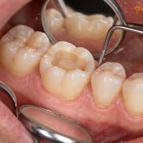
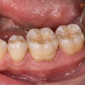
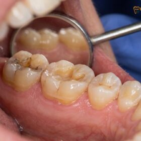
The management of deep subgingival carious lesions presents one of the most profound technical challenges in modern restorative dentistry. When tooth decay, structural fractures, or failing amalgam restorations extend apically below the gingival margin, the resulting defect compromises the clinician’s ability to achieve adequate moisture control, optical visibility, and precise marginal adaptation. Historically, the default treatment for such severely compromised teeth involved aggressive surgical crown lengthening—a periodontal procedure requiring the irreversible removal of healthy alveolar bone to expose the deep margin—followed by the placement of a traditional full-coverage porcelain-fused-to-metal (PFM) crown. However, the advent of advanced adhesive protocols and biomimetic dentistry has introduced a highly predictable, minimally invasive alternative: Deep Margin Elevation (DME). By utilizing specialized matrix systems and advanced resin technologies under high-magnification microscopy, specialists can now relocate deep cervical margins supragingivally, preserving critical periodontal architecture and saving cracked decayed molars without extraction in Vietnam.
Clinical Summary:
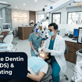
The clinical decision between deep margin elevation vs crown lengthening subgingival caries hinges on the ability to achieve absolute isolation and the preservation of the supracrestal tissue attachment (biologic width). Deep Margin Elevation (DME), also known as cervical margin relocation, is a biomimetic technique that utilizes flowable and pre-heated composite resins to elevate a subgingival defect to a supragingival level, facilitating the subsequent bonding of a ceramic inlay or onlay. Conversely, surgical crown lengthening involves ostectomy (bone removal) to apically reposition the periodontal tissues. At HCMC Dental Clinic in District 1, Ho Chi Minh City, Dr. Nguyen Van Cuong, DDS, employs 25x Carl Zeiss microscopic precision, 100% latex-free nitrile rubber dam isolation, and Immediate Dentin Sealing (IDS) to execute highly successful DME procedures, offering international patients a conservative, enamel-preserving alternative to invasive periodontal surgery.
Key Takeaways:
- Biomimetic Preservation: Deep Margin Elevation (DME) allows for the supragingival relocation of deep carious margins without the need for irreversible surgical bone removal (ostectomy).
- Biologic Width Compliance: Successful DME requires strict adherence to the 2.0mm bone-to-margin rule to prevent supracrestal tissue attachment biologic width violation and chronic periodontal inflammation.
- Absolute Isolation: The procedure mandates a 100% latex-free nitrile rubber dam and advanced matrix-in-matrix teflon isolation subgingival onlay techniques to ensure zero moisture contamination.
- Adhesive Superiority: Utilizing Pascal Magne’s Immediate Dentin Sealing (IDS) protocol increases microtensile dentin bond strength by up to 400% (achieving 45–55 MPa).
- Cost-Effective Excellence: International patients can access world-class biomimetic restorations, including IPS E.max All-Ceramic Onlays, at a transparent -40% online booking rate at HCMC Dental Clinic in District 1.
The Clinical Challenge of Deep Subgingival Interproximal Caries: Why Traditional Crowns Fail Below the Gumline
Deep subgingival interproximal caries typically develops silently, often originating below the contact point of adjacent posterior teeth and migrating apically toward the cementoenamel junction (CEJ) and the root surface. Because the root surface lacks the protective, highly mineralized enamel layer, dentin and cementum are rapidly degraded by the acidic byproducts of anaerobic cariogenic bacteria. By the time the patient experiences thermal sensitivity or localized gingival bleeding, the carious lesion has often progressed deep into the periodontal pocket, presenting a severe restorative dilemma.
The primary clinical challenge in treating these lesions lies in the hostile environment of the gingival sulcus. The continuous exudation of gingival crevicular fluid, the high probability of hemorrhage from inflamed sulcular epithelium, and the presence of saliva make it exceptionally difficult to achieve the dry, uncontaminated field required for modern adhesive dentistry. When traditional full-coverage crowns are employed to treat these defects, the clinician is forced to place the crown margin deep subgingivally to capture sound tooth structure. This approach is fraught with long-term complications. Traditional cements (such as zinc phosphate or glass ionomer) are highly soluble in the acidic oral environment; when placed subgingivally, they are prone to micro-washout, leading to microleakage, secondary recurrent caries, and eventual catastrophic failure of the abutment tooth. Furthermore, aggressive subgingival crown preparations frequently violate the delicate periodontal attachment apparatus, triggering a cascade of chronic localized periodontitis, unpredictable crestal bone resorption, and persistent gingival erythema.[1]
In the context of saving a cracked decayed molar without extraction in Vietnam, the traditional amputational approach—which involves aggressive core buildups, cast post-and-cores, and heavy circumferential reduction—often weakens the remaining radicular structure, predisposing the tooth to vertical root fractures (VRF). A VRF is clinically characterized by a J-shaped halo lesion on a periapical radiograph and a narrow, isolated periodontal probing defect. Once a vertical root fracture occurs, the prognosis is hopeless, and extraction followed by implant placement becomes the only viable option. Therefore, shifting the paradigm toward defect-driven, minimally invasive biomimetic protocols is essential for maximizing the lifespan of the natural dentition.
What Is Biomimetic Deep Margin Elevation (DME / Cervical Margin Relocation) and How Does It Relocate the Margin Supragingivally?
Deep Margin Elevation (DME), frequently referred to in the scientific literature as cervical margin relocation subgingival caries, is a foundational technique in biomimetic restorative dentistry. First conceptualized by Dietschi and Spreafico, and later refined by Dr. Pascal Magne, DME is designed to non-surgically reposition a deep, inaccessible subgingival cavity margin to a more coronal, supragingival level. This is achieved by meticulously isolating the deep defect, applying advanced dentin bonding agents, and layering a highly filled composite resin base directly over the subgingival margin. Once the margin is elevated, the subsequent steps of optical 3D scanning, isolation, and adhesive cementation of the final indirect restoration (such as a ceramic inlay, onlay, or overlay) become highly predictable and technically straightforward.
The success of deep margin elevation dme biomimetic dentistry relies heavily on the Immediate Dentin Sealing (IDS) protocol. When dentin is freshly cut or excavated, the dentinal tubules are exposed, making them highly susceptible to bacterial microleakage, pulpal irritation, and collapse of the collagen fibril network. By applying a 3-step etch-and-rinse or a high-quality universal adhesive system immediately after caries removal—prior to taking any impressions or digital scans—the clinician effectively seals the dentin complex. This IDS layer is then protected by a low-viscosity flowable resin biobase. According to Pascal Magne’s research, this protocol allows the dentin bond to mature stress-free, increasing microtensile dentin bond strength by up to 400%, consistently achieving values of 45–55 MPa. This robust hybrid layer mimics the natural dentinoenamel junction (DEJ), providing exceptional resistance to occlusal shear forces and virtually eliminating postoperative sensitivity.[2]
“The philosophy of biomimetic dentistry is rooted in maximum tissue preservation and the replication of natural biomechanics. By utilizing Deep Margin Elevation in conjunction with Immediate Dentin Sealing, we can predictably restore severely compromised molars that would have historically been condemned to extraction or aggressive surgical crown lengthening. Our goal is to seal the tooth biologically and structurally, utilizing advanced adhesive protocols under high-magnification microscopy to ensure a flawless, gap-free interface.”
— Dr. Nguyen Van Cuong, DDS (Clinical Director — MOH License 0003265/BL-CCHN)
Following the elevation of the margin, the tooth is restored using indirect ceramic restorations. In biomimetic practice, there is a distinct preference for materials that closely match the modulus of elasticity of natural enamel. While pressed or cut-back IPS E.max lithium disilicate (offering flexural strengths of 450–500 MPa) is excellent for high-stress areas, hand-layered refractory Feldspathic porcelain (90–110 MPa) is often utilized for highly esthetic, minimally invasive anterior restorations. For posterior subgingival defects, an IPS E.max onlay bonded over a DME biobase provides an optimal balance of fracture toughness and biomimetic stress distribution.
Respecting the Supracrestal Tissue Attachment (Biologic Width): 2.0mm Bone-to-Margin Rules on Bitewing Radiographs
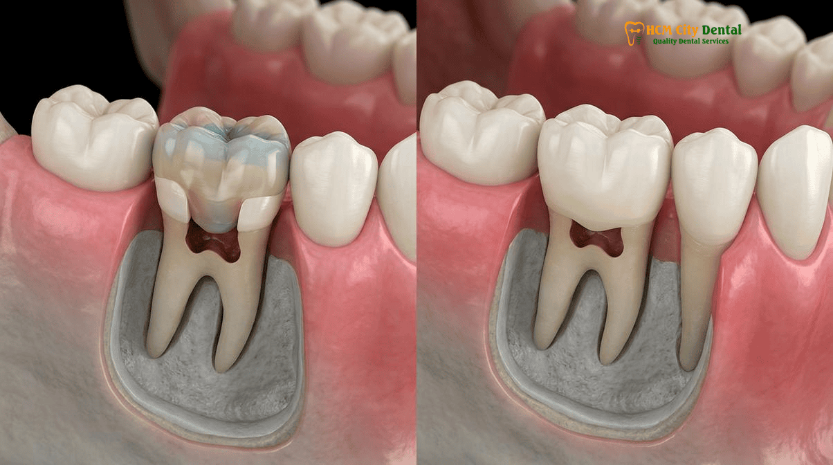
A critical anatomical boundary that dictates the success or failure of any subgingival restorative procedure is the supracrestal tissue attachment, historically known as the biologic width. This complex comprises the junctional epithelium (approximately 0.97mm in height) and the supracrestal connective tissue attachment (approximately 1.07mm in height), which together form a protective physiological seal around the neck of the tooth, defending the underlying alveolar bone from the microbial flora of the oral cavity.
When evaluating a tooth for deep margin elevation vs surgical crown lengthening, the clinician must rigorously assess the distance from the deepest extent of the carious lesion to the crest of the alveolar bone. The fundamental periodontal rule mandates a minimum distance of 2.0mm (ideally 3.0mm to account for the sulcus depth) between the restorative margin and the alveolar bone crest. If a composite resin or ceramic margin is placed within this restricted zone, it results in a supracrestal tissue attachment biologic width violation. The body responds to this foreign body intrusion with a chronic, localized inflammatory response. Clinically, this manifests as persistent gingival hyperplasia, bleeding on probing, cyanotic (bluish-red) tissue discoloration, and ultimately, unpredictable osteoclastic activity leading to localized alveolar bone loss as the body attempts to recreate space for the attachment apparatus.[3]
To prevent this catastrophic outcome, precise diagnostic protocols are mandatory. Prior to initiating treatment, the clinician performs “bone sounding” by passing a periodontal probe through the anesthetized gingival sulcus until it contacts the alveolar crest, allowing for an accurate measurement of the available restorative space. Furthermore, high-resolution digital bitewing radiographs are indispensable. A bitewing radiograph provides an undistorted, orthogonal view of the interproximal bone crest relative to the cavity margin. If the radiograph reveals that the carious defect is at or below the bone crest, DME is strictly contraindicated, and surgical intervention is required. Conversely, if the 2.0mm rule is satisfied, DME can proceed. Crucially, a post-operative bitewing radiograph must be taken immediately after the DME procedure to verify the absolute marginal integrity of the composite elevation, ensuring there are no overhangs or voids before proceeding with the optical 3D scan for the final Dental Crowns, Fixed Bridges & Biomimetic Ceramic Restorations in Vietnam.
Deep Margin Elevation (DME) vs. Ostectomy Surgical Crown Lengthening: Furcation Exposure and Crown-to-Root Ratio Risks
The debate between deep margin elevation vs crown lengthening subgingival caries is one of the most heavily discussed topics in contemporary restorative periodontics. Surgical Crown Lengthening (SCL) is a traditional periodontal surgery designed to increase the clinical crown length by apically repositioning the gingival margin and recontouring the underlying alveolar bone. The procedure involves raising a full-thickness mucoperiosteal flap, performing an ostectomy (removal of supporting bone) and osteoplasty (reshaping of non-supporting bone) using rotary instruments and bone chisels, and suturing the tissue at a more apical level. While SCL effectively resolves biologic width violations and exposes sound tooth structure, it carries significant biological costs and anatomical risks.
One of the primary risks of SCL in the posterior maxilla and mandible is the inadvertent exposure of the root furcation (the area where the roots of a multi-rooted tooth divide). Once a furcation is exposed to the oral environment, it becomes a highly retentive niche for plaque biofilm, drastically increasing the risk of furcation involvement, chronic periodontitis, and eventual tooth loss. Additionally, removing alveolar bone inherently alters the crown-to-root ratio. By increasing the length of the clinical crown and decreasing the length of the root embedded in bone, the tooth becomes subjected to unfavorable biomechanical leverage during mastication, increasing the risk of mobility and occlusal trauma. Furthermore, SCL requires a healing period of 6 to 8 weeks before final impressions can be taken, delaying the restorative timeline, and often results in unesthetic “black triangles” due to the loss of interdental papillae.[4]
Clinical Case Study: Avoiding Surgery for an Expat in District 1
A 45-year-old Australian expatriate presented to HCMC Dental Clinic with a severely decayed lower right first molar (Tooth 46). The previous amalgam filling had fractured, and secondary caries had progressed 1.5mm below the gingival margin on the distal aspect. A local clinic had recommended extraction and implant placement, citing the depth of the decay. Upon clinical and radiographic evaluation by Dr. Nguyen Van Cuong, DDS, it was determined that while the margin was deep, it remained 2.5mm coronal to the alveolar crest, satisfying the biologic width requirement. Utilizing a matrix-in-matrix isolation technique under a 25x Carl Zeiss microscope, Dr. Cuong successfully performed a Deep Margin Elevation using a pre-heated nanohybrid composite. The tooth was subsequently restored with a biomimetic IPS E.max onlay. The patient avoided invasive periodontal surgery, preserved their natural tooth, and completed the entire treatment within 5 days, utilizing their international insurance coverage as outlined in our Expat Insurance Claim for Inlays & Onlays in Vietnam: Clinical Guide.
In stark contrast, Deep Margin Elevation is a non-surgical, additive procedure. It preserves the existing alveolar bone, maintains the natural crown-to-root ratio, avoids furcation exposure, and preserves the interdental papillae. Because there is no surgical wound, there is no prolonged healing time; the final optical scan can often be taken in the same appointment or shortly thereafter. However, DME is exceptionally technique-sensitive. It is only indicated when absolute isolation can be achieved. If the bleeding cannot be controlled or the matrix band cannot seal the deep margin, DME will fail, and surgical crown lengthening becomes the mandatory default.
Absolute Nitrile Dam Isolation, Modified Tofflemire / Garrison Matrices, and Pre-Heated Composite Layering Protocol
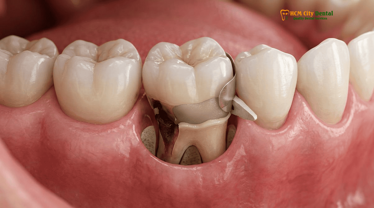
The Achilles heel of Deep Margin Elevation is moisture contamination. Even microscopic amounts of blood, saliva, or gingival crevicular fluid will instantly degrade the adhesive interface, leading to bond failure, microleakage, and recurrent decay. Therefore, the execution of a matrix-in-matrix teflon isolation subgingival onlay protocol is not merely a recommendation; it is an absolute clinical mandate.
The procedure begins with the placement of a 100% latex-free nitrile rubber dam. Nitrile is preferred over latex as it provides superior retraction of the gingival tissues and eliminates the risk of latex-induced allergic reactions. Heavy-gauge nitrile dams are inverted into the sulcus and secured with specialized clamps to physically retract the gingival tissue and create a hermetic seal around the operative field. When dealing with extreme subgingival margins, standard sectional matrix systems (like Garrison or Palodent) often fail to reach the depth of the defect. In these scenarios, the clinician employs a modified Tofflemire matrix or a specialized “matrix-in-matrix” technique. This involves placing a rigid, circumferential matrix band to retract the tissue, and then sliding a smaller, highly contoured sectional matrix inside it to perfectly adapt to the deep cervical margin. To ensure a flawless seal at the gingival floor, sterile polytetrafluoroethylene (Teflon) tape is meticulously packed between the matrix band and the adjacent tooth or wedge. Teflon is highly adaptable, chemically inert, and provides an exceptional barrier against sulcular fluid seepage.
Once isolation is verified and the Immediate Dentin Sealing (IDS) protocol is complete, the elevation is performed using a pre-heated composite layering protocol. Heating highly filled nanohybrid composite resins to approximately 68°C (155°F) using a specialized composite warmer temporarily lowers the material’s viscosity. This allows the composite to flow seamlessly into the micro-irregularities of the deep margin, ensuring intimate adaptation without the risk of incorporating air voids, while maintaining superior mechanical properties compared to traditional flowable resins. The composite is layered in thin increments (maximum 1.5mm) to minimize polymerization shrinkage stress (the C-factor). This entire meticulous process is performed under a 25x Carl Zeiss Extaro 300 operating microscope, allowing the specialist to visually confirm the marginal seal at a microscopic level.[5]
Clinical Precaution: Endodontic Considerations in Deep Caries
When subgingival caries approaches the pulp chamber, advanced endodontic diagnostics are required. If the pulp is vital but exposed, Vital Pulp Therapy (achieving hemostasis within 5 minutes using 2.5% NaOCl followed by Septodont Biodentine or ProRoot MTA) may be indicated to preserve tooth vitality. If the pulp is necrotic, Microscopic Endodontic Root Canal Therapy must be completed prior to Deep Margin Elevation. In cases of previous failing root canals with apical pathology, a Microscopic Apicoectomy (utilizing a 0-degree 3mm root-end resection, methylene blue staining, 3mm Satelec ultrasonic retro-prep, and a bioceramic root-end plug) may be necessary. Patients experiencing severe, lingering thermal pain or spontaneous throbbing should contact Dr. Nguyen Van Cuong immediately for a comprehensive 3D CBCT evaluation.
2026 Deep Margin Elevation, Crown Lengthening & Biomimetic Onlay Fee Schedule at HCMC Dental Clinic (District 1)
For international patients, dental travelers, and expatriates residing in Vietnam, navigating the costs of advanced biomimetic dentistry and periodontal procedures can be complex. At HCMC Dental Clinic, located in the heart of District 1 (just minutes from the Pullman Saigon Centre and Ben Thanh Market), we prioritize absolute financial transparency. By utilizing our Official 2026 Dental Price List & -40% Online Booking Fee Schedule, patients can access world-class, enamel-preserving treatments at a fraction of the cost of comparable care in the United States, Canada, or Australia.
The table below outlines the 2026 fee schedule for procedures related to the restoration of deeply decayed teeth, including Deep Margin Elevation, biomimetic ceramic restorations, and microscopic endodontics. Please note that the -40% online booking rates are exclusively available to patients who schedule their consultations in advance through our digital portal.
| Clinical Procedure (Biomimetic & Restorative) | 2026 Online Booking Rate (-40% USD) | Standard Walk-In Rate (USD) | US / Australian Benchmark (USD) |
|---|---|---|---|
| IPS E.max All-Ceramic Crown / Inlay / Onlay / Overlay | $231 USD (6,000,000 VND) | $385 USD | $1,200 – $1,800+ |
| Full Zirconia Crown (Cercon Standard / HT Multilayer) | $154 – $192 USD (4,000,000 – 5,000,000 VND) | $256 – $321 USD | $1,500 – $2,200+ |
| Biomimetic Quartz/Glass Fiber Post & Core Buildup (Often combined with DME) | $38 – $58 USD | $64 – $96 USD | $350 – $500+ |
| Microscopic Endodontic Root Canal Therapy (Complex Molar) | $115 – $154 USD (3,000,000 – 4,000,000 VND) | $192 – $256 USD | $1,200 – $1,800+ |
| Bioactive ProRoot MTA / Septodont Biodentine Perforation Repair | $58 – $96 USD | $96 – $160 USD | $400 – $600+ |
| 3D CBCT Scan (Diagnostic Imaging) | $18 USD (480,000 VND) | $30 USD | $150 – $300+ |
When planning your treatment, it is important to understand the clinical timeline. Unlike traditional surgical crown lengthening, which requires weeks of healing, DME allows for immediate scanning and fabrication of the final restoration. For a detailed breakdown of the required appointments and laboratory fabrication times, please review our Turnaround Time for Inlays & Onlays in Vietnam: Clinical Guide. Furthermore, patients comparing the long-term benefits of different restorative materials can consult our comprehensive Composite vs Ceramic Inlays in Vietnam: Expert Dental Guide to make an informed decision regarding their oral health investment.
“Our commitment at HCMC Dental Clinic is to provide uncompromising clinical excellence. By integrating 25x Carl Zeiss microscopy, 3D CBCT diagnostics, and strict biomimetic protocols, we ensure that every Deep Margin Elevation and ceramic onlay meets the highest international standards of precision and longevity, all while offering transparent, accessible pricing for our global patients.”
— Dr. Nguyen Van Cuong, DDS
Frequently Asked Questions
Can Deep Margin Elevation (DME) save a deeply decayed molar without surgical gum and bone cutting?
Yes. Deep Margin Elevation (DME) is a minimally invasive biomimetic technique that uses advanced adhesive protocols and specialized matrix systems to relocate a deep subgingival cavity margin to a supragingival position. As long as absolute isolation can be achieved with a nitrile rubber dam and the restorative margin does not violate the supracrestal tissue attachment (biologic width), DME can successfully save a deeply decayed molar without the need for surgical ostectomy or crown lengthening.
Does a subgingival composite Deep Margin Elevation cause gum inflammation or periodontal pockets?
When executed correctly under high-magnification microscopy with absolute isolation, a subgingival composite Deep Margin Elevation does not inherently cause chronic gum inflammation. Clinical studies indicate that highly polished, well-adapted composite resins are well-tolerated by the periodontium. However, if the composite overhangs, is poorly polymerized due to moisture contamination, or violates the 2.0mm biologic width, it can lead to localized gingivitis, bleeding on probing, and eventual periodontal pocket formation.
When is surgical crown lengthening mandatory instead of non-surgical Deep Margin Elevation?
Surgical crown lengthening becomes mandatory when absolute isolation for DME is impossible to achieve, or when the carious lesion extends so deeply that restoring it would inevitably violate the supracrestal tissue attachment (leaving less than 2.0mm between the restorative margin and the alveolar bone crest). In such cases, surgical ostectomy is required to apically reposition the bone and soft tissue, re-establishing a healthy biologic width before any restorative work can begin.
Why is a bitewing radiograph taken immediately after Deep Margin Elevation before optical 3D scanning?
A post-operative bitewing radiograph is a critical quality control step taken immediately after the DME procedure to verify the marginal integrity of the composite elevation. It ensures there are no subgingival overhangs, voids, or gaps between the composite and the root surface. Confirming a flawless seal radiographically is mandatory before proceeding with the optical 3D scan for the final ceramic onlay or crown.
How much does Deep Margin Elevation + an IPS E.max Ceramic Onlay cost at HCMC Dental Clinic in District 1?
At HCMC Dental Clinic in District 1, Ho Chi Minh City, an IPS E.max All-Ceramic Onlay costs $231 USD per tooth when booked online (a 40% savings compared to the $385 USD walk-in rate). The Deep Margin Elevation procedure and biomimetic core buildup are typically integrated into the restorative treatment plan, offering international patients and expats world-class, enamel-preserving dentistry at a fraction of the cost found in the US or Australia.
References
- Magne, P., & Spreafico, R. (2012). Deep margin elevation: a paradigm shift. The American Journal of Esthetic Dentistry, 2(2), 86-96.
- Samartzi, T. K., Papalexopoulos, D., & Sarafianou, A. (2022). Deep Margin Elevation: A Literature Review. Dentistry Journal, 10(3), 48.
- Bresser, R. A., Gerdolle, D., van den Heuvel, K. E., Sluiter-Pouwels, L. M. A., & Cune, M. S. (2019). Up to 12 years clinical evaluation of 197 partial indirect restorations with deep margin elevation in the posterior region. Journal of Dentistry, 91, 103227.
- Ferrari, M., Koken, S., Grandini, S., Ferrari Cagidiaco, E., Joda, T., & Discepoli, N. (2018). Influence of cervical margin relocation (CMR) on periodontal health: 12-month results of a controlled trial. Journal of Dentistry, 69, 70-76.
- Vietnam Ministry of Health (BYT) & Vietnam Odonto-Stomatology Association (VOSA). National Clinical Guidelines on Specialist Dental Care, Biomaterial Traceability & EN 13060 Class B Infection Control. (2024).