A fixed dental bridge is a permanent prosthetic restoration designed to replace one or more missing teeth. It consists of an artificial tooth, known as a pontic, which is securely anchored to adjacent natural teeth or dental implants, restoring both masticatory function and aesthetic harmony.
Clinical Summary:
A fixed dental bridge is a cornerstone of restorative prosthodontics, utilized to span an edentulous gap created by missing teeth. By utilizing adjacent healthy teeth (abutments) for support, a custom-fabricated artificial tooth (pontic) is permanently cemented into place. This non-removable prosthesis prevents the shifting of remaining teeth, restores proper occlusal alignment, and rehabilitates the patient’s chewing capacity. Modern bridges are crafted from advanced biocompatible materials such as zirconia, lithium disilicate, or porcelain-fused-to-metal (PFM), offering exceptional durability and lifelike aesthetics. The clinical workflow requires precise tooth preparation, accurate impressions, and meticulous cementation protocols to ensure long-term structural integrity and periodontal health.
Key Takeaways:
- A fixed bridge permanently replaces missing teeth by anchoring an artificial tooth to adjacent natural teeth or implants.
- It restores chewing efficiency, speech clarity, and prevents surrounding teeth from migrating into the empty space.
- Common materials include high-strength zirconia, aesthetic ceramics, and traditional porcelain-fused-to-metal alloys.
- The procedure typically requires two to three clinical visits, involving tooth preparation, temporization, and final cementation.
- Long-term success depends heavily on meticulous interdental hygiene and the structural health of the supporting abutment teeth.
The Clinical Definition: What is a Fixed Dental Bridge?
A fixed dental bridge is a non-removable prosthesis that bridges an edentulous gap by permanently cementing a custom-fabricated artificial tooth to the prepared adjacent abutment teeth.
In the realm of restorative dentistry, tooth loss presents significant biomechanical and aesthetic challenges. When a tooth is extracted or lost due to trauma or advanced periodontal disease, the integrity of the entire dental arch is compromised. To address this, a fixed dental bridge serves as a highly effective, non-removable solution. Unlike removable partial dentures, which a patient can take out of their mouth, a fixed bridge is permanently bonded into place, offering a stable and secure restoration that closely mimics the feel and function of natural dentition [1].
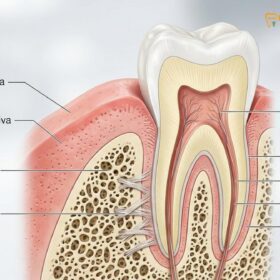
To fully understand this restorative option, it is essential to break down its anatomical components. A standard fixed bridge consists of two primary elements: the abutments and the pontic. The abutments are the anchoring structures—typically the natural teeth located immediately on either side of the empty space. These teeth must be structurally sound, possess adequate bone support, and have healthy periodontal ligaments to withstand the additional occlusal (chewing) forces they will bear. The pontic is the artificial tooth that fills the void. It is fused to the abutment crowns, creating a single, continuous prosthetic unit.
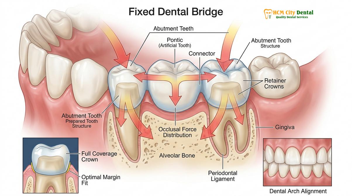
The biomechanical principles governing fixed bridges are rigorous. Prosthodontists rely on clinical guidelines, such as Ante’s Law, which dictates that the combined root surface area of the abutment teeth must equal or exceed that of the teeth being replaced [1]. If this principle is violated, the abutment teeth may suffer from occlusal overload, leading to mobility, bone loss, and eventual failure of the prosthesis. Therefore, a comprehensive clinical evaluation, including radiographic imaging and periodontal probing, is mandatory before prescribing a bridge.
Dr. Nguyen Van Cuong, a leading restorative expert, emphasizes that the success of a fixed bridge is not merely about filling a gap, but about harmonizing the restoration with the patient’s dynamic bite. “The prosthesis must be engineered to distribute masticatory forces evenly across the abutments while maintaining a passive fit that respects the biological width of the gingival tissues,” notes Dr. Cuong. This level of precision ensures that the surrounding gums remain healthy and free from chronic inflammation.
Furthermore, the design of the pontic itself plays a crucial role in the health of the underlying soft tissue. Clinicians carefully select the pontic’s gingival surface design—such as a modified ridge lap, ovate, or sanitary design—based on the location of the missing tooth and the aesthetic demands of the patient [3]. An ovate pontic, for instance, is often used in the highly visible anterior (front) region because it creates the illusion that the artificial tooth is emerging naturally from the gum line, providing superior aesthetic outcomes.
Types of Fixed Dental Bridges in Modern Prosthodontics
Prosthodontists utilize four primary bridge designs—traditional, cantilever, Maryland, and implant-supported—each selected based on the specific location of the missing tooth and the condition of adjacent abutments.
Not all edentulous spaces are identical, and therefore, the architectural design of a fixed bridge must be tailored to the patient’s unique anatomical presentation. The selection of the appropriate bridge type depends on factors such as the number of missing teeth, the location in the dental arch, the health of the adjacent teeth, and the patient’s occlusal forces. For a comprehensive overview of restorative options, patients can refer to our comprehensive general dental guide.
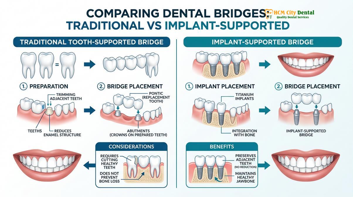
1. Traditional Fixed Bridge: This is the most common and biomechanically robust design. It involves creating a crown for the tooth or implant on either side of the missing tooth, with a pontic suspended in between. Traditional bridges are highly versatile and can be used to replace teeth in both the anterior (front) and posterior (back) regions of the mouth. However, they require the irreversible removal of healthy enamel from the adjacent abutment teeth to accommodate the retaining crowns.
2. Cantilever Bridge: A cantilever bridge is utilized when there is an adjacent natural tooth on only one side of the missing tooth gap. The pontic is supported by one or more abutments on just one side. Because the pontic acts as a lever arm, this design is subjected to significant torque and rotational forces during chewing. Consequently, cantilever bridges are generally contraindicated in the posterior regions where bite forces are heaviest, but they can be a viable, conservative option for replacing missing lateral incisors.
3. Maryland Bonded Bridge: Also known as a resin-bonded bridge, this conservative alternative minimizes the need for extensive tooth preparation. Instead of full-coverage crowns, a Maryland bridge utilizes a metal or porcelain framework with “wings” that are bonded to the lingual (back) surfaces of the adjacent teeth. While this preserves natural tooth structure, the bond strength is generally lower than that of a traditional crown, making Maryland bridges more susceptible to debonding. They are primarily indicated for replacing anterior teeth where occlusal forces are minimal.
4. Implant-Supported Bridge: When multiple teeth are missing in a row, or when the adjacent natural teeth are not healthy enough to serve as abutments, an implant-supported bridge is the gold standard. Instead of resting on natural teeth, the bridge is anchored to titanium dental implants surgically placed into the jawbone. This design provides unparalleled stability, prevents the need to alter healthy natural teeth, and helps preserve alveolar bone volume [4]. Patients exploring extensive restorations often consider full dental bridge restorations supported by implants for full-arch rehabilitation.
| Bridge Type | Primary Indication | Abutment Preparation | Biomechanical Strength |
|---|---|---|---|
| Traditional | Single or multiple missing teeth with healthy adjacent teeth | Extensive (Full crown preparation) | High (Excellent force distribution) |
| Cantilever | Missing tooth with only one adjacent abutment available | Moderate to Extensive | Moderate (Susceptible to rotational forces) |
| Maryland | Anterior missing teeth; conservative approach | Minimal (Lingual surface etching only) | Low to Moderate (Relies on resin bond strength) |
| Implant-Supported | Multiple missing teeth; lack of healthy natural abutments | None (Implants act as abutments) | Very High (Independent structural support) |
The Clinical Workflow: Step-by-Step Procedure
The fabrication of a fixed bridge involves precise abutment preparation, accurate digital or physical impressions, temporization, and the final adhesive cementation of the permanent prosthesis.
The process of receiving a fixed dental bridge is a meticulous clinical endeavor that typically spans two to three appointments. The workflow requires a deep understanding of dental anatomy, occlusion, and material science to ensure the final prosthesis integrates seamlessly into the patient’s stomatognathic system. The journey begins with a comprehensive diagnostic phase, including vitality testing of the abutment teeth and radiographic analysis to confirm adequate bone support.
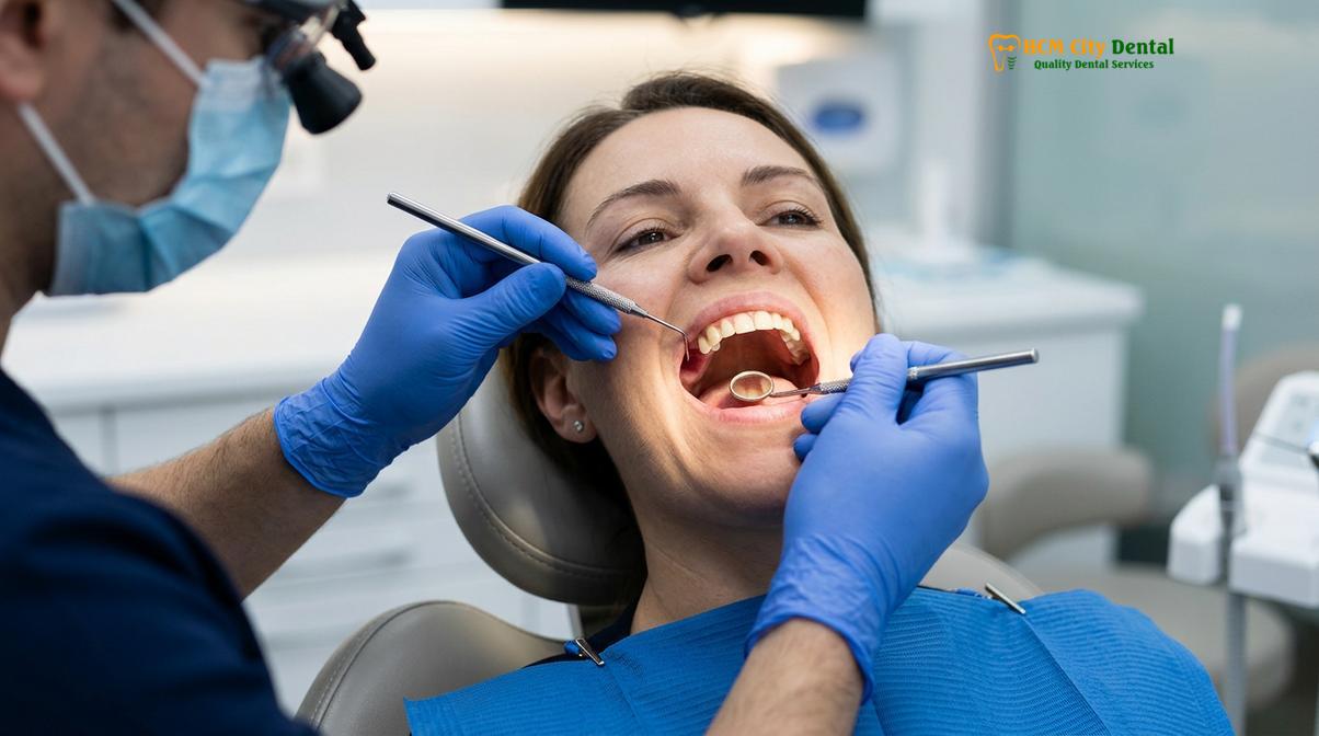
Phase 1: Abutment Preparation. Once the patient is profoundly anesthetized using local anesthetics, the clinician begins the preparation of the abutment teeth. Using specialized high-speed diamond burs, the dentist carefully reduces the enamel and dentin to create space for the restorative material. The geometry of this preparation is critical; the clinician must establish a precise “path of insertion” so the rigid bridge can slide into place without binding. Furthermore, the margins (the junction where the crown meets the tooth) must be flawlessly smooth—typically designed as a chamfer or shoulder margin—to ensure a watertight seal that prevents bacterial microleakage [5].
Phase 2: Tissue Management and Impressions. Capturing an exact replica of the prepared teeth is paramount. To achieve this, the dentist may use a retraction cord—a tiny thread packed gently into the gingival sulcus—to temporarily push the gum tissue away from the tooth margins. This allows the impression material or digital intraoral scanner to capture the full extent of the preparation. Modern clinics increasingly rely on 3D digital scanning, which offers superior accuracy and patient comfort compared to traditional polyvinyl siloxane (PVS) putty impressions.
n “The longevity of a fixed bridge is directly proportional to the marginal integrity achieved during the impression phase. A discrepancy of even a few microns can lead to cement dissolution, recurrent decay, and the eventual failure of the entire restoration.”
— Dr. Nguyen Van Cuong, Clinical Director
Phase 3: Temporization. Because the permanent bridge takes several days to weeks to be fabricated in a dental laboratory, the prepared teeth must be protected. A temporary (provisional) bridge is fabricated chairside using an acrylic or bis-acryl composite resin. This temporary restoration protects the exposed dentin from thermal sensitivity, prevents the abutment teeth from shifting, and allows the patient to evaluate the proposed aesthetics and function. Patients undergoing complex cases can view examples of dental reconstruction before and after to understand the transformative impact of provisional and final restorations.
Phase 4: Final Cementation. During the final visit, the temporary bridge is carefully removed, and the abutment teeth are cleaned of any residual temporary cement. The permanent bridge is then tried in. The clinician meticulously evaluates the marginal fit, interproximal contacts (how tight it is against neighboring teeth), and the occlusion (how it meets the opposing teeth). Adjustments are made as necessary. Once perfection is achieved, the bridge is permanently bonded using advanced resin or glass ionomer cements. The occlusion is verified one final time to ensure no premature contacts exist, which could cause trauma to the abutment teeth.
Material Selection for Fixed Bridges
Modern fixed bridges are fabricated from high-strength ceramics like monolithic zirconia, traditional porcelain-fused-to-metal (PFM), or gold alloys, balancing aesthetic demands with occlusal load requirements.
The choice of material for a fixed dental bridge is a critical decision that impacts the restoration’s strength, aesthetics, and biocompatibility. The oral cavity is a harsh environment, subjecting dental materials to extreme temperature fluctuations, acidic pH levels, and immense biting forces. Therefore, the selected material must possess high fracture toughness while mimicking the optical properties of natural tooth enamel.
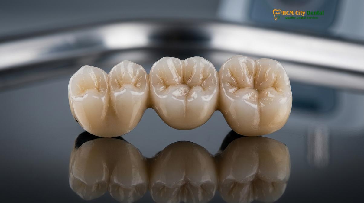
Zirconia (Yttria-Stabilized Tetragonal Zirconia Polycrystal): Zirconia has revolutionized modern prosthodontics. It is a highly biocompatible, tooth-colored ceramic known for its exceptional flexural strength, often exceeding 1000 MPa. Monolithic zirconia bridges are milled from a single block of material using CAD/CAM technology, making them virtually indestructible under normal chewing forces [2]. They are particularly indicated for posterior bridges where strength is paramount. For anterior regions, highly translucent zirconia or layered zirconia (where aesthetic porcelain is baked over a strong zirconia core) provides a lifelike appearance.
Porcelain-Fused-to-Metal (PFM): For decades, PFM bridges were the standard of care. They consist of a cast metal alloy substructure that provides rigidity, overlaid with tooth-colored feldspathic porcelain for aesthetics. While PFM bridges have a long track record of clinical success, they have some aesthetic drawbacks. Over time, if the gums recede, the dark metal margin may become visible at the gum line. Additionally, the overlying porcelain is susceptible to chipping under heavy occlusal stress.
Lithium Disilicate (E-max): This is a highly aesthetic glass-ceramic material renowned for its beautiful translucency and ability to bond strongly to tooth structure. While not as strong as zirconia, it is an excellent choice for short-span anterior bridges where cosmetic demands are the highest. The optical properties of lithium disilicate closely resemble those used in premium Porcelain Veneers, making it ideal for patients seeking a flawless smile.
Gold Alloys: Although less common today due to aesthetic preferences, high-noble gold alloys remain one of the most durable and biocompatible materials in dentistry. Gold is gentle on opposing natural teeth, does not fracture, and allows for highly precise marginal adaptation. It is occasionally requested for posterior bridges in patients with severe bruxism (teeth grinding).
Fixed Bridge vs. Dental Implants: A Clinical Comparison
While fixed bridges offer a faster, non-surgical solution for replacing missing teeth, dental implants provide independent structural support and actively preserve underlying alveolar bone volume.
When a patient presents with a missing tooth, the two primary fixed restorative options are a traditional dental bridge and a single dental implant. Both modalities have high success rates, but they operate on fundamentally different biomechanical principles. Understanding these differences is crucial for making an informed clinical decision.
A fixed bridge is often preferred when the adjacent teeth already require crowns due to extensive decay or large existing fillings. In such cases, preparing these teeth to serve as abutments for a bridge “kills two birds with one stone”—restoring the damaged teeth while simultaneously replacing the missing one. Furthermore, a bridge can be completed in a matter of weeks, making it a faster solution. It is also a non-surgical procedure, which is advantageous for patients with medical contraindications to surgery or those who lack sufficient bone volume for implant placement.
However, the primary disadvantage of a traditional bridge is the biological cost to the adjacent teeth. If the abutment teeth are perfectly healthy, removing significant amounts of pristine enamel to accommodate the bridge is a highly invasive compromise. Additionally, because the pontic simply rests above the gums, it does not stimulate the underlying jawbone. According to Wolff’s Law, bone requires mechanical stimulation to maintain its density. Without a tooth root, the alveolar bone in the edentulous space will gradually resorb (shrink) over time.
Clinical Warning: Bone Resorption Risk
Patients must be aware that a traditional fixed bridge does not halt the natural process of alveolar bone resorption in the area of the missing tooth. Over years, this bone loss can create a visible gap between the pontic and the gum line, potentially compromising aesthetics and creating a trap for food debris.
Conversely, a dental implant acts as an artificial tooth root. It is surgically placed into the jawbone, where it undergoes osseointegration—fusing directly with the bone. This independent support means that adjacent natural teeth remain completely untouched. More importantly, the implant transmits chewing forces directly into the bone, providing the necessary stimulation to prevent bone atrophy. While implants require a longer treatment timeline and surgical intervention—often discussed when planning wisdom teeth removal settings and subsequent extractions—they are generally considered the superior long-term investment for single-tooth replacement.
Longevity and Maintenance of Your Dental Bridge
A fixed bridge can function optimally for over a decade with meticulous oral hygiene, including the use of specialized interdental cleaners to remove plaque beneath the pontic.
The longevity of a fixed dental bridge is not solely dependent on the materials used or the skill of the prosthodontist; it relies heavily on the patient’s commitment to daily oral hygiene and regular professional maintenance. While the ceramic or metal components of the bridge cannot decay, the natural tooth structure of the abutments remains highly susceptible to bacterial attack, particularly at the margins where the crown meets the root.
Because the pontic is fused to the adjacent crowns, traditional dental floss cannot be passed down between the teeth. Patients must use specialized tools to clean the critical area underneath the pontic and around the abutment margins. Floss threaders or pre-cut Superfloss allow the patient to thread the floss horizontally under the bridge. Interdental brushes (such as TePe brushes) and oral water irrigators (water flossers) are also highly effective at flushing out plaque and food debris from these hard-to-reach areas. Maintaining excellent hygiene is just as critical here as it is when learning maintaining white teeth during orthodontic treatment, as plaque accumulation leads to gingivitis and secondary caries.
Clinical Case Study: Bridge Rehabilitation
A 45-year-old patient presented to HCMC Dental Clinic in Ho Chi Minh City with a failing 15-year-old PFM bridge. The patient complained of food impaction and a dark line near the gums. Clinical examination revealed secondary decay under the posterior abutment due to inadequate interdental cleaning. The old bridge was sectioned and removed, the decay was excavated, and the abutment was rebuilt with a core buildup. A new, highly aesthetic monolithic zirconia bridge was fabricated with an ovate pontic design. The patient was thoroughly instructed on the use of water flossers and interdental brushes, ensuring the long-term success of the new restoration.
Routine dental prophylaxis (professional cleanings) every six months is mandatory. During these visits, the clinician will use ultrasonic scalers to remove calculus around the bridge margins and evaluate the occlusion to ensure the bridge is not being subjected to excessive traumatic forces. If a patient suffers from bruxism, a custom occlusal guard (night guard) is strongly recommended to protect the ceramic from chipping and to reduce stress on the abutment teeth.
When to See a Doctor: Important Clinical Considerations
While fixed dental bridges are highly reliable, complications can arise that require immediate professional intervention. Patients must be vigilant in monitoring their restoration and the surrounding tissues. Ignoring early warning signs can lead to the catastrophic failure of the bridge and the potential loss of the supporting abutment teeth.
You should schedule an immediate evaluation if you experience any of the following symptoms:
- Mobility or Looseness: A fixed bridge should feel completely rigid. If you detect any movement, it may indicate that the cement seal has broken on one or both abutments, or that the underlying tooth structure has fractured.
- Localized Pain or Sensitivity: Sharp pain upon biting or lingering sensitivity to hot or cold suggests that the nerve of an abutment tooth may be inflamed (pulpitis) or that recurrent decay has penetrated the dentin.
- Swelling or Bleeding Gums: Chronic inflammation, bleeding upon brushing, or a localized swelling (abscess) near the bridge margins indicates a periodontal infection or a failing root canal.
- Foul Taste or Odor: A persistent bad taste or halitosis can be a sign of bacterial microleakage under the crowns or food impaction beneath a poorly adapted pontic.
n “A loose bridge is a clinical emergency. If the cement washes out on just one abutment, the bridge may still feel somewhat secure due to the other anchor, but bacteria will rapidly colonize the unsealed tooth, leading to rampant, silent decay that can destroy the abutment within months.”
— Clinical Prosthodontic Guidelines
Early diagnosis is critical. In many cases, if a cement seal has simply failed without underlying decay, the bridge can be carefully removed, cleaned, and recemented. However, if decay is extensive, the bridge must be remade, and the abutment teeth may require endodontic therapy or, in severe cases, extraction. Proper planning and timely intervention, much like the foresight required in correcting overjets with braces, prevent minor issues from escalating into complex reconstructive failures.
Frequently Asked Questions
Is the procedure for getting a fixed dental bridge painful?
The procedure is generally not painful because local anesthesia is administered to numb the abutment teeth and surrounding gingival tissues. Patients may experience mild sensitivity or gingival tenderness for a few days following the preparation and cementation phases, which typically resolves quickly. Over-the-counter anti-inflammatory medications are usually sufficient to manage any post-operative discomfort.
How long does a permanent fixed dental bridge last?
A well-maintained fixed dental bridge typically lasts between 10 to 15 years, though many can function longer with excellent care. Longevity depends heavily on meticulous oral hygiene, the structural integrity of the abutment teeth, and the patient’s occlusal habits. Regular dental checkups are essential to monitor the margins and ensure the cement seal remains intact.
Can a fixed dental bridge be removed for cleaning?
No, a fixed dental bridge cannot be removed by the patient for cleaning. It is permanently cemented onto the abutment teeth or implants, requiring specialized interdental cleaning tools like floss threaders or water irrigators to clean beneath the artificial tooth. Only a dental professional can safely remove a fixed bridge, usually only if replacement or repair is necessary.
What is the difference between a traditional bridge and a Maryland bridge?
A traditional bridge requires significant reshaping of adjacent teeth to accommodate full crowns, whereas a Maryland bridge uses metal or porcelain wings bonded to the back of adjacent teeth. Maryland bridges are more conservative but generally less capable of withstanding heavy chewing forces. Therefore, Maryland bridges are primarily indicated for replacing front teeth, while traditional bridges can be used anywhere in the mouth.
Will a fixed dental bridge look like my natural teeth?
Yes, modern fixed bridges are highly aesthetic and custom-fabricated to match the exact shade, shape, and translucency of your natural dentition. Materials like layered zirconia or lithium disilicate provide lifelike optical properties that blend seamlessly with your surrounding teeth. The dental laboratory carefully contours the pontic to mimic the natural emergence profile from the gums.
References
- Journal of Prosthetic Dentistry. Biomechanical considerations and Ante’s Law in fixed partial dentures. (2021).
- International Journal of Prosthodontics. Marginal adaptation and fracture resistance of monolithic zirconia bridges. (2020).
- Journal of Clinical Periodontology. Periodontal response to various pontic designs in fixed prosthodontics. (2019).
- American Dental Association. Clinical guidelines for the restoration of edentulous spaces. (2022).
- Dental Materials Journal. Adhesion protocols and resin cements for all-ceramic fixed dental prostheses. (2018).
If you are experiencing tooth loss and wish to explore your restorative options, a comprehensive clinical evaluation is the first step. Contact the expert team at HCMC Dental Clinic in Ho Chi Minh City to schedule a consultation and discover how a custom-crafted fixed dental bridge can restore the function and beauty of your smile.
| Bridge Material | Durability | Aesthetic Level | Price in Vietnam (Per Unit) |
|---|---|---|---|
| Porcelain-Fused-to-Metal (PFM) | 10–15 Years | Good (Posterior) | $150 – $220 |
| All-Ceramic Zirconia | 15–20+ Years | Excellent (Natural) | $280 – $400 |
| E-max Lithium Disilicate | 12–15 Years | Highest Translucency | $350 – $500 |
n{n “@context”: “https://schema.org”,n “@type”: “MedicalProcedure”,n “name”: “Fixed Dental Bridge”,n “procedureType”: “Prosthodontic”,n “description”: “Permanent prosthetic restoration to replace missing teeth anchored by abutment teeth or implants.”,n “howPerformed”: “Custom fabrication of pontic and abutment crowns using PFM, Zirconia, or E-max ceramic materials.”n}n