A dental crown typically lasts between 10 and 15 years, though with meticulous oral hygiene, precise clinical fabrication, and favorable bite dynamics, many crowns can function successfully for decades. The exact lifespan heavily depends on the chosen material, the underlying tooth structure, and daily maintenance habits.
Clinical Summary:
The longevity of a dental crown is a multifactorial outcome determined by material science, clinical precision, and patient compliance. Modern restorative dentistry utilizes advanced materials such as monolithic zirconia, lithium disilicate, and high-noble gold alloys, each offering distinct biomechanical advantages. While the average lifespan ranges from a decade to 15 years, restorations can endure significantly longer when supported by healthy periodontal tissues and protected from excessive occlusal forces. Clinical success relies heavily on achieving an impeccable marginal seal to prevent secondary caries, utilizing advanced adhesive protocols, and maintaining rigorous daily oral hygiene. Regular professional evaluations are critical to identifying early signs of microleakage or mechanical stress, ensuring the long-term viability of the prosthetic.
Key Takeaways:
- The average clinical lifespan of a dental crown is 10 to 15 years, but many last 20 years or more with proper care.
- Material selection plays a crucial role; gold and zirconia offer superior durability, while lithium disilicate provides unmatched aesthetics.
- Secondary tooth decay at the crown margin is the leading cause of premature restoration failure.
- Excessive bite forces, such as bruxism (teeth grinding), can significantly reduce the lifespan of ceramic crowns.
- Routine professional cleanings and meticulous daily oral hygiene are essential to protect the underlying natural tooth structure.
The Clinical Lifespan of Dental Crowns by Material
The longevity of a dental restoration is intrinsically linked to its material composition, with gold and zirconia offering the highest durability, while porcelain options prioritize aesthetic integration.
When patients ask how long a dental crown will last, the first clinical variable to consider is the material used to fabricate the prosthesis. Modern dentistry offers a spectrum of restorative materials, each engineered to balance biomechanical strength with optical properties that mimic natural enamel. The selection of material is not merely a matter of preference but a clinical decision based on the tooth’s location, the patient’s occlusal (bite) forces, and aesthetic demands[1].
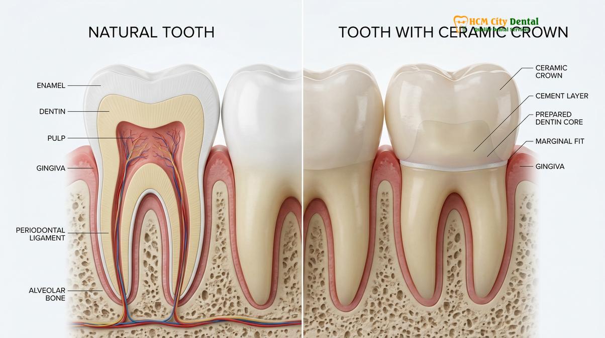
Zirconia Crowns: The Pinnacle of Durability
Zirconia (zirconium dioxide) has revolutionized restorative dentistry due to its exceptional flexural strength and fracture toughness. Categorized as a polycrystalline ceramic, zirconia undergoes a unique phase transformation when subjected to stress, effectively stopping crack propagation—a phenomenon known as transformation toughening. Because of this structural resilience, monolithic zirconia restorations are highly resistant to chipping and breaking, making them an ideal choice for posterior (back) teeth that endure heavy masticatory forces.
Clinically, a well-fabricated zirconia crown can easily last 15 to 20 years, and in many cases, a lifetime. However, the extreme hardness of zirconia requires precise occlusal adjustment to ensure it does not cause excessive wear on the opposing natural teeth. Advances in material science have also led to the development of highly translucent zirconia, which bridges the gap between the immense strength of traditional zirconia and the aesthetic requirements of anterior (front) teeth.
Porcelain-Fused-to-Metal (PFM) Crowns
For decades, porcelain-fused-to-metal (PFM) crowns were the gold standard in restorative dentistry, offering a reliable compromise between the strength of a metal substructure and the natural appearance of layered feldspathic porcelain. The underlying metal coping provides robust support, while the porcelain veneer is color-matched to the adjacent dentition.
The average lifespan of a PFM crown is typically 10 to 15 years. While the metal substructure is virtually indestructible under normal physiological conditions, the overlying porcelain is susceptible to cohesive fractures or chipping over time. Furthermore, as patients age and natural gingival recession occurs, the dark metal margin of a PFM crown may become visible at the gumline, leading to aesthetic failure even if the crown remains functionally intact. Patients often inquire about porcelain-fused-to-metal safety and longevity, and while they remain a viable option, many clinicians now prefer all-ceramic alternatives for highly visible areas.
Lithium Disilicate (E-max) Crowns
Lithium disilicate, commonly known by the brand name E-max, is a glass-ceramic material celebrated for its unparalleled optical properties. It mimics the natural translucency, opalescence, and fluorescence of human enamel better than almost any other material. Beyond aesthetics, lithium disilicate boasts an impressive flexural strength (approximately 400-500 MPa), making it significantly stronger than traditional layered porcelain.
When bonded correctly using advanced adhesive protocols, lithium disilicate crowns have a highly predictable lifespan of 10 to 15 years. They are the premier choice for anterior restorations and premolars. However, for patients with severe bruxism or for second molars subjected to extreme load, clinicians may recommend comparing zirconia and porcelain options to ensure mechanical survival.
Gold and High-Noble Metal Alloys
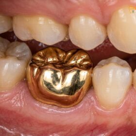
From a purely biomechanical and biological standpoint, cast gold alloys remain the ultimate restorative material. Gold is highly biocompatible, meaning it elicits minimal inflammatory response from the surrounding gingival tissues. More importantly, the wear characteristics of gold are nearly identical to natural human enamel, ensuring that it will not abrade opposing teeth. Gold is also highly ductile, allowing for an incredibly precise marginal fit that seals the underlying tooth against bacterial microleakage.
It is not uncommon for a gold crown to last 30 to 40 years, or even a lifetime. The primary limitation of gold is, of course, its highly unnatural appearance, which restricts its use almost exclusively to non-visible molar teeth.
| Material Type | Average Lifespan | Flexural Strength | Aesthetic Quality | Primary Clinical Indication |
|---|---|---|---|---|
| Monolithic Zirconia | 15 – 20+ Years | 900 – 1200 MPa | Good to Excellent | Posterior molars, heavy bite forces |
| Lithium Disilicate (E-max) | 10 – 15 Years | 400 – 500 MPa | Superior | Anterior teeth, premolars, high aesthetics |
| Porcelain-Fused-to-Metal (PFM) | 10 – 15 Years | Variable (Metal base) | Moderate | Bridges, posterior restorations |
| High-Noble Gold Alloy | 20 – 40+ Years | High Yield Strength | Poor (Metallic) | Non-visible molars, severe bruxers |
Biological and Mechanical Factors Influencing Crown Longevity
A crown’s survival depends not only on the material but also on the health of the underlying tooth structure, the precision of the marginal seal, and the patient’s bite forces.
While material science dictates the theoretical limits of a restoration, the actual clinical lifespan of a dental crown is governed by a complex interplay of biological and mechanical factors within the oral cavity. The mouth is a harsh environment, characterized by extreme temperature fluctuations, acidic challenges, and immense physical forces. Understanding these variables is essential for both the clinician and the patient to maximize the longevity of the treatment[2].
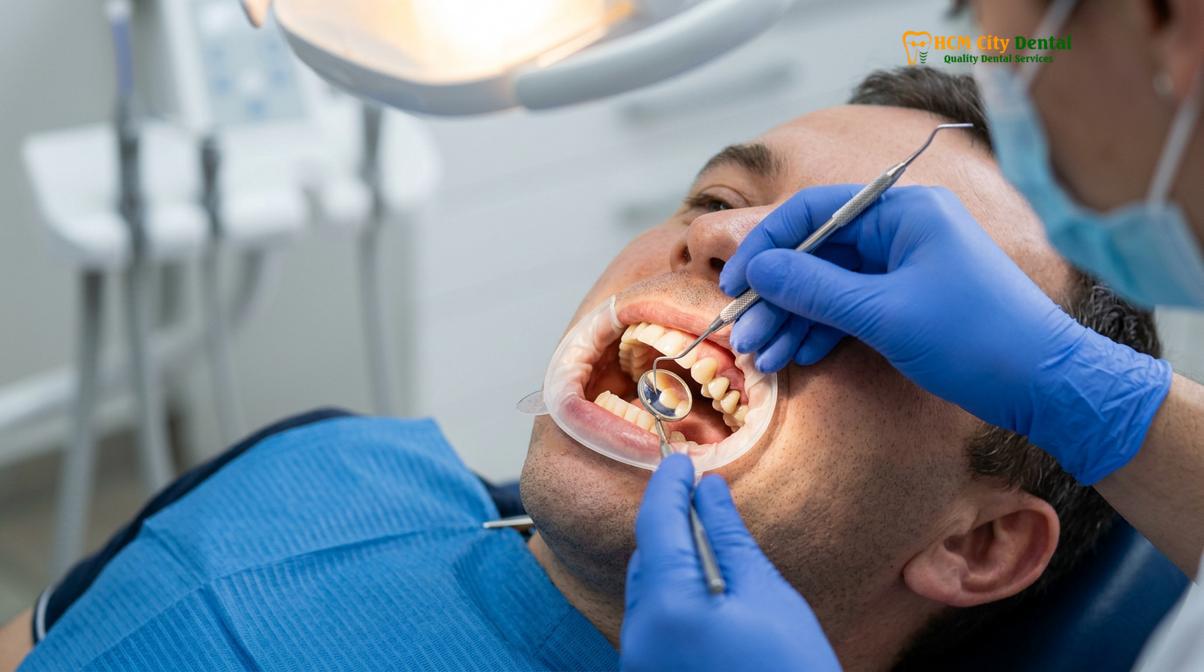
Marginal Fit and Microleakage
The most critical factor in the long-term success of any fixed prosthesis is the integrity of the margin—the microscopic junction where the artificial crown meets the natural tooth structure. If this margin is not perfectly sealed, a phenomenon known as microleakage occurs. Saliva, laden with cariogenic (cavity-causing) bacteria, seeps into the microscopic gap between the crown and the tooth, dissolving the dental cement and initiating secondary caries (decay) underneath the restoration.
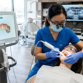
Because the crown itself covers the tooth, this underlying decay is often hidden from visual inspection and may progress silently until it reaches the dental pulp, necessitating a root canal or causing catastrophic structural failure of the tooth. Achieving a flawless marginal fit requires meticulous tooth preparation by the dentist, highly accurate impressions (often utilizing digital intraoral scanners), and precise fabrication by the dental laboratory.
Occlusal Forces and Bruxism
The human jaw is capable of exerting tremendous force, particularly in the posterior region. During normal mastication (chewing), teeth are subjected to forces ranging from 50 to 250 pounds. However, patients who suffer from bruxism—the subconscious clenching and grinding of teeth, often during sleep—can generate forces exponentially higher, sometimes exceeding 500 pounds of pressure.
These parafunctional habits place immense stress on dental restorations. Over time, excessive occlusal loading can lead to the cohesive fracture of ceramic materials, the decementation (loosening) of the crown, or even the vertical fracture of the underlying tooth root. For patients with a history of bruxism, clinicians must carefully select high-strength materials and often prescribe a custom-fabricated occlusal guard (night guard) to protect the investment.
“The mechanical survival of a dental crown is constantly tested by the dynamic forces of the stomatognathic system. We must engineer restorations not just to look beautiful, but to withstand decades of functional loading without compromising the underlying biological structures.”
— Dr. Nguyen Van Cuong, Clinical Director
Endodontic Status: Restoring Root Canal Treated Teeth
A significant percentage of teeth that require crowns have previously undergone endodontic therapy (root canal treatment). While a root canal saves an infected tooth, it also alters the tooth’s biomechanical properties. The removal of the internal pulp tissue and the necessary access cavity preparation significantly reduce the structural volume of the tooth. Furthermore, endodontically treated teeth lose their proprioceptive feedback mechanism, meaning patients may unknowingly bite down harder on these teeth than on vital ones.
To ensure longevity when restoring a tooth after endodontic therapy, the clinician must carefully evaluate the remaining ferrule—a band of solid tooth structure above the gumline that the crown can grasp. If the ferrule is insufficient, a post and core build-up may be required to provide adequate retention and resistance form for the final crown. The structural integrity of this foundation is paramount; a crown is only as strong as the tooth supporting it[3].
Clinical Workflows for Maximizing Crown Survival
Advanced digital scanning, conservative tooth preparation, and meticulous adhesive bonding protocols are critical steps that significantly extend the functional life of a dental crown.
The execution of the clinical procedure is just as vital as the material selected. Modern restorative dentistry relies on highly standardized, evidence-based workflows designed to minimize human error, preserve healthy tissue, and optimize the biomechanical integration of the prosthesis. At leading institutions, the process of delivering a custom dental crown involves several critical phases.
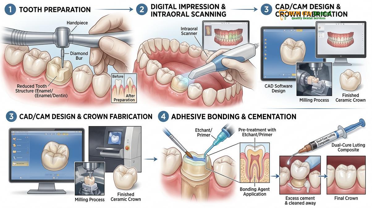
Precision Tooth Preparation and Tissue Management
The foundation of a long-lasting crown begins with the preparation of the natural tooth. The clinician must reduce the tooth circumferentially and occlusally to create adequate restorative space for the chosen material. However, this reduction must be as conservative as possible to preserve the vitality of the dental pulp and maintain structural strength. The geometry of the preparation—specifically the taper of the axial walls and the design of the finish line (e.g., chamfer or modified shoulder)—dictates the mechanical retention of the crown.
Equally important is the management of the gingival tissues. To capture an accurate impression of the margin, the clinician must temporarily retract the gums using specialized cords or astringent pastes. This ensures that the final crown will have a smooth, seamless transition with the root surface, preventing plaque accumulation and subsequent periodontal inflammation.
Digital Impressions and CAD/CAM Technology
The transition from traditional elastomeric impression materials to digital intraoral scanning has profoundly impacted the accuracy of dental restorations. Digital scanners capture the three-dimensional topography of the prepared tooth with micron-level precision. This data is then utilized in Computer-Aided Design and Computer-Aided Manufacturing (CAD/CAM) systems to design and mill the crown.
Digital workflows eliminate the dimensional instability associated with traditional impression materials and stone models. The resulting crowns exhibit superior marginal adaptation, reducing the risk of microleakage and extending the clinical lifespan of the restoration.
Advanced Adhesive Bonding Protocols
The method used to attach the crown to the tooth has evolved significantly. While traditional metal and PFM crowns are often cemented using luting agents that rely on mechanical retention, modern glass-ceramics like lithium disilicate require advanced adhesive bonding. This process involves etching both the internal surface of the ceramic and the natural tooth structure, followed by the application of silane coupling agents and dual-cure resin cements.
When executed under strict isolation (often using a rubber dam to prevent moisture contamination), these advanced veneer and crown adhesives create a micromechanical and chemical bond that essentially fuses the crown to the tooth. This monolithic complex distributes stress more evenly and dramatically reduces the likelihood of decementation.
Clinical Case Review: Maximizing Longevity
A 45-year-old patient presented to HCMC Dental Clinic in Ho Chi Minh City with a failing, 15-year-old PFM crown on a lower first molar. Clinical examination revealed secondary decay at the margin due to microleakage. The treatment protocol involved careful removal of the old restoration, complete caries excavation, and the placement of a composite core build-up. Utilizing a digital intraoral scanner, a highly precise impression was captured. A monolithic zirconia crown was fabricated and delivered using a dual-cure resin cement. By upgrading the material and ensuring a flawless marginal seal, the new restoration is prognosed to provide decades of functional stability.
Warning Signs of Crown Failure: When to See a Doctor
Despite the best materials and clinical execution, dental crowns can fail. Early detection of complications is crucial to saving the underlying tooth. Patients must remain vigilant and seek professional evaluation if they experience any abnormal symptoms associated with a crowned tooth. Ignoring these signs can lead to irreversible pulp damage, severe periodontal infections, or the complete loss of the natural tooth root[4].
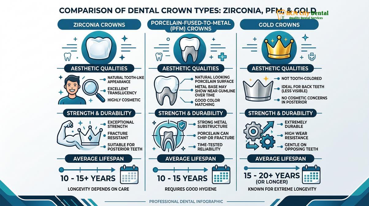
Important Clinical Considerations: Signs of Failure
Immediate clinical evaluation is recommended if you experience any of the following symptoms around a dental crown:
- Localized Pain or Sensitivity: Spontaneous throbbing pain, or lingering sensitivity to hot, cold, or sweet stimuli, strongly indicates secondary decay or pulpal inflammation beneath the crown.
- Gingival Inflammation: Persistent redness, swelling, or bleeding of the gums specifically around the crowned tooth may signal a violation of the biological width, an overhanging margin, or localized periodontal disease.
- Mobility or Shifting: A crown that feels loose, clicks when chewing, or shifts out of alignment has suffered a failure of the cement seal and requires immediate recementation to prevent bacterial ingress.
- Foul Odor or Taste: A persistent bad taste or odor originating from a specific tooth is a classic sign of bacterial accumulation and decay under a compromised crown margin.
- Visible Damage: Any visible chipping, cracking, or perforation of the crown material compromises its structural integrity and necessitates professional assessment.
If a crown fails, the treatment depends on the extent of the damage. If the crown has simply decemented but the underlying tooth is healthy, it can often be cleaned and rebonded. If secondary decay is present, the crown must be sectioned and removed, the decay excavated, and a new crown fabricated. In cases where the decay has progressed extensively, endodontic therapy or even extraction may be the only viable clinical options.
Maintenance and Oral Hygiene Protocols
Consistent daily plaque removal and biannual professional evaluations are the most effective strategies to prevent secondary decay and ensure the long-term success of your restoration.
The lifespan of a dental crown is ultimately in the hands of the patient. While the ceramic or metal material itself cannot decay, the natural tooth structure at the margin and the surrounding periodontal tissues remain highly vulnerable to bacterial attack. A comprehensive maintenance protocol is non-negotiable for long-term success[5].

Meticulous Daily Plaque Control
Patients must brush their teeth at least twice daily using a soft-bristled toothbrush and fluoridated toothpaste. The Modified Bass brushing technique, which angles the bristles at 45 degrees toward the gumline, is particularly effective at disrupting plaque at the critical crown margin. Furthermore, interdental cleaning is essential. Dental floss must be used daily to clean the proximal surfaces between the teeth where a toothbrush cannot reach. For patients with larger interdental spaces or those who struggle with traditional floss, water flossers (oral irrigators) and interdental brushes are excellent adjuncts for maintaining marginal hygiene.
Protection Against Parafunctional Forces
As previously discussed, mechanical stress is a primary cause of crown failure. Patients who engage in contact sports must wear a custom-fitted athletic mouthguard to protect their restorations from acute trauma. More commonly, patients who exhibit signs of nocturnal bruxism should be fitted with a hard acrylic occlusal splint (night guard). This device acts as a shock absorber, distributing bite forces evenly and preventing the catastrophic fracture of ceramic materials like Porcelain Veneers or crowns.
“The placement of a dental crown is not the end of treatment; it is the beginning of a lifelong commitment to maintenance. The most beautifully crafted restoration will inevitably fail if the biological environment surrounding it is neglected.”
— Dr. Nguyen Van Cuong, Clinical Director
Biannual Professional Evaluations
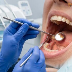
Routine dental visits every six months are critical. During these appointments at HCMC Dental Clinic, the clinical team performs a comprehensive evaluation of all restorations. This includes tactile examination of the margins using a dental explorer to detect microleakage, periodontal probing to ensure gingival health, and periodic radiographic imaging (X-rays) to visualize the internal tooth structure and surrounding bone. Professional prophylaxis (cleaning) removes hardened calculus that cannot be eliminated by home care alone, ensuring the biological environment remains conducive to the longevity of the crown.
By combining advanced clinical techniques with rigorous patient compliance, the modern dental crown serves as a highly predictable, long-term solution for restoring dental function and aesthetics.
Frequently Asked Questions
Can a dental crown last a lifetime?
While some dental crowns can last a lifetime with exceptional care, the average clinical lifespan is between 10 and 15 years. Longevity depends heavily on the material used, the patient’s oral hygiene, bite forces, and the structural integrity of the underlying natural tooth. Gold and zirconia restorations have the highest probability of lasting several decades, provided the marginal seal remains intact and the patient avoids excessive occlusal trauma.
Why do dental crowns fail over time?
Dental crowns typically fail due to secondary tooth decay at the margin, structural fractures of the crown material, or underlying periodontal disease. Additionally, excessive occlusal forces from bruxism (teeth grinding) can cause the cement seal to break or the porcelain to chip. Poor initial tooth preparation or inadequate adhesive bonding protocols can also lead to premature decementation and failure.
Does a crowned tooth still need to be brushed?
Yes, a crowned tooth must be brushed and flossed daily just like a natural tooth. While the artificial crown material cannot decay, the natural tooth structure beneath it and the surrounding gum tissue are still highly susceptible to bacterial plaque, cavities, and periodontal disease. Neglecting oral hygiene around a crown is the leading cause of secondary caries and subsequent restoration failure.
How do I know if there is decay under my crown?
Decay under a crown often presents as localized sensitivity to hot, cold, or sweet stimuli, spontaneous pain, or a foul odor around the tooth. However, early decay may be asymptomatic, which is why routine clinical examinations and dental X-rays are essential for early detection. A dentist can use an explorer to check the integrity of the margin and identify microleakage before it progresses to the dental pulp.
Is it normal for a new crown to feel sensitive?
Mild sensitivity to temperature and bite pressure is normal for a few days to a few weeks after a new crown is placed. This occurs due to nerve irritation during the preparation process. If the pain is severe, throbbing, or persists beyond a few weeks, an occlusal adjustment or endodontic evaluation may be necessary, as it could indicate an uneven bite or irreversible pulpal inflammation.
References
- Journal of Prosthetic Dentistry. Long-term survival rates of various dental crown materials: A systematic review. (2021).
- International Journal of Prosthodontics. Biomechanical factors influencing the longevity of fixed dental prostheses. (2020).
- Journal of the American Dental Association. Clinical evaluation of monolithic zirconia vs. porcelain-fused-to-metal crowns. (2019).
- Clinical Oral Investigations. The impact of marginal fit and microleakage on the success of dental restorations. (2022).
- Journal of Endodontics. Survival outcomes of endodontically treated teeth restored with full-coverage crowns. (2018).