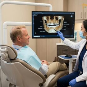
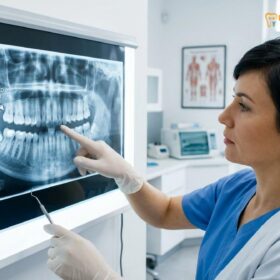
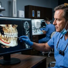
A tooth extraction in Saigon is a safe outpatient oral surgery when performed under strict clinical protocols. Modern clinics utilize advanced diagnostics, minimally invasive techniques, and rigorous sterilization to ensure painless removal of damaged or impacted teeth while promoting rapid tissue recovery.
Clinical Summary:
Tooth extraction is a fundamental oral surgery procedure indicated for non-restorable decay, advanced periodontal disease, severe trauma, or symptomatic impactions. In modern clinical settings, the procedure emphasizes atraumatic techniques to preserve the surrounding alveolar bone and gingival architecture, which is critical for future prosthetic rehabilitation such as dental implants. The workflow integrates high-resolution 3D CBCT imaging for precise anatomical mapping, profound local anesthesia for optimal pain management, and strict adherence to international sterilization standards to mitigate infection risks. Post-operative success relies heavily on stabilizing the surgical site, managing localized inflammation, and following evidence-based aftercare guidelines to ensure seamless tissue regeneration and prevent complications like alveolar osteitis.
Key Takeaways:
- Extractions are clinically indicated when teeth cannot be saved by endodontic or restorative therapies.
- Advanced 3D imaging ensures precise surgical planning and protects vital anatomical structures.
- Minimally invasive techniques, including Piezosurgery, reduce trauma and accelerate post-operative healing.
- Strict Class B sterilization protocols guarantee a safe, infection-free surgical environment.
- Proper blood clot stabilization is the most critical factor in immediate post-operative recovery.
When is a Tooth Extraction Necessary?
Tooth extraction is clinically indicated when a tooth is non-restorable due to severe decay, advanced periodontal disease, traumatic fracture, or when impacted wisdom teeth threaten adjacent anatomical structures.
The decision to remove a natural tooth is never taken lightly in modern dentistry. Clinicians prioritize tooth preservation through endodontic therapy and restorative techniques whenever biologically feasible. However, certain pathological conditions render a tooth non-restorable, making extraction the most responsible medical intervention to prevent systemic infection and protect surrounding oral tissues [1]. Understanding the specific clinical indications for a comprehensive general dentistry extraction helps patients make informed decisions about their oral health.

Severe Dental Caries and Pulpal Necrosis
When dental caries (tooth decay) progresses untreated, bacteria penetrate the protective enamel and dentin layers, eventually reaching the dental pulp. This inner chamber houses the tooth’s vital nerves and blood vessels. Bacterial infiltration leads to irreversible pulpitis and subsequent pulpal necrosis. If the structural integrity of the tooth is so severely compromised that it cannot support a dental crown, or if the infection has caused a massive periapical abscess that does not respond to root canal therapy, extraction becomes necessary to eradicate the source of infection and prevent the spread of bacteria into the jawbone and bloodstream.
Advanced Periodontal Disease
Periodontal disease is a chronic inflammatory condition affecting the periodontium—the specialized tissues that surround and support the teeth, including the gingiva, periodontal ligament, and alveolar bone. In its advanced stages (severe periodontitis), the chronic immune response to bacterial plaque causes extensive destruction of the alveolar bone. As the bone recedes, teeth lose their foundational support, resulting in severe mobility (looseness). When bone loss is too profound to be stabilized by periodontal splinting or regenerative surgeries, extracting the mobile tooth is required to halt the localized inflammatory process and prepare the site for future rehabilitation.
Traumatic Dental Injuries
Facial trauma can result in various types of dental fractures. While minor chips or coronal fractures can often be repaired with composite bonding or crowns, vertical root fractures or fractures extending deep beneath the alveolar bone crest generally carry a poor prognosis. A vertical root fracture creates a pathway for bacteria to continuously infect the surrounding bone, leading to chronic pain and localized bone destruction. In these scenarios, a prompt dental extraction Saigon is indicated to prevent further complications.
Impacted Wisdom Teeth
Third molars, commonly known as wisdom teeth, often lack sufficient space to erupt fully into functional alignment within the dental arch. This lack of space leads to impaction, where the tooth remains partially or completely encased in bone and gingival tissue. Impacted wisdom teeth are highly susceptible to pericoronitis (infection of the overlying gum flap), cyst formation, and causing resorption or decay in the adjacent second molars. Prophylactic or therapeutic removal of impacted third molars is a standard surgical procedure designed to prevent these long-term pathological consequences.
Orthodontic Considerations
In cases of severe dental crowding, where the discrepancy between tooth size and jaw size is significant, orthodontic treatment may require the strategic extraction of specific teeth—most commonly the first premolars. This creates the necessary anatomical space to align the remaining teeth properly, correct malocclusions, and achieve a stable, functional, and aesthetic bite. Furthermore, extraction before implant denture preparation is often a critical first step for patients transitioning to full-arch prosthetics.
The Clinical Extraction Process: Step-by-Step
The extraction workflow involves comprehensive radiographic assessment, precise luxation or surgical sectioning of the tooth, and meticulous socket debridement to ensure optimal bone preservation and healing.
A successful tooth extraction is a highly orchestrated surgical procedure that demands a deep understanding of maxillofacial anatomy, biomechanics, and tissue management. The goal is not merely to remove the tooth, but to do so atraumatically, preserving the maximum amount of surrounding bone and soft tissue to facilitate rapid healing and future restorative procedures.
Pre-Operative Diagnostics and Treatment Planning
The foundation of a safe oral surgery Ho Chi Minh City experience begins with meticulous diagnostics. Clinicians utilize digital panoramic radiographs (OPG) and 3D Cone Beam Computed Tomography (CBCT) to visualize the exact morphology of the tooth roots and their proximity to critical anatomical landmarks. For mandibular extractions, identifying the precise location of the Inferior Alveolar Nerve is paramount to prevent post-operative paresthesia. For maxillary extractions, assessing the relationship between the tooth roots and the maxillary sinus floor is crucial to avoid creating an oroantral communication (a hole between the mouth and the sinus cavity).
Simple vs. Surgical Extractions
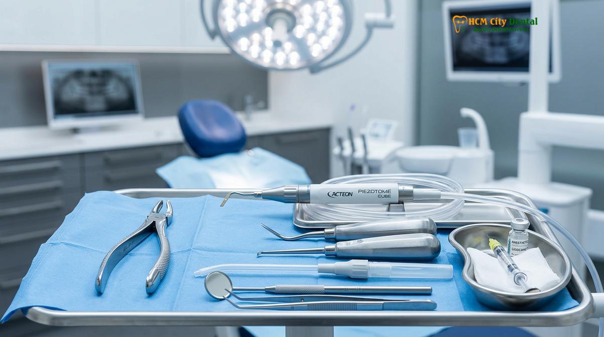
The complexity of the procedure dictates the surgical approach. A simple extraction is performed on a tooth that is fully erupted and visible within the oral cavity. The clinician uses specialized instruments called elevators to sever the periodontal ligament fibers and gently expand the alveolar bone socket. Once the tooth is sufficiently mobile, extraction forceps are used to deliver the tooth using controlled, continuous force. You can learn more about the specific simple vs surgical extraction differences explained by our clinical team.
Conversely, a surgical extraction is required for teeth that are impacted, severely decayed at the gum line, or possess complex, divergent roots. This approach involves raising a mucoperiosteal flap (incising and reflecting the gum tissue) to gain direct visual access to the underlying bone and tooth structure. The surgeon may need to perform an ostectomy (removal of a small amount of bone) to free the tooth. Frequently, the tooth is sectioned into smaller fragments using a high-speed surgical handpiece, allowing each root to be removed individually with minimal force, thereby preserving the surrounding alveolar ridge.
Clinical Case Study:
A 28-year-old patient presented to HCMC Dental Clinic in Ho Chi Minh City with severe localized pain radiating from the lower right jaw. A 3D CBCT scan revealed a horizontally impacted mandibular third molar whose roots were intimately wrapped around the Inferior Alveolar Nerve canal. Dr. Nguyen Van Cuong designed a precise surgical approach utilizing a Piezotome device. By carefully sectioning the crown and elevating the roots individually, the tooth was successfully removed without any neurological complications, demonstrating the efficacy of advanced surgical planning.
Minimally Invasive Techniques: The Role of Piezosurgery
Modern oral surgery increasingly relies on minimally invasive extraction techniques. One of the most significant advancements is the use of Piezosurgery (ultrasonic bone surgery). Unlike traditional rotary burs, a Piezotome uses micro-vibrations of ultrasonic frequency to cut bone precisely while leaving soft tissues—such as nerves, blood vessels, and the sinus membrane—completely unharmed [2]. This selective cutting capability significantly reduces surgical trauma, minimizes post-operative swelling, and accelerates the biological healing cascade.
Socket Debridement and Hemostasis
Following the removal of the tooth, the surgical site must be meticulously managed. The clinician uses a surgical curette to debride the socket, removing any infected granulation tissue, cysts, or debris. Thorough debridement is essential to establish a healthy environment for bone regeneration. The socket is then irrigated with sterile saline. To achieve hemostasis (control bleeding), the surgeon may place a hemostatic agent or a collagen sponge into the socket before suturing the gingival flap back into its anatomical position.
Sterilization & Infection Control Standards in Saigon
Reputable clinics in Saigon adhere to international infection control protocols, utilizing Class B autoclaves and strict cross-contamination barriers to guarantee patient safety during oral surgery.
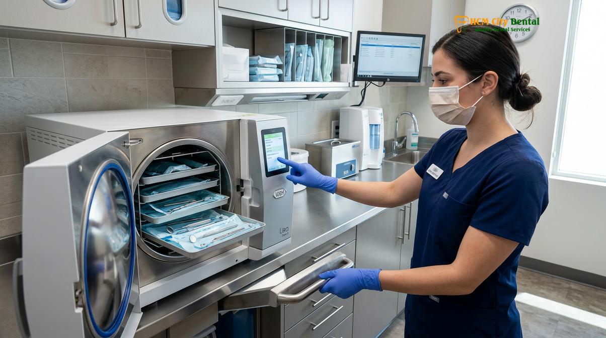
Infection control is the cornerstone of any surgical discipline. When undergoing an extraction, patients must be confident that the clinical environment operates under the highest standards of asepsis. A rigorous sterilization check saigon clinic protocol ensures that the risk of cross-contamination and post-operative infection is virtually eliminated [3].
The Sterilization Workflow
The journey of a surgical instrument from the operatory back to the sterile field involves multiple highly controlled steps. Immediately after use, instruments are transported to a dedicated sterilization center. They first undergo ultrasonic cleaning, where high-frequency sound waves create microscopic cavitation bubbles that implode, effectively scrubbing biological debris from even the most intricate hinges and serrations of extraction forceps.
Following ultrasonic cleaning, the instruments are thoroughly rinsed, dried, and sealed in specialized sterilization pouches. These pouches feature chemical indicators that change color only when specific parameters of heat, pressure, and time have been met. The packaged instruments are then placed into a Class B medical autoclave. Class B autoclaves utilize a fractionated pre-vacuum system, which completely removes air from the chamber before introducing pressurized steam. This ensures that the steam penetrates every surface of the instruments, including the internal lumens of surgical handpieces, achieving absolute sterility.
“Strict adherence to Class B sterilization protocols and the utilization of single-use disposable barriers are non-negotiable elements in modern oral surgery, ensuring that every patient is protected against the transmission of bloodborne pathogens.”
Operatory Asepsis and Cross-Contamination Barriers
Beyond instrument sterilization, the surgical operatory itself must be meticulously prepared. Between every patient, all clinical contact surfaces—including the dental chair, light handles, delivery units, and countertops—are disinfected using hospital-grade, broad-spectrum virucidal and bactericidal agents. Furthermore, high-touch areas are covered with single-use disposable plastic barriers that are replaced for each procedure. The surgical team strictly adheres to personal protective equipment (PPE) protocols, utilizing sterile surgical gloves, masks, and protective eyewear to maintain a sterile field throughout the extraction process.
Pain Management: Local Anesthesia & Sedation Options
Modern extractions are virtually painless through the precise administration of profound local anesthesia, supplemented by conscious sedation options for patients experiencing dental anxiety.
The anticipation of pain is the primary source of anxiety for patients requiring oral surgery. However, advancements in pharmacological pain management have transformed tooth extraction into a highly tolerable, comfortable procedure. The strategic use of local anesthetics and sedation ensures that patients experience no sharp pain during the surgery [4].
Profound Local Anesthesia
The foundation of intraoperative pain control is local anesthesia. Dental surgeons utilize highly effective anesthetic agents, most commonly Articaine or Lidocaine, formulated with a vasoconstrictor such as epinephrine. The vasoconstrictor serves two critical purposes: it prolongs the duration of the anesthetic effect by keeping the medication localized in the tissues, and it significantly reduces bleeding at the surgical site, providing the surgeon with a clear field of vision.
Before the injection, a topical anesthetic gel (usually Benzocaine) is applied to the oral mucosa to numb the surface tissue, minimizing the pinch of the needle. The clinician then employs specific injection techniques based on the tooth’s location. For maxillary teeth, local infiltration is often sufficient, as the porous nature of the upper jawbone allows the anesthetic to diffuse easily to the tooth roots. For mandibular teeth, the denser bone requires an Inferior Alveolar Nerve Block, which numbs the entire quadrant of the lower jaw, including the teeth, gums, and lower lip.
Clinical Precaution: Patients with a history of severe cardiovascular disease, uncontrolled hypertension, or specific arrhythmias must inform their dental surgeon prior to the procedure. The epinephrine contained in standard local anesthetics may be contraindicated, and the surgeon will select an alternative, epinephrine-free anesthetic formulation to ensure systemic safety.
Conscious Sedation for Dental Anxiety
While local anesthesia blocks pain, it does not alleviate the psychological stress or the sensation of pressure and vibration during the extraction. For patients with high dental anxiety, a strong gag reflex, or those undergoing complex surgical extractions, conscious sedation is highly beneficial. You can explore our comprehensive guide on painless tooth extraction sedation options Vietnam to understand the available modalities.
Options range from mild sedation using Nitrous Oxide (laughing gas), which induces a state of deep relaxation while allowing the patient to remain fully responsive, to oral conscious sedation (prescribed anxiolytic medications taken prior to the appointment). For extensive procedures, intravenous (IV) sedation may be administered by a qualified anesthesiologist, placing the patient in a state of profound tranquility where they have little to no memory of the surgery, yet can still breathe independently and respond to verbal cues.
Immediate Post-Operative Care Guidelines
Successful recovery relies on stabilizing the blood clot, managing localized inflammation with cold therapy, and maintaining strict oral hygiene without disrupting the surgical site.
The surgical removal of a tooth is only the first phase of the treatment; the subsequent biological healing process is equally critical. Proper post tooth extraction care is essential to prevent complications, minimize discomfort, and ensure the rapid regeneration of gingival and osseous tissues. The immediate post-operative period focuses heavily on hemostasis and the protection of the newly formed blood clot [5].
The Importance of the Blood Clot
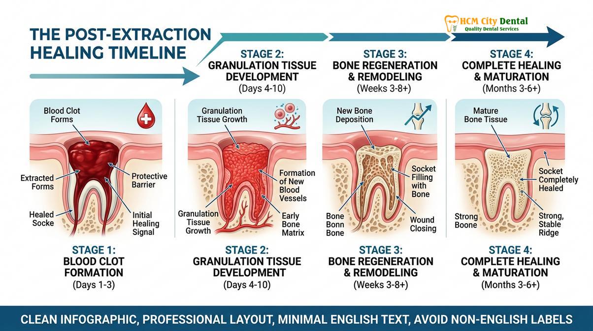
Immediately following the extraction, a blood clot forms within the empty socket. This clot acts as a biological bandage, protecting the underlying exposed bone and nerve endings from the oral environment. It also serves as the foundational matrix into which fibroblasts and osteoblasts will migrate to begin forming new connective tissue and bone. Dislodging this clot prematurely leads to a painful condition known as alveolar osteitis, or “dry socket.”
To protect the clot, patients must strictly avoid any actions that create negative pressure in the oral cavity. This includes avoiding the use of straws, vigorous spitting, or smoking for at least 72 hours post-surgery. Smoking not only risks dislodging the clot but also introduces toxins that severely impair local blood flow, drastically delaying the healing process.
Managing Inflammation and Discomfort
Post-operative swelling and mild to moderate discomfort are normal physiological responses to surgical trauma. Swelling typically peaks at 48 to 72 hours before gradually subsiding. To manage inflammation, patients are instructed to apply an ice pack to the outside of the face adjacent to the surgical site for 15-minute intervals during the first 24 hours. Cold therapy induces vasoconstriction, reducing fluid accumulation in the tissues.
Pain management is typically achieved through a prescribed regimen of non-steroidal anti-inflammatory drugs (NSAIDs), such as Ibuprofen, which address both pain and inflammation. In cases of complex surgical extractions, the surgeon may prescribe a short course of stronger analgesics or prophylactic antibiotics to prevent infection.
“The first 24 hours post-extraction are critical. Patients must prioritize rest, maintain firm biting pressure on the sterile gauze to ensure hemostasis, and strictly adhere to a soft diet to prevent mechanical trauma to the surgical site.”
Dietary Modifications and Oral Hygiene
Nutrition plays a vital role in tissue repair, but the physical consistency of the diet must be modified. For the first few days, patients should consume a soft, cool, or lukewarm diet—such as yogurt, applesauce, scrambled eggs, and smoothies. Hot liquids and spicy foods should be avoided, as they can dissolve the blood clot and irritate the healing tissues. As healing progresses, patients can gradually reintroduce solid foods, chewing on the side opposite the extraction site.
Maintaining oral hygiene is crucial to prevent infection, but it must be done cautiously. Patients should continue to brush and floss their remaining teeth normally but must avoid brushing directly over the extraction site. After 24 hours, gentle rinsing with warm salt water (half a teaspoon of salt in a cup of warm water) is recommended after meals to keep the socket clean and promote healing.
| Healing Phase | Biological Process | Patient Action Required |
|---|---|---|
| 0 – 24 Hours | Hemostasis and initial blood clot formation. | Bite on gauze, apply ice packs, rest, avoid spitting/straws. |
| 24 – 72 Hours | Peak inflammatory response (swelling). Clot stabilization. | Begin gentle warm salt water rinses, maintain soft diet, take prescribed NSAIDs. |
| 3 – 7 Days | Granulation tissue begins replacing the clot. Swelling subsides. | Gradually return to normal diet, continue gentle hygiene around the site. |
| 1 – 4 Weeks | Gingival tissue closes over the socket. Early osteoid formation. | Resume normal brushing, attend follow-up appointment for evaluation. |
For international patients planning their treatment journey, understanding the tooth extraction recovery travel timeline is essential for scheduling flights and activities. Furthermore, specific flying after tooth extraction safety rules must be followed to avoid complications related to cabin pressure changes.
When to See a Doctor: Clinical Red Flags
While the vast majority of tooth extractions heal uneventfully, patients must be vigilant in monitoring their recovery. Differentiating between normal post-operative sequelae and signs of clinical complications is vital for timely intervention. Dr. Nguyen Van Cuong advises all patients to contact the clinic immediately if they experience any of the following red flags.
Prolonged or heavy bleeding that does not subside after applying firm pressure with a gauze pad for 45 minutes is abnormal and requires clinical attention. While mild oozing that tints the saliva pink is expected, active, bright red bleeding is not. Severe, radiating pain that begins or worsens 3 to 5 days after the extraction, and is unresponsive to prescribed analgesics, is a classic symptom of alveolar osteitis (dry socket) and requires the placement of a medicated dressing by the dentist.
Furthermore, signs of systemic infection—such as a fever, chills, or the presence of purulent exudate (pus) draining from the surgical site—demand immediate evaluation and likely antibiotic therapy. Finally, if a patient experiences prolonged numbness or a tingling sensation (paresthesia) in their lip, chin, or tongue that persists well beyond the expected duration of the local anesthetic, they must be evaluated to assess potential nerve irritation. For patients undergoing extensive procedures, adhering to our multiple extractions one visit safety protocol minimizes these risks significantly.
Frequently Asked Questions
Is tooth extraction in Vietnam painful?
Tooth extraction in Vietnam is virtually painless due to the administration of advanced local anesthetics and modern sedation techniques. Before the procedure begins, the dental surgeon applies a topical numbing gel followed by a precise local anesthetic injection to completely block nerve pain signals. Patients may feel pressure or movement during the extraction, but sharp pain is eliminated. For those with severe dental anxiety, conscious sedation options are available to ensure a completely relaxed and comfortable experience.
How do I verify the qualifications of my dental surgeon?
You can verify a dental surgeon’s qualifications by checking their medical license, specialized maxillofacial training credentials, and affiliations with recognized dental boards. Reputable clinics will transparently display their specialists’ educational backgrounds, including degrees from top medical universities and international certifications in oral surgery or implantology. Additionally, reading patient reviews and requesting a consultation to discuss their clinical experience with complex extractions can provide further assurance of their expertise.
How long should I stay in Saigon after a tooth extraction?
Patients should plan to stay in Saigon for at least 3 to 5 days following a routine or surgical tooth extraction. This window allows the initial blood clot to stabilize and gives your dental surgeon the opportunity to conduct a post-operative follow-up examination to ensure proper healing. For complex surgical extractions involving bone removal or multiple impactions, extending your stay to 7 days is highly recommended to monitor for any signs of infection or delayed recovery before flying.
What is the difference between a simple and surgical extraction?
A simple extraction removes a visible, accessible tooth using forceps, while a surgical extraction involves incising the gum and sometimes removing bone to extract an impacted or broken tooth. Simple extractions are typically performed on teeth that have fully erupted and have straight roots. Surgical extractions are necessary for wisdom teeth, teeth broken at the gum line, or teeth with complex root anatomies, requiring specialized instruments and techniques to safely section and remove the tooth fragments.
Can I get a dental implant immediately after an extraction?
Yes, immediate dental implant placement is often possible if there is sufficient healthy jawbone and an absence of active acute infection at the extraction site. This technique, known as immediate loading or immediate placement, preserves the natural gum contours and reduces overall treatment time. However, candidacy must be determined through a comprehensive 3D CBCT scan. If severe bone loss or infection is present, the surgeon will recommend bone grafting and a healing period of several months before implant placement.
References
- Journal of Oral and Maxillofacial Surgery. Biomechanical principles of atraumatic tooth extraction and alveolar bone preservation. (2021).
- International Journal of Oral and Maxillofacial Surgery. Efficacy of ultrasonic piezosurgery in impacted third molar extractions. (2020).
- British Dental Journal. Cross-infection control and sterilization protocols in modern outpatient dental surgery. (2019).
- Journal of the American Dental Association. Pharmacological management of post-extraction dental pain and inflammation. (2022).
- Clinical Oral Investigations. Incidence and management of alveolar osteitis following surgical extractions. (2018).